

Abstract
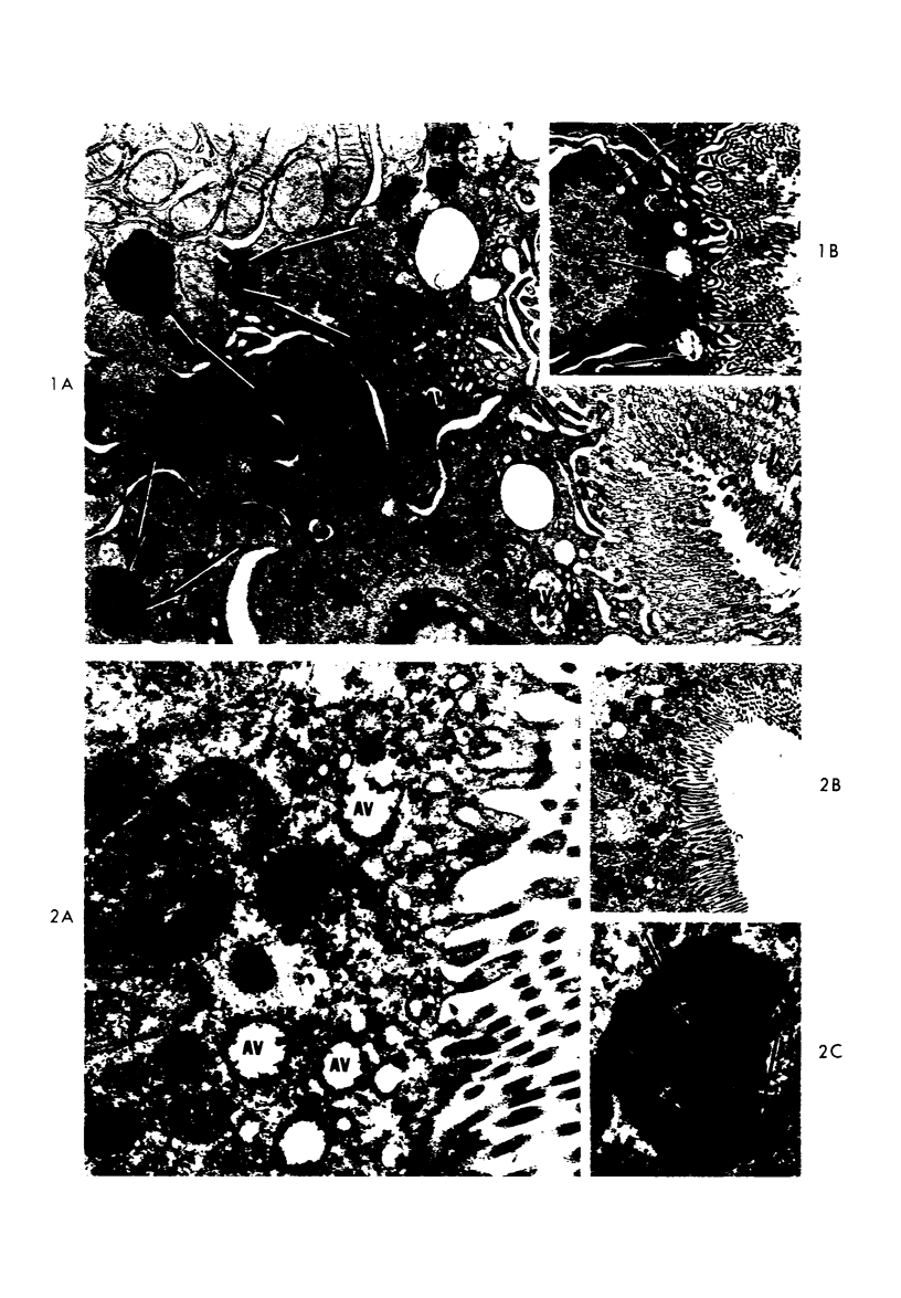
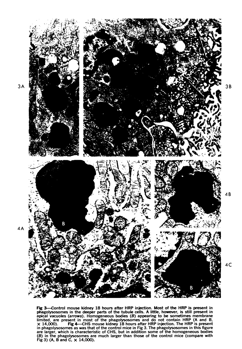
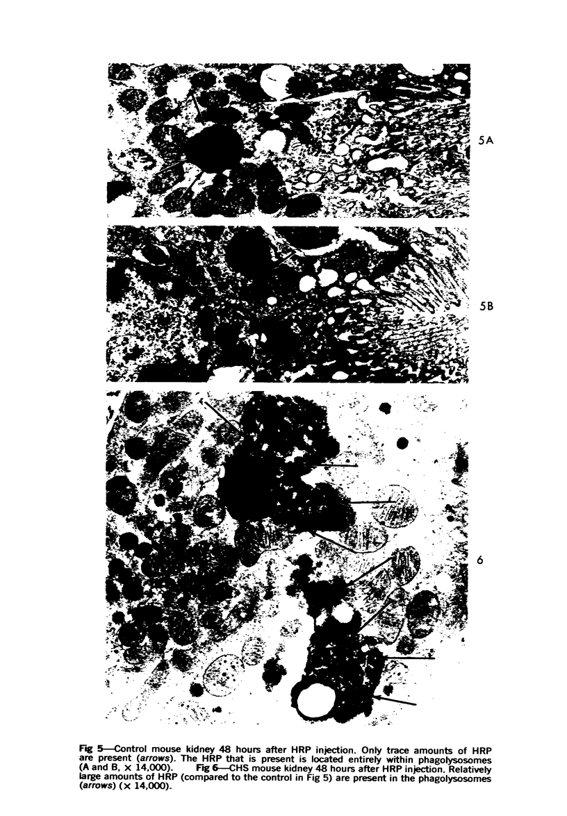
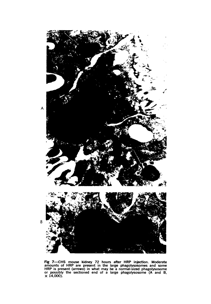
Morphologically abnormal lysosomes demonstrated in individuals with the Chediak-Higashi syndrome (CHS) suggested a defect in the function of these abnormal lysosomes. To gain direct experimental evidence of such a defect, horseradish peroxidase (HRP) was injected intravenously into CHS and control mice, the mice killed at varying intervals and the kidneys studied by ultrastructural cytochemistry. No morophologic difference was observed in the absorption and uptake of HRP by proximal convoluted tubules in the two groups of mice. In CHS mice, however, some of the HRP fused with enlarged lysosomes. By 48 hours after injection, the lysosomes of normal mice had digested all but trace amounts of HRP, whereas large amounts were still present in CHS mice at this time. In CHS mice, moderate amounts were still present at 72 hours and trace amounts 96 hours post injection. This slowed rate of digestion of HRP by lysosomes of the proximal convoluted tubule cells of CHS mice suggests a similar defect in all cells in CHS individuals in which there is a lysosomal degradation of protein or other matter obtained by endocytosis. Such a defect may explain some manifestations of impaired host defense observed in CHS.
Full text
PDF
Images in this article
Selected References
These references are in PubMed. This may not be the complete list of references from this article.
- Danes B. S., Bearn A. G. Correction of celluar metachromasia in cultured fibroblasts in several inherited mucopolysaccharidoses. Proc Natl Acad Sci U S A. 1970 Sep;67(1):357–364. doi: 10.1073/pnas.67.1.357. [DOI] [PMC free article] [PubMed] [Google Scholar]
- Davis W. C., Spicer S. S., Greene W. B., Padgett G. A. Ultrastructure of bone marrow granulocytes in normal mink and mink with the homolog of the Chediak-Higashi trait of humans. I. Origin of the abnormal granules present in the neutrophils of mink with the C-HS trait. Lab Invest. 1971 Apr;24(4):303–317. [PubMed] [Google Scholar]
- Davis W. C., Spicer S. S., Greene W. B., Padgett G. A. Ultrastructure of cells in bone marrow and peripheral blood of normal mink and mink with the homologue of the Chediak-Higashi trait of humans. II. Cytoplasmic granules in eosinophils, basophils, mononuclear cells and platelets. Am J Pathol. 1971 Jun;63(3):411–432. [PMC free article] [PubMed] [Google Scholar]
- Ehrenreich B. A., Cohn Z. A. The fate of peptides pinocytosed by macrophages in vitro. J Exp Med. 1969 Jan 1;129(1):227–245. doi: 10.1084/jem.129.1.227. [DOI] [PMC free article] [PubMed] [Google Scholar]
- Graham R. C., Jr, Karnovsky M. J. Glomerular permeability. Ultrastructural cytochemical studies using peroxidases as protein tracers. J Exp Med. 1966 Dec 1;124(6):1123–1134. doi: 10.1084/jem.124.6.1123. [DOI] [PMC free article] [PubMed] [Google Scholar]
- Graham R. C., Jr, Karnovsky M. J. The early stages of absorption of injected horseradish peroxidase in the proximal tubules of mouse kidney: ultrastructural cytochemistry by a new technique. J Histochem Cytochem. 1966 Apr;14(4):291–302. doi: 10.1177/14.4.291. [DOI] [PubMed] [Google Scholar]
- Henson J. B., Gorham J. R., Padgett G. A., Davis W. C. Pathogenesis of the glomerular lesions in aleutian disease of mink. Immunofluorescent studies. Arch Pathol. 1969 Jan;87(1):21–28. [PubMed] [Google Scholar]
- Kvam D. C. Adrenergic receptors for metabolic effects in muscle. Fed Proc. 1970 Jul-Aug;29(4):1379–1380. [PubMed] [Google Scholar]
- LEADER R. W., PADGETT G. A., GORHAM J. R. STUDIES OF ABNORMAL LEUKOCYTE BODIES IN THE MINK. Blood. 1963 Oct;22:477–484. [PubMed] [Google Scholar]
- Lodmell D. L., Bergman R. K., Hadlow W. J., Munoz J. J. Cellular and humoral antibody responses of normal pastel and sapphire mink to goat erythrocytes. Infect Immun. 1971 Feb;3(2):221–227. doi: 10.1128/iai.3.2.221-227.1971. [DOI] [PMC free article] [PubMed] [Google Scholar]
- Lodmell D. L., Hadlow W. J., Munoz J. J., Whitford H. W. Hemagglutinin antibody response of normal and Aleutian disease-affected mink to keyhole limpet hemocyanin. J Immunol. 1970 Apr;104(4):878–887. [PubMed] [Google Scholar]
- Lutzner M. A., Lowrie C. T., Jordan H. W. Giant granules in leukocytes of the beige mouse. J Hered. 1967 Nov-Dec;58(6):299–300. doi: 10.1093/oxfordjournals.jhered.a107620. [DOI] [PubMed] [Google Scholar]
- MOLLENHAUER H. H. PLASTIC EMBEDDING MIXTURES FOR USE IN ELECTRON MICROSCOPY. Stain Technol. 1964 Mar;39:111–114. [PubMed] [Google Scholar]
- PADGETT G. A., LEADER R. W., GORHAM J. R., O'MARY C. C. THE FAMILIAL OCCURRENCE OF THE CHEDIAK-HIGASHI SYNDROME IN MINK AND CATTLE. Genetics. 1964 Mar;49:505–512. doi: 10.1093/genetics/49.3.505. [DOI] [PMC free article] [PubMed] [Google Scholar]
- Padgett G. A. Neutrophilic function in animals with the Chediak-Higashi syndrome. Blood. 1967 Jun;29(6):906–915. [PubMed] [Google Scholar]
- Padgett G. A., Reiquam C. W., Henson J. B., Gorham J. R. Comparative studies of susceptibility to infection in the Chediak-Higashi syndrome. J Pathol Bacteriol. 1968 Apr;95(2):509–522. doi: 10.1002/path.1700950224. [DOI] [PubMed] [Google Scholar]
- STRAUS W. CYTOCHEMICAL OBSERVATIONS ON THE RELATIONSHIP BETWEEN LYSOSOMES AND PHAGOSOMES IN KIDNEY AND LIVER BY COMBINED STAINING FOR ACID PHOSPHATASE AND INTRAVENOUSLY INJECTED HORSERADISH PEROXIDASE. J Cell Biol. 1964 Mar;20:497–507. doi: 10.1083/jcb.20.3.497. [DOI] [PMC free article] [PubMed] [Google Scholar]
- STRAUS W. OCCURRENCE OF PHAGOSOMES AND PHAGO-LYSOSOMES IN DIFFERENT SEGMENTS OF THE NEPHRON IN RELATION TO THE REABSORPTION, TRANSPORT, DIGESTION, AND EXTRUSION OF INTRAVENOUSLY INJECTED HORSERADISH PEROXIDASE. J Cell Biol. 1964 Jun;21:295–308. doi: 10.1083/jcb.21.3.295. [DOI] [PMC free article] [PubMed] [Google Scholar]
- Shortman K., Diener E., Russell P., Armstrong W. D. The role of nonlymphoid accessory cells in the immune response to different antigens. J Exp Med. 1970 Mar 1;131(3):461–482. doi: 10.1084/jem.131.3.461. [DOI] [PMC free article] [PubMed] [Google Scholar]
- Straus W. Changes in intracellular location of small phagosomes (micropinocytic vesicles) in kidney and liver cells in relation to time after injection and dose of horseradish peroxidase. J Histochem Cytochem. 1967 Jul;15(7):381–393. doi: 10.1177/15.7.381. [DOI] [PubMed] [Google Scholar]
- White J. G. The Chediak-Higashi syndrome: a possible lysosomal disease. Blood. 1966 Aug;28(2):143–156. [PubMed] [Google Scholar]
- Windhorst D. B., Zelickson A. S., Good R. A. Chediak-Higashi syndrome: hereditary gigantism of cytoplasmic organelles. Science. 1966 Jan 7;151(3706):81–83. doi: 10.1126/science.151.3706.81. [DOI] [PubMed] [Google Scholar]