

Abstract
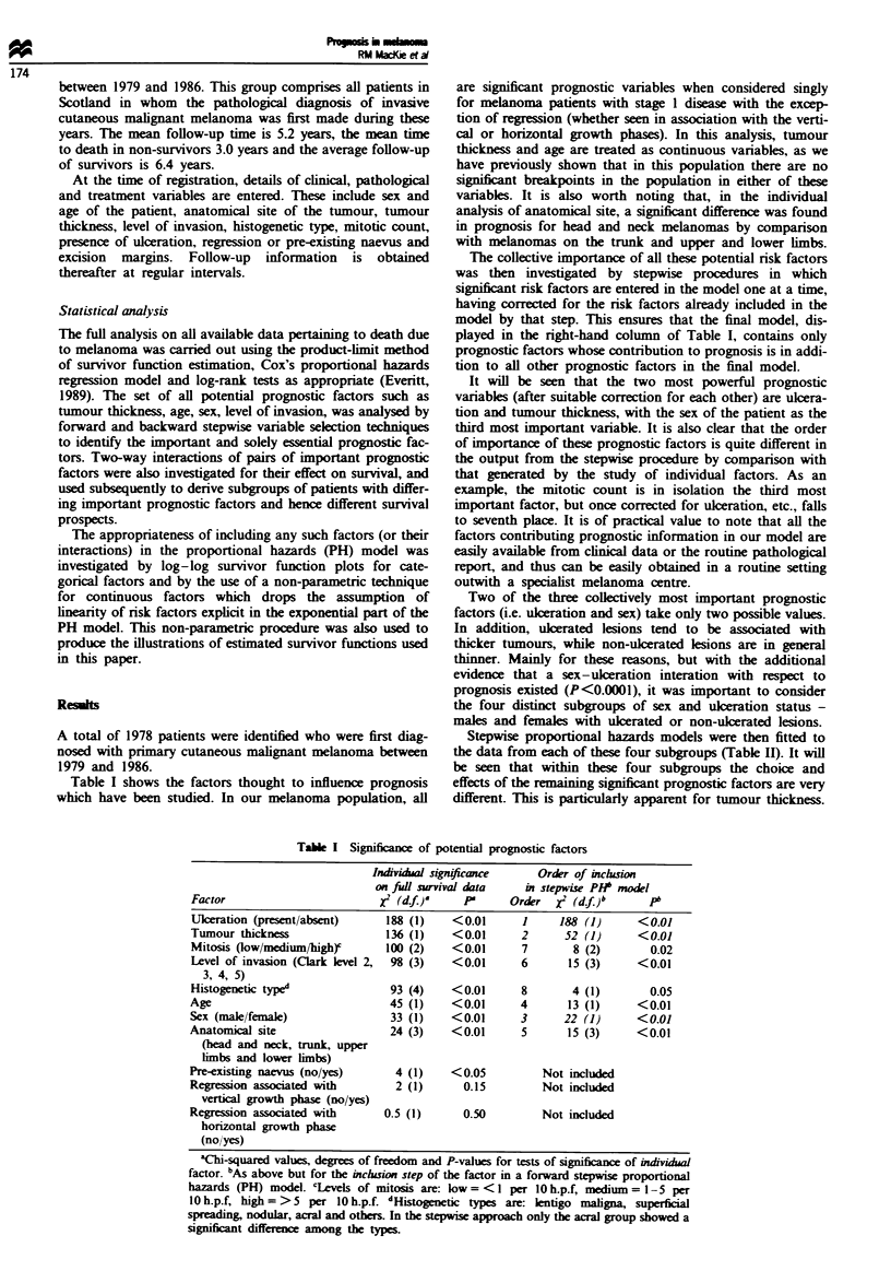
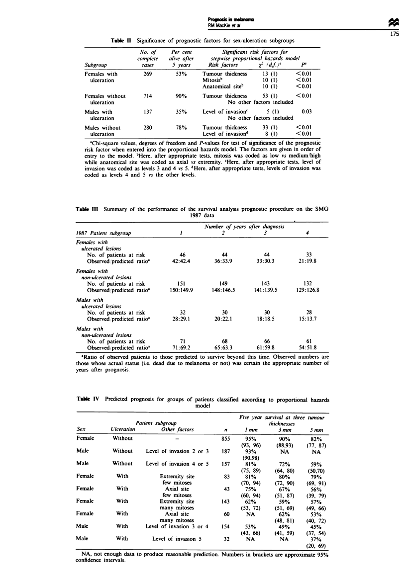
For the past 20 years thickness of the primary tumour has been accepted as the most important guide to prognosis for patients with primary cutaneous malignant melanoma. The changing epidemiology of melanoma with an increasing number of patients with thin tumours has necessitated a reappraisal of this, with particular reference to interactions among tumour thickness, the patients' sex and the presence or absence of ulceration of the primary tumour. All primary cutaneous malignant melanomas diagnosed in Scotland between 1979 and 1986 were used as the test group (1978 patients). The proportional hazards model was used on all potential risk factors in the database and their two-way interactions, and the resulting models based on stepwise procedures were subsequently validated on 289 melanoma patients first diagnosed in 1987 in the same geographic area. Four distinct subgroups of males and females with ulcerated or non-ulcerated lesions were identified. For females with ulcerated lesions, tumour thickness, mitotic count and anatomical site of primary all gave valuable prognostic information, whereas for females with non-ulcerated lesions only tumour thickness was of prognostic value. For males with ulcerated lesions, level of invasion was the only prognostic guide, while for males with non-ulcerated lesions both tumour thickness and level of invasion contributed significantly to prediction of prognosis. Prognosis markedly different across subgroups of the melanoma population, even to the extent that essential prognostic factors are not the same in the distinct subgroups. Verification of these prognostic guides derived from 1979-86 patients has been achieved for all patients diagnosed with melanoma in 1987 from the same geographic area. These data will therefore be useful aids for clinicians managing patients.
Full text
PDF
Selected References
These references are in PubMed. This may not be the complete list of references from this article.
- Breslow A. Thickness, cross-sectional areas and depth of invasion in the prognosis of cutaneous melanoma. Ann Surg. 1970 Nov;172(5):902–908. doi: 10.1097/00000658-197011000-00017. [DOI] [PMC free article] [PubMed] [Google Scholar]
- Breslow A. Tumor thickness, level of invasion and node dissection in stage I cutaneous melanoma. Ann Surg. 1975 Nov;182(5):572–575. doi: 10.1097/00000658-197511000-00007. [DOI] [PMC free article] [PubMed] [Google Scholar]
- Clark W. H., Jr, Elder D. E., Guerry D., 4th, Braitman L. E., Trock B. J., Schultz D., Synnestvedt M., Halpern A. C. Model predicting survival in stage I melanoma based on tumor progression. J Natl Cancer Inst. 1989 Dec 20;81(24):1893–1904. doi: 10.1093/jnci/81.24.1893. [DOI] [PubMed] [Google Scholar]
- MacKie R., Hunter J. A., Aitchison T. C., Hole D., Mclaren K., Rankin R., Blessing K., Evans A. T., Hutcheon A. W., Jones D. H. Cutaneous malignant melanoma, Scotland, 1979-89. The Scottish Melanoma Group. Lancet. 1992 Apr 18;339(8799):971–975. doi: 10.1016/0140-6736(92)91539-k. [DOI] [PubMed] [Google Scholar]
- MacLennan R., Green A. C., McLeod G. R., Martin N. G. Increasing incidence of cutaneous melanoma in Queensland, Australia. J Natl Cancer Inst. 1992 Sep 16;84(18):1427–1432. doi: 10.1093/jnci/84.18.1427. [DOI] [PubMed] [Google Scholar]
- Soong S. J., Shaw H. M., Balch C. M., McCarthy W. H., Urist M. M., Lee J. Y. Predicting survival and recurrence in localized melanoma: a multivariate approach. World J Surg. 1992 Mar-Apr;16(2):191–195. doi: 10.1007/BF02071520. [DOI] [PubMed] [Google Scholar]
- Vollmer R. T. Malignant melanoma. A multivariate analysis of prognostic factors. Pathol Annu. 1989;24(Pt 1):383–407. [PubMed] [Google Scholar]