

Abstract
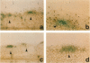
To investigate the site-specific implantation of cancer cells in peritoneal dissemination, we inoculated CDF1 mice intraperitoneally with mouse P388 leukaemia cells labelled with bromodeoxyuridine (BrdU) and then observed immunohistochemically the distribution of the cells in the greater omentum taken from the mice using an anti-BrdU antibody. We found the BrdU-labelled cells infiltrating selectively into the milky spots in the omentum. Furthermore, we intraperitoneally inoculated the BrdU-labelled P388 cells at 10(5), 10(6) and 10(7) cells per mouse into three groups of ten CDF1 mice and then quantified the distribution of the BrdU-labelled cells by counting the number of the labelled cells per unit area at each milky spot and non-milky spot site in the omentum. Inoculations of 10(5), 10(6) and 10(7) BrdU-labelled P388 cells per mouse resulted in 15.8 +/- 13.3, 120 +/- 46.5 and 504 +/- 208 cells mm-2 respectively in the milky spot sites and 9.14 x 10(-3) +/- 1.58 x 10(-2), 1.14 x 10(-1) +/- 7.82 x 10(-2) and 7.07 x 10(-1) +/- 5.98 x 10(-1) cells mm-2 respectively in the non-milky spot sites. The ratios of the mean labelled cell numbers in the milky spot sites vs those in the non-milky spot sites were 1728:1, 1049:1 and 713:1 respectively. In all cases, there were statistically significant differences in the number of BrdU-labelled cells mm-2 between milky spot sites and non-milky spot sites. However, the ratios decreased as the numbers of inoculated cells increased. In addition, we inoculated C57/BL mice intraperitoneally with B-16 PC melanoma cells, which were easily differentiated from the other cells by the intrinsic black melanin, and examined the distribution of the cells macro- and microscopically. The B-16 PC melanoma cells were also found to be infiltrating preferentially into the milky spots in the omentum. These results suggest that cancer cells seeded intraperitoneally specifically infiltrate the milky spots in the early stages of peritoneal dissemination.
Full text
PDF


Images in this article

Selected References
These references are in PubMed. This may not be the complete list of references from this article.
- Beelen R. H., Fluitsma D. M., Hoefsmit E. C. The cellular composition of omentum milky spots and the ultrastructure of milky spot macrophages and reticulum cells. J Reticuloendothel Soc. 1980 Dec;28(6):585–599. [PubMed] [Google Scholar]
- Dux K. Role of the greater omentum in the immunological response of mice and rats to the intraperitoneal inoculation of Ehrlich ascites tumor. Arch Immunol Ther Exp (Warsz) 1969;17(4):425–432. [PubMed] [Google Scholar]
- Dux K., Shimotsuma M., Simpson-Morgan M. W. Technique for in situ excision of distended samples of greater omentum from small laboratory animals. Biotech Histochem. 1993 Jan;68(1):46–49. doi: 10.3109/10520299309105577. [DOI] [PubMed] [Google Scholar]
- Gratzner H. G. Monoclonal antibody to 5-bromo- and 5-iododeoxyuridine: A new reagent for detection of DNA replication. Science. 1982 Oct 29;218(4571):474–475. doi: 10.1126/science.7123245. [DOI] [PubMed] [Google Scholar]
- Green J. A., Williams A. E. The relationship between inflammatory responses and WBP1 tumour cell attachment to the rat omentum. Eur J Cancer. 1978 Oct;14(10):1153–1155. doi: 10.1016/0014-2964(78)90072-5. [DOI] [PubMed] [Google Scholar]
- Hagiwara A., Takahashi T., Sawai K., Taniguchi H., Shimotsuma M., Okano S., Sakakura C., Tsujimoto H., Osaki K., Sasaki S. Milky spots as the implantation site for malignant cells in peritoneal dissemination in mice. Cancer Res. 1993 Feb 1;53(3):687–692. [PubMed] [Google Scholar]
- Shimotsuma M., Takahashi T., Kawata M., Dux K. Cellular subsets of the milky spots in the human greater omentum. Cell Tissue Res. 1991 Jun;264(3):599–601. doi: 10.1007/BF00319049. [DOI] [PubMed] [Google Scholar]
- Tsuruo T., Naganuma K., Iida H., Tsukagoshi S. Lymph node metastasis and effects of 1-beta-D-arabinofuranosylcytosine, 5-fluorouracil, and their lipophilic derivatives in an experimental model system using P388 leukemia. Cancer Res. 1980 Dec;40(12):4758–4763. [PubMed] [Google Scholar]