

Abstract
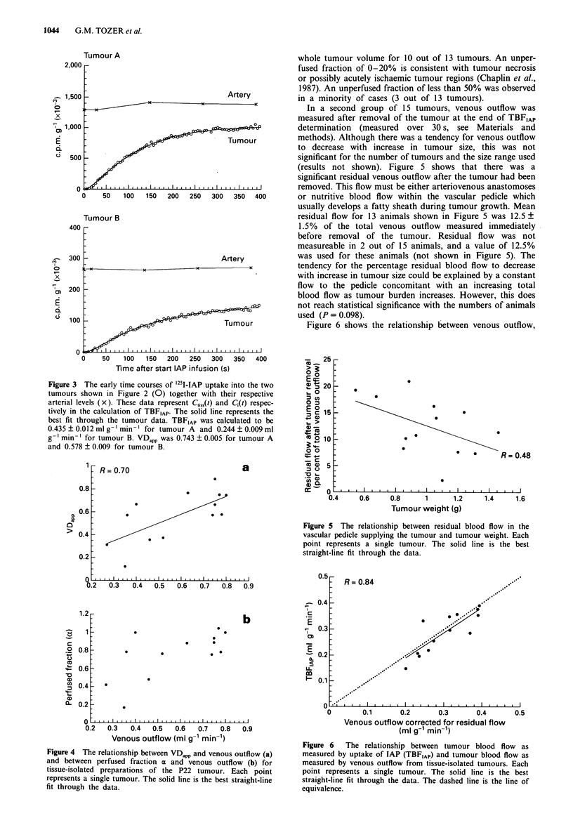
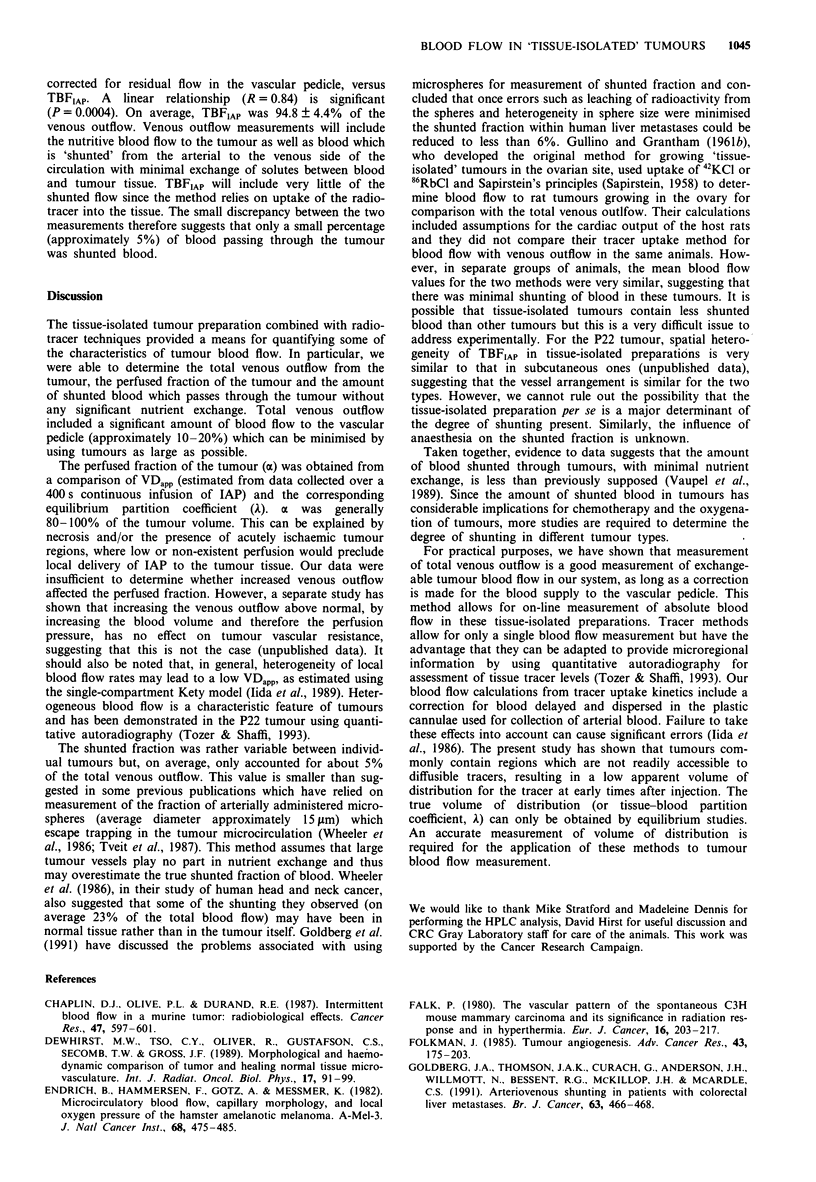
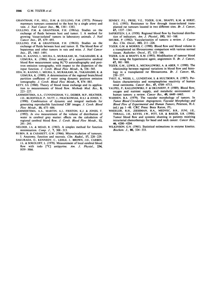
Tumour blood flow was characterised in a 'tissue-isolated' rat tumour model, in which the vascular supply is derived from a single artery and vein. Tumours were perfused in situ and blood flow was calculated from simultaneous measurement of (1) venous outflow from the tumour and (2) uptake into the tumour of radiolabelled iodo-antipyrine (IAP). Comparison of results from the two measurements enabled assessment of the amount of blood 'shunted' through the tumours with minimal exchange between blood and tissue. Kinetics of IAP uptake were also used to determine the apparent volume of distribution (VDapp) for the tracer and the equilibrium tissue-blood partition coefficient (lambda). lambda was also measured by in vitro techniques and checks were made for binding and metabolism of IAP using high-pressure liquid chromatography. VDapp and lambda were used to calculate the perfused fraction (alpha) of the tumours. Tumour blood flow, as measured by IAP (TBFIAP), was 94.8 +/- 4.4% of the blood flow as measured by venous outflow, indicating only a small amount of non-exchanging flow. This level of shunting is lower than some previous estimates in which the percentage tumour entrapment of microspheres was used. The unperfused fraction ranged from 0 to 20% of the tumour volume in the majority of tumours. This could be due to tumour necrosis and/or acutely ischaemic tumour regions. For practical purposes, measurement of the total venous outflow of tumours is a reasonable measure of exchangeable tumour blood flow in this system and allows for on-line measurements. Tracer methods can be used to obtain additional information on the distribution of blood flow within tumours.
Full text
PDF
Selected References
These references are in PubMed. This may not be the complete list of references from this article.
- Chaplin D. J., Olive P. L., Durand R. E. Intermittent blood flow in a murine tumor: radiobiological effects. Cancer Res. 1987 Jan 15;47(2):597–601. [PubMed] [Google Scholar]
- Dewhirst M. W., Tso C. Y., Oliver R., Gustafson C. S., Secomb T. W., Gross J. F. Morphologic and hemodynamic comparison of tumor and healing normal tissue microvasculature. Int J Radiat Oncol Biol Phys. 1989 Jul;17(1):91–99. doi: 10.1016/0360-3016(89)90375-1. [DOI] [PubMed] [Google Scholar]
- Endrich B., Hammersen F., Götz A., Messmer K. Microcirculatory blood flow, capillary morphology and local oxygen pressure of the hamster amelanotic melanoma A-Mel-3. J Natl Cancer Inst. 1982 Mar;68(3):475–485. [PubMed] [Google Scholar]
- Falk P. The vascular pattern of the spontaneous C3H mouse mammary carcinoma and its significance in radiation response and in hyperthermia. Eur J Cancer. 1980 Feb;16(2):203–217. doi: 10.1016/0014-2964(80)90152-8. [DOI] [PubMed] [Google Scholar]
- Folkman J. Tumor angiogenesis. Adv Cancer Res. 1985;43:175–203. doi: 10.1016/s0065-230x(08)60946-x. [DOI] [PubMed] [Google Scholar]
- GULLINO P. M., GRANTHAM F. H. Studies on the exchange of fluids between host and tumor. I. A method for growing "tissue-isolated" tumors in laboratory animals. J Natl Cancer Inst. 1961 Sep;27:679–693. [PubMed] [Google Scholar]
- GULLINO P. M., GRANTHAM F. H. Studies on the exchange of fluids between host and tumor. II. The blood flow of hepatomas and other tumors in rats and mice. J Natl Cancer Inst. 1961 Dec;27:1465–1491. [PubMed] [Google Scholar]
- Goldberg J. A., Thomson J. A., McCurrach G., Anderson J. H., Willmott N., Bessent R. G., McKillop J. H., McArdle C. S. Arteriovenous shunting in patients with colorectal liver metastases. Br J Cancer. 1991 Mar;63(3):466–468. doi: 10.1038/bjc.1991.109. [DOI] [PMC free article] [PubMed] [Google Scholar]
- Grantham F. H., Hill D. M., Gullino P. M. Primary mammary tumors connected to the host by a single artery and vein. J Natl Cancer Inst. 1973 May;50(5):1381–1383. doi: 10.1093/jnci/50.5.1381. [DOI] [PubMed] [Google Scholar]
- Iida H., Kanno I., Miura S., Murakami M., Takahashi K., Uemura K. A determination of the regional brain/blood partition coefficient of water using dynamic positron emission tomography. J Cereb Blood Flow Metab. 1989 Dec;9(6):874–885. doi: 10.1038/jcbfm.1989.121. [DOI] [PubMed] [Google Scholar]
- Iida H., Kanno I., Miura S., Murakami M., Takahashi K., Uemura K. Error analysis of a quantitative cerebral blood flow measurement using H2(15)O autoradiography and positron emission tomography, with respect to the dispersion of the input function. J Cereb Blood Flow Metab. 1986 Oct;6(5):536–545. doi: 10.1038/jcbfm.1986.99. [DOI] [PubMed] [Google Scholar]
- Lammertsma A. A., Cunningham V. J., Deiber M. P., Heather J. D., Bloomfield P. M., Nutt J., Frackowiak R. S., Jones T. Combination of dynamic and integral methods for generating reproducible functional CBF images. J Cereb Blood Flow Metab. 1990 Sep;10(5):675–686. doi: 10.1038/jcbfm.1990.121. [DOI] [PubMed] [Google Scholar]
- Lammertsma A. A., Martin A. J., Friston K. J., Jones T. In vivo measurement of the volume of distribution of water in cerebral grey matter: effects on the calculation of regional cerebral blood flow. J Cereb Blood Flow Metab. 1992 Mar;12(2):291–295. doi: 10.1038/jcbfm.1992.39. [DOI] [PubMed] [Google Scholar]
- Rubin P., Casarett G. Microcirculation of tumors. I. Anatomy, function, and necrosis. Clin Radiol. 1966 Jul;17(3):220–229. doi: 10.1016/s0009-9260(66)80027-2. [DOI] [PubMed] [Google Scholar]
- SAPIRSTEIN L. A. Regional blood flow by fractional distribution of indicators. Am J Physiol. 1958 Apr;193(1):161–168. doi: 10.1152/ajplegacy.1958.193.1.161. [DOI] [PubMed] [Google Scholar]
- Sakurada O., Kennedy C., Jehle J., Brown J. D., Carbin G. L., Sokoloff L. Measurement of local cerebral blood flow with iodo [14C] antipyrine. Am J Physiol. 1978 Jan;234(1):H59–H66. doi: 10.1152/ajpheart.1978.234.1.H59. [DOI] [PubMed] [Google Scholar]
- Sensky P. L., Prise V. E., Tozer G. M., Shaffi K. M., Hirst D. G. Resistance to flow through tissue-isolated transplanted rat tumours located in two different sites. Br J Cancer. 1993 Jun;67(6):1337–1341. doi: 10.1038/bjc.1993.247. [DOI] [PMC free article] [PubMed] [Google Scholar]
- Shubik P. Vascularization of tumors: a review. J Cancer Res Clin Oncol. 1982;103(3):211–226. doi: 10.1007/BF00409698. [DOI] [PMC free article] [PubMed] [Google Scholar]
- Tozer G. M., Lewis S., Michalowski A., Aber V. The relationship between regional variations in blood flow and histology in a transplanted rat fibrosarcoma. Br J Cancer. 1990 Feb;61(2):250–257. doi: 10.1038/bjc.1990.46. [DOI] [PMC free article] [PubMed] [Google Scholar]
- Tozer G. M., Morris C. C. Blood flow and blood volume in a transplanted rat fibrosarcoma: comparison with various normal tissues. Radiother Oncol. 1990 Feb;17(2):153–165. doi: 10.1016/0167-8140(90)90103-4. [DOI] [PubMed] [Google Scholar]
- Tozer G. M., Shaffi K. M. Modification of tumour blood flow using the hypertensive agent, angiotensin II. Br J Cancer. 1993 May;67(5):981–988. doi: 10.1038/bjc.1993.180. [DOI] [PMC free article] [PubMed] [Google Scholar]
- Tveit E., Weiss L., Lundstam S., Hultborn R. Perfusion characteristics and norepinephrine reactivity of human renal carcinoma. Cancer Res. 1987 Sep 1;47(17):4709–4713. [PubMed] [Google Scholar]
- Vaupel P., Kallinowski F., Okunieff P. Blood flow, oxygen and nutrient supply, and metabolic microenvironment of human tumors: a review. Cancer Res. 1989 Dec 1;49(23):6449–6465. [PubMed] [Google Scholar]
- WILKINSON G. N. Statistical estimations in enzyme kinetics. Biochem J. 1961 Aug;80:324–332. doi: 10.1042/bj0800324. [DOI] [PMC free article] [PubMed] [Google Scholar]
- Wheeler R. H., Ziessman H. A., Medvec B. R., Juni J. E., Thrall J. H., Keyes J. W., Pitt S. R., Baker S. R. Tumor blood flow and systemic shunting in patients receiving intraarterial chemotherapy for head and neck cancer. Cancer Res. 1986 Aug;46(8):4200–4204. [PubMed] [Google Scholar]