Abstract
There are several independent sets of findings concerning the neural basis of reading. One set demonstrates a powerful relationship between phonological processing and reading skills. Another set reveals a relationship between visual responses in the motion pathways and reading skills. It is widely assumed that these two findings are unrelated. We tested the hypothesis that phonological awareness is related to motion responsivity in children’s MT+. We measured BOLD signals to drifting gratings as a function of contrast. Subjects were 35 children ages 7–12y with a wide range of reading skills. Contrast responsivity in MT+, but not V1, was correlated with phonological awareness and to a lesser extent with two other measures of reading. No correlation was found between MT+ signals and rapid naming, age or general IQ measures. These results establish an important link between visual and phonological processing in children and suggest that MT+ responsivity is a marker for healthy reading development.
Introduction
Phonological processing abilities are essential to reading development. The relation between phonology and reading acquisition has been studied extensively using behavioral methods (Bradley and Bryant, 1978; Liberman et al., 1989; Stanovich, 1988; Wagner and Torgesen, 1987). In addition, numerous neuroimaging studies examined the relation between phonology and reading using tasks such as word and non-word rhyming (see studies from 10 different laboratories: (Aylward et al., 2003; Backes et al., 2002; Booth et al., 2004; Corina et al., 2001; Eden et al., 2004; Georgiewa et al., 2002; Milne et al., 2002; Rumsey et al., 1997; Shaywitz et al., 2003; Temple et al., 2003). The causal link between phonological and reading skills has been established in training and longitudinal studies (Bradley and Bryant, 1983; Bus and IJzendoorn, 1999; Torgesen et al., 1994; but see Castles and Coltheart, 2004). Standardized tests for the evaluation of phonological awareness and memory are used routinely to diagnose developmental reading disorders (e.g., Torgesen and Bryant, 1994; Wagner et al., 1999) and phonological skills are explicitly targeted in intervention methods.
It is further proposed that reading depends crucially on the proper functioning of certain fast-processing sensory pathways in audition (Tallal, 1980, 2004) and vision (Livingstone et al., 1991; Stein and Walsh, 1997). In vision, this hypothesis predicts a specific deficit in the magnocellular pathway, which originates in the retinal parasol cells, projects to the inferior layers of the lateral geniculate nucleus, to layer IVCα in primary visual cortex, and dominates the response of neurons in motion selective cortex, MT (Maunsell et al., 1990; Wandell, 1995 ). But, there is no consensus regarding the connection between reading abilities and fast sensory processes.
In favor of the fast-processing deficit hypothesis, developmental dyslexics show reduced contrast sensitivity at low spatial frequencies and low luminance (Lovegrove et al., 1982; Martin and Lovegrove, 1984; Martin and Lovegrove, 1988), as well as reduced sensitivity detecting coherent motion (Cornelissen et al., 1995; Talcott et al., 2002) and velocity discrimination (Demb et al., 1998a; Wilmer et al., 2004). There are also published reports showing differences in auditory performance and anatomical structures (Dougherty et al., 1998; Galaburda et al., 1994; Tallal, 1980; Witton et al., 1998) as well as fixation stability (Fischer and Hartnegg, 2000). Several of these variables are related to reading skills in the general (non-dyslexic) population as well (Patching and Jordan, 2005; Talcott et al., 2002; Witton et al., 1998).
However, the generality of the empirical and the theoretical claims is debated (Bishop et al., 1999; Hutzler et al., 2006; Ramus, 2003; Skottun, 2000b) with some authors reporting difficulties in replicating the visual performance effects (Williams et al., 2003) or their specificity to fast processing conditions (Amitay et al., 2002; Sperling et al., 2005). Paradigm choice (e.g. sequential vs. simultaneous comparison) and screening methods (e.g. including or excluding language impaired dyslexics) may partially account for the different findings (Ben-Yehudah et al., 2001, Sperling et al., 2005).
The majority of the measurements testing the magnocellular hypothesis are behavioral. Behavioral measurements may depend on signals carried by one of many visual pathways; it is impossible to determine with great confidence that a particular behavioral task is limited entirely by the signals within the magnocellular pathway. Hence, physiological measurements that reliably probe the magnocellular pathway are necessary.
There are only a few studies elucidating the neural basis of the magnocellular deficit. One report describes a significant difference in the size of magnocellular neurons in the lateral geniculate nucleus (LGN) as well as a deficit in the early portion of visually evoked potentials (Livingstone et al., 1991). To our knowledge only two groups have measured fMRI signals for moving patterns in adult good and poor readers (Demb et al., 1997, 1998b; Eden et al., 1996). Eden et al. suggested that MT+ was unresponsive in dyslexics (N=5). Subsequently, Demb et al. (1998a; 1997) showed that MT+ responds, but responses are weaker in dyslexic adults (N=6) compared to normal controls. An MEG study also found a responsive MT+ in 6 adult dyslexics, but with different latencies compared to 11 controls (Vanni et al., 1997). Hence, the biological literature uniformly supports some difference between good and poor readers in the adult magnocellular pathway.
In the current study we tested the hypothesis that reading skills in children are related to contrast responsivity in MT+. Responsivity, or contrast gain, measures the slope of the contrast-response function; higher responsivity implies greater discriminability between contrast levels. This is an important measurement to make in a child population. By adulthood, group differences between dyslexics and controls may reflect changes in cortical properties caused by a lifelong exposure to print. A further aim of the current study was to examine the specificity of the relationship between MT+ signals and various components of reading.
To accomplish these aims, we measured BOLD signals in a group of healthy children ages 7–12y, with a wide range of reading skills (see Table 1). In behavioral testing, each child was administered an extensive battery of standardized neuropsychological tests that included several measures of reading and phonological skills. In fMRI scans, we parametrically varied the contrast of moving luminance gratings while subjects were engaged in a sequential speed discrimination task (see Figure 1A). We measured BOLD responses as a function of stimulus contrast in motion sensitive regions (MT+, V1 and V3A). Contrast responsivity in MT+ covaried specifically with certain reading measures, such as phonological awareness and basic reading, but not with rapid naming or processing speed. These results establish an important link between visual and phonological processing in children, and show that MT+ responsivity is a marker for healthy reading development.
Table 1.
Characteristics of the analyzed sample (N=35)a
| Mean (SD) | Range | |
|---|---|---|
| Age | 9.8 (±1.4) | 7.1 – 12.1 |
| Full Scale IQ (WISC-IV) | 110 (±13.4) | 87 – 144 |
| Verbal IQ (WISC-IV) | 111.1 (±14.7) | 81–136 |
| Perceptual reasoning (WISC-IV) | 113.8 (±15) | 88–155 |
| Basic reading (WJ-III) | 103.5 (±12.5) | 77 – 129 |
| Oral reading quotient (GORT4) | 103.2 (±18.5) | 67 – 142 |
| Phonological awareness (CTOPP) | 98.4 (±12.3) | 76 – 121 |
| Rapid naming (CTOPP) | 96.7 (±11) | 82 – 130 |
Distributions of these and other measures are shown in Supplemental Figure S5.
Figure 1. Stimulus and behavioral results.
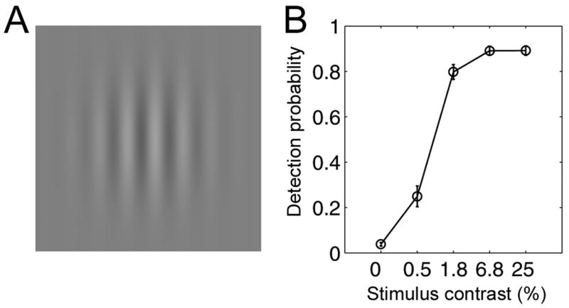
A. Stimuli were grayscale gratings moving right or left at four contrast levels. The stimuli were presented sequentially in pairs, both moving in the same direction and with the same contrast. Subjects judged which of the stimuli moved faster. A 25% contrast grating is illustrated. B. In-scanner detection performance (N = 31). Y-axis shows proportion of responses (detection rate) for each contrast level including fixation trials (zero contrast). Stimulus contrast (x-axis) is represented on a logarithmic scale. Error bars: ± 1 SEM.
Materials and Methods
Participants
Data are reported from 35 children, 7–12 years old, who were part of a larger group (N=50) that participated in a study of reading development. Subjects were recruited from local area public schools. We sought subjects from the normal population with a wide range of reading skills. Written informed consent/assent was obtained from all parents and children. All subjects were physically healthy and had no history of neurological disease, head injury, ADD/ADHD, language disability, or psychiatric disorder. Screening was further based on the “Conners’ Parent Rating Scales-Revised: Short Form” (an ADHD screening form completed by parent) and the “Children’s Depression Inventory” (completed by child). All subjects were native English speakers and had normal or corrected to normal vision and normal hearing. The Stanford Panel on Human Subjects in Medical and Non-Medical Research approved all procedures.
Subjects took part in three separate sessions in the following order: neuropsychological assessment, anatomical scan and functional MRI. 15 additional participants (same age range) completed all three sessions but their data were excluded from analysis based on excessive within-scan motion in the fMRI measurement (see fMRI data analysis).
Behavioral assessment
All participants were administered a comprehensive battery of behavioral tests as part of a longitudinal study of reading. The battery took approximately four hours to complete. Tests were chosen in order to provide measures of general intelligence, phonological processing, oral reading of single words and pseudo-words (timed and untimed), paragraph reading and reading comprehension skills. Of main importance to the current study are the subtests that comprise the phonological awareness and RaN composite scores, measured by the Comprehensive Test of Phonological Processing (CTOPP) (Wagner et al., 1999). Phonological awareness is the composite of two oral tests: Blending (‘What word do these sounds make? /b/-/oi/’. Correct response: ‘boy’) and Elision (‘Say brand. Now say brand without saying /r/’. Correct response: ‘band’). Importantly, both of these tests are oral, with no explicit visual component. RaN is a composite of two speeded naming tests. The score is based on the time it takes to say aloud two arrays of 36 letters, and two arrays of 36 digits, assuming less than 4 errors are made in each array. General intelligence was assessed by the Wechsler Intelligence Scale for Children-IV (WISC-IV) (Wechsler, 1991), which yielded the Full Scale IQ (FSIQ), as well as four index scores: Verbal Comprehension, Perceptual Organization, Working Memory, and Processing Speed. A FSIQ of at least 85 was used as a screening criterion for inclusion. Standardized tests of reading related abilities included: Word Attack (reading aloud pseudowords), Letter-Word Identification (reading aloud words), Passage Comprehension, and Reading Fluency subtests from the Woodcock-Johnson-III Test of Achievement (Woodcock, 1987), the Gray Oral Reading Test-4 (GORT-4) (Wiederholt and Bryant, 2001), and the Test of Word Reading Efficiency (TOWRE) (Torgesen et al., 1999). Language abilities were assessed with the Clinical Evaluation of Language Fundamentals-3 Screening Test (CELF-III) (Semel et al., 1995). There was a wide range of reading and phonological processing skills demonstrated by the participants (see supplemental Figure S5). Given the considerable overlap between subtests included in these neuropsychological batteries, we relied primarily on 8 composite scores that span various aspects of reading and cognitive processing (see Table 2 and Table S1). We rely on the composite scores developed and validated by the authors of the test batteries, which have higher reliability and external validity than the component scores. All scores are standardized and do not correlate with subjects’ age (e.g., for PA: r2 = −0.03, p = 0.36).
Table 2.
Correlation coefficients (Pearson’s r) between MT+ contrast responsivity and behavioral measures
| Behavioral measure | MT+ responsivity | 95% Confidence interval 3 |
|---|---|---|
| Age | −0.26 | <−0.53, 0.04> |
| Basic Reading | 0.44 2 | <0.07, 0.68> |
| Oral reading quotient | 0.42 2 | <0.04, 0.67> |
| Phonological Awareness | 0.59 1 | <0.31, 0.79> |
| Phonological Memory | 0.22 | <−0.09, 0.49> |
| Rapid Naming | −0.01 | <−0.35, 0.28> |
| WISC Verbal comprehension | 0.23 | <−0.11, 0.53> |
| WISC Processing speed | 0.02 | <−0.25, 0.23> |
| WISC Perceptual reasoning | 0.34 | <0, 0.57> |
Statistically significant (p<0.0005, uncorrected, p<0.05, Bonferroni corrected; N=30).
Statistically significant at a level of p<0.05, uncorrected.
Computed on the sampling distribution of the Pearson correlation coefficients using a bootstrap procedure (Efron and Tibshirani, 1993).
fMRI stimulus paradigm
Stimuli were presented in 12s activation blocks (4 trials in each block), interleaved with 12s fixation blocks. In each trial we presented two consecutive moving sinusoidal gratings (Figure 1A), each presented for 1s, with a 100ms inter-stimulus interval. One grating moved at 8 deg/sec and the other moved at 6 deg/sec. Participants were asked to decide which of the gratings moved faster and to press an appropriate response button. The grating’s spatial frequency was 0.5 cycles/degree, its mean luminance was 86 cd/m2, and it subtended 20 degrees visual angle (FWHM = 9.6mm; temporally windowed by a raised cosine). Four contrast levels of 0.5%, 1.8%, 6.8% and 25% were presented in separate blocks with each level repeated 10 or 11 times (though only 9 repetitions per condition were included in analysis, see below).
In addition to the speed discrimination task, a fixation mark (red dot) was present in all trials (including fixation epochs), and participants were asked to fixate throughout the experimental runs. The experiment was divided into 6 short runs of 2 minutes and 48 seconds, to minimize within scan motion and loss of concentration. Each run started with an activation block, and included 7 activation blocks and 7 fixation periods. The first activation block (6 images) in each run was excluded from analysis, to let magnetization stabilize and cognitive arousal return to baseline. Thus, the analysis included 9 repeated activation blocks per contrast condition.
Visual stimulus presentation and response collection was controlled by an in-house Python script (using the VisionEgg library from http://visionegg.org) and a Macintosh G3 iBook computer. Stimuli were presented using a NEC LT158 projector at its native resolution of 1024×768. Stimuli were presented through a backlit projection screen, visible to the subject by a mirror mounted on the top of the head coil. The luminance output was linearized by calibrating the entire system using a PR650 spectral photometer. Responses were collected using an MRI compatible response box (NeuroScan, Sterling, USA).
MRI data acquisition
FMRI measurements were performed on a 3T General Electric scanner with a custom-built volume head coil. Head movements were minimized by padding and tape. Functional MR data were acquired with a spiral pulse sequence (Glover, 1999). Twenty-six oblique slices were prescribed approximately along the ac-pc plane, to cover the whole occipital and temporal lobes (3mm thick, no gap). The sampling rate of the BOLD signal (TR) was 2s. The voxel size was 2.5 × 2.5 × 3mm.
In-plane anatomical images were acquired before the functional scans using a fast SPGR sequence. These T1-weighted slices were taken at the same location of the functional slices, and were used to align the functional data with high-resolution anatomical data acquired in a separate session. We further collected high-resolution whole brain anatomical images for each subject. These images were acquired on a GE 1.5-T Signa LX scanner using a 3D SPGR pulse sequence. We acquired and averaged several T1-weighted anatomical datasets in the axial and sagittal planes for each participant.
fMRI data analysis
Preprocessing
FMRI data were analyzed using the mrVista tools (http://white.stanford.edu/software/). Data were analyzed voxel-by-voxel in individual subjects with no spatial smoothing. Baseline drifts were removed from the time series by high pass temporal filtering.
Motion detection and compensation
Images acquired through the functional scans were inspected for motion by an experienced individual. In a single slice, each image was compared to the first image in the scan, by computing the root mean square error and the mutual information between these images. Following this procedure, 15 subjects were excluded from analysis due to excessive within scan motion in more than 2 of the 6 scans. The remaining 35 datasets were corrected for between scan motion by registering the mean images of each scan. We find that this registration is more robust than the registration of single images, which are much noisier. We applied 3 different transformations that minimized the mutual information between the mean maps of the 6 scans. The transformation could be rigid, non-linear, or both applied successively. Different data sets benefited differently from different transformations. The best transformation was chosen manually by an experienced individual, based on the resulting correspondence between the mean maps. Motion parameters were automatically estimated by running the FSL motion correction algorithm (MCFLIRT, see http://www.fmrib.ox.ac.uk/fsl/). For each subject, we computed the maximum translation in every scan, and made sure that this parameter never exceeded 1 voxel (2 single scans were excluded by this criterion). The mean of these values across scans was correlated with phonological awareness and the slopes in MT+, to exclude the role of subject motion as a confounding factor.
Anatomical MRI processing
High-resolution whole-brain T1 anatomies were aligned, averaged and re-sampled into a 1×1×1mm resolution three dimensional ac-pc aligned anatomical volume (Dougherty et al., 2005). Functional data were registered to the high-resolution anatomical volume for ROI definition, MNI coordinate computation and visualization. Alignment was computed between the inplane anatomicals and the high-resolution volume anatomy using a mutual-information co-registration algorithm (from SPM2). This transformation was then applied to the functional data. For the purpose of reporting MNI coordinates, a non-linear transformation was computed between the volume anatomy and the MNI template (ICBM152) using the spatial normalization tools from SPM2 (http://www.fil.ion.ucl.ac.uk/spm/).
The high resolution volume anatomy of 10 participants was segmented into gray and white matter using custom software and then hand edited to minimize segmentation errors (Teo et al., 1997). The surface defined by the white/gray boundary was smoothed and rendered as a three dimensional object using custom C++ software (available from http://white.stanford.edu/software/), the VTK library (http://www.vtk.org/), and cortical inflation tools from SurfRelax (Larsson, 2001). Functional data from all gray matter layers were projected to each vertex of the surface mesh by taking the maximum value of those layers that were assigned to that vertex. For visualization purposes only, spatial smoothing along the cortical surface was applied using an iterative neighborhood average that is roughly equivalent to a 4 mm FWHM Gaussian kernel.
ROI analysis
We used an ROI analysis for the following reasons: (a) Based on adult studies, we aimed to test a specific hypothesis with respect to MT+ signals and their relation to reading skills; (b) Previous studies have documented considerable variability in the anatomical location of MT+ in adults (Dumoulin et al., 2000; Huk et al., 2002). Thus, an individual definition of ROIs is essential; (c) No previous study has looked at MT+ signals in children. Considering the age and reading skill ranges, individual ROI definition is important to overcome variability in brain size and shape. We defined the ROIs in individual subject brains according to anatomical guidelines and activation evoked by a functional localizer, as described below.
Anatomical definitions
We looked for MT+ activation clusters in the junction between the inferior temporal sulcus (ITS), the ascending limb of ITS (ALITS) and the posterior continuation of the ITS (PCITS) (Dumoulin et al., 2000; Huk et al., 2002). Talairach coordinates reported in four MT studies (Dumoulin et al., 2000; Kourtzi et al., 2002; Tootell et al., 1995; Watson et al., 1993) were used as landmarks in cases of ambiguous sulcal patterns.
Anatomical V1 regions of interest were placed in the depth of the calcarine sulcus near the occipital pole. The anatomical location for V3A regions of interest were identified within the posterior portion of the intra-parietal sulcus (Tootell et al., 1997). As with MT+, we identified activation clusters within these anatomical boundaries.
Functional localizer
A general linear model (GLM) was fit to each voxel’s time course, estimating the relative contribution of each experimental condition (contrast level) to the time course. GLM predictors were constructed as a boxcar for each condition convolved with a standard hemodynamic response model (Boynton et al., 1996). Additional predictors (simple boxcars, 1 within a run and zero elsewhere) were added for each run, to model between run variations. Contrast maps were computed as voxel-wise t-tests between the weights of the predictors of the relevant experimental conditions. ROI definition was done in the volume, by contrasting the three highest contrast conditions with fixation (threshold at 10−3, uncorrected). ROIs were then transformed back to the original (inplane) functional slices, such that time courses from the clusters of functional activation were collected in the original, non-interpolated data.
The percent BOLD contrast values plotted in the contrast response functions (Fig. 2) and used for computing the linear slope (Fig. 3–5) are computed as the average projected amplitude of the signal in a given condition, with respect to the average time course across all conditions. We first obtain the responses to all experimental blocks, Bi. A block is defined as including all of the BOLD measurements from 2 seconds prior to the first stimulus and extending 12 seconds past it. We average across all experimental blocks, yielding one “template” block time course:
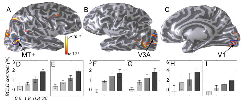
Figure 2. Contrast responsivity in MT+, V3A and V1.
Top: Regions of interest shown on partially inflated surface of the right (A, C) or left (B) hemisphere of an 8 year-old good reader. Voxels where activation increased with stimulus contrast are indicated by the color overlay (p<,0.001 uncorrected). Blue lines denote the ROI borders, defined by the functional localizer (moving patterns at 1.8%, 6.8%, 25% contrast vs. fixation; see also Fig. S1). Bottom: Mean BOLD contrast levels within each ROI in the same individual. Bar plots show mean percent BOLD contrast (y-axis) as a function of stimulus contrast (x-axis) in left and right MT+ (D,E), left and right V3A (F,G), and left and right V1 (H,I). Note the different scale on the y-axis in panels H,I. Error bars are ± 1 SEM.
Figure 3. Contrast responsivity in MT+ correlates with Phonological Awareness scores.
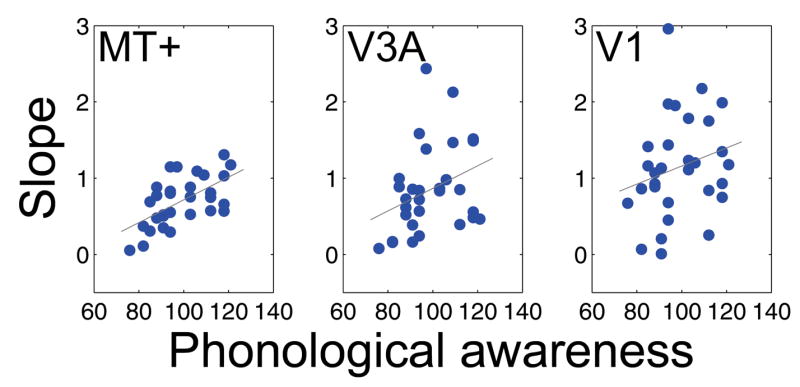
Each point is a measurement from a single subject. The X-axis measures Phonological Awareness (CTOPP) standardized scores. The Y-axis measures the slope of a linear function through the mean BOLD contrast values plotted against log stimulus contrast. Slopes are averaged across hemispheres for each subject (N=30) for each ROI. The three panels are measurements in three different cortical regions: MT+ (left panel), V3A (center) and V1 (right panel). The correlation between responsivity and phonological awareness in MT+ is r = 0.597 (r2 = 0.36, p<0.0005). The correlations within left and right MT+ are significant as well (see supplemental Figure S2). The correlation in V1 is not significant (r = 0.23; p = 0.2) and in V3A it is marginal (r = 0.33; p = 0.08). Shown in gray are the regression lines fit to data points to minimize the mean square error.
Figure 5. MT+ contrast response curves grouped by phonological awareness and rapid naming scores.
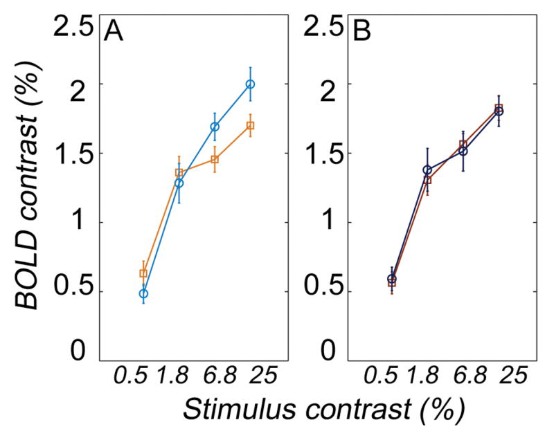
Mean percent BOLD contrast in MT+ (averaged across left and right MT+) is plotted as a function of stimulus contrast. Data are grouped by Phonological Awareness standard scores (A) or by Rapid Naming standard scores (B). In both cases the cutoff is the standard score of 100. Orange squares: PA < 100 (N=21), Light blue circles: PA ≥ 100 (N=14). Red squares: RaN < 100 (N=24), Dark blue circles: RaN ≥ 100 (N=11). Error bars are ± 1 SEM.
We normalize this template into a unit length vector . This quantity represents the average, normalized, block time course across all experimental blocks. By using the average block time course as the reference for the projected amplitudes we account for variance in the shape of the hemodynamic response function across individuals and ROIs.
We then compute the mean time course of the responses in the jth experimental condition, uj The BOLD contrast for this condition is given by the inner product between the mean time course of that condition and the average block time course:
cj has units of percent BOLD modulation.
We summarize the contrast response function by a single value, the slope of the contrast response function, which measures contrast responsivity. We first plot the 4 mean percent BOLD modulation values as a function of log10(% contrast). Empirically, these data are well-fitted by a line. We used the slope of the fitted line (least mean-squared error fit) as a measure of contrast responsivity in that ROI for a given subject. These slopes are then correlated with standardized behavioral scores.
Results
Behavioral results
Table 1 lists the mean, standard deviation and range of the subjects’ age, IQ and behavioral measures. For a full description of the neuropsychological tests included in our analysis see Table S1.
Task performance measurements during MR data acquisition were obtained from 31/35 subjects (in 4 cases data files were corrupted). Probability of detection of the drifting grating (Figure 1A), as measured by the proportion of all responses (correct + incorrect) out of all trials, increased with contrast (Figure 1B). To ensure that the children remained alert during the fixation blocks, there was no explicit delineation of the task and fixation blocks. As a result, subjects sometimes erroneously responded during the fixation blocks (false-alarms) and failed to respond during task blocks (misses), especially at low contrasts. There was no significant correlation between phonological awareness and probability of detection (p≫0.1). The detection levels for high and low phonological awareness groups did not differ, again suggesting that there was no difference in the groups’ alertness levels (see supplementary Figure S4a).
At low contrast levels, subjects’ speed discrimination between the 6 deg/sec and 8 deg/sec stimuli was poor, as expected (28% detection rate; ~45% correct discrimination given detection; this performance level is not significantly different than chance). Detection and speed discrimination accuracy significantly increased with contrast, but even at the highest contrast (detection ratio ~90%), speed discrimination given detection was only 61% correct.
Children are worse than adults at speed discrimination. For example, five-year-old children require a 40% change in speed before they reliably detect a speed difference in a stimulus moving with a velocity of 6 deg/sec (Ahmed et al., 2005). Adults perform reliably with a 10% change in speed. As expected, the children in this study were below adult performance.
There was a weak but measurable relation between discrimination accuracy and phonological awareness at high contrast levels: children with high phonological awareness (age standardized) scores were more accurate on the discrimination task (see Figure S4b). Notice however that the task, speed discrimination between a pair of stimuli at a given contrast level, is not obviously related to contrast responsivity. We expect contrast responsivity to enhance individual’s ability to distinguish between two stimuli at different contrast levels; but it is not clear that this measure should predict sensitivity to small speed differences.
Thus, task performance in this case is a measure of alertness. The equal detection probability supports the assumption that children in the two groups were equally alert.
fMRI results
Figure 2 demonstrates the location, extent and average BOLD contrast in areas MT+, V3A and V1 in an 8 year old girl. Dark blue lines show the borders of these regions of interest (ROIs), defined by a functional localizer (activation for moving patterns at 1.8%, 6.8% and 25% contrast vs. fixation). Colored overlays are regions where BOLD responses increased with stimulus contrast, detected by a parametric contrast map with weights [−0.75, −0.25, +0.25, +0.75] for the different conditions in order of increasing contrast (p<10−3, uncorrected). The bar graphs show increasing BOLD signals within all 3 ROIs in both hemispheres (Figure 2D–I). This response pattern is not enforced by voxel selection, as the ROIs were defined by contrasting motion blocks vs. fixation, without reference to the parametric contrast manipulation. The statistical maps show that a large proportion of the voxels within each ROI were indeed sensitive to the contrast manipulation.
Left and right MT+ were activated by the functional localizer in all 35 subjects analyzed (see Figure S1 for ROI locations in 20 individuals). Mean (±SD) MNI coordinates of LMT+: [−50 ±5, −74 ±5, 10 ±6]; RMT+: [48 ±5, −71 ±6, 7 ±6]. In addition, activation clusters were collected in left and right V1 (N=30; mean MNI coordinates of LV1: [−9 ±5, −97 ±6, 0 ±8]; RV1: [11 ±5, −96 ±6, 4 ±7]), and left and right V3A (N=30; mean MNI coordinates of LV3A: [−25 ±7, −88 ±6, 31 ±10]; RV3A: [25 ±6, −88 ±7, 32 ±7]). The location and magnitude of the signals were similar to those recorded in adults, in agreement with findings from Conner et al. (2004) in early retinotopic areas.
In the vast majority of subjects, the BOLD responses were proportional to the log of stimulus contrast (see Figure 2D–I; Figure 6). To test the statistical relation between cortical contrast responsivity and phonological awareness (PA), we found the best-fitting line relating log contrast and mean BOLD response amplitude in MT+, V3A and V1 for each subject. The slope of this line measures contrast responsivity in each ROI. Since we did not find any differences between the hemispheres in these ROIs, we report group data averaged across the left and right homologues to simplify presentation (Figures 3–5).
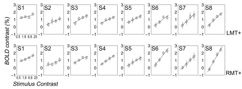
Figure 6. MT+ contrast response curves in 8 individual subjects.
Mean percent BOLD contrast is plotted as a function of stimulus contrast in left (top) and right (bottom) MT+. Data are shown for 8 subjects of various ages, sorted from left to right by phonological awareness standard scores (ranging from 85 (S1) to 121 (S8)). Lines are fit by minimizing least square error. See Table S2 for the behavioral characteristics and slopes measured in each of these individuals. Error bars are ± 1 SEM across repetitions of each condition throughout the experiment.
Figure 3 shows the relationship between contrast responsivity and the phonological awareness age-standardized score (CTOPP, see Methods and Table S1 for a precise definition). Each panel shows this relationship for a different cortical ROI. Data are shown for 30 subjects who had motion-related signals in these three ROIs.
There is a strong correlation between MT+ contrast responsivity and phonological awareness (r = 0.59; p<0.0005; n=30; Fig 3, left panel). The correlations between PA and MT+ slopes within each hemisphere were significant as well (see supplemental Figure S2). There was no significant correlation between phonological awareness and the intercept of the fitted line through the measurements in left and right MT+ (p≫0.1). Neither PA nor MT+ slopes correlated with the estimated motion parameters (p≫0.1). Further, there was no significant correlation with PA in V1 (r = 0.23; p=0.2; n=30; Fig 3, right panel). V3A shows a non-significant trend for a positive relation (r = 0.33; p=0.08; n=30; Fig. 3, center panel). The difference between the correlations in MT+ and V1 was significant (t(27) = 2.89, p<0.01; this measure takes into account the dependence between the measures, see (Howell, 1997, pp. 264). We verified, in a separate bootstrap analysis, that the correlation in MT+ fell outside the 95% confidence interval on the correlation in V1. There was no significant difference between the correlations in MT and V3A, nor between the correlations in V1 and V3A.
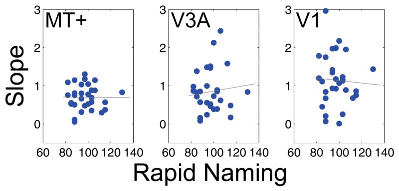
We also analyzed the relationship between Rapid Naming (RaN) standardized scores and contrast responsivity (Figure 4). There was no significant correlation between RaN and contrast responsivity or intercept in any of the ROIs (p≫0.1). The difference between the correlations MT+ ~PA and MT+ ~ RaN (computed as above) was significant (t(27) = 2.57, p<0.01).
Figure 4. Contrast responsivity in MT+, V3A and V1 does not correlate with Rapid Naming scores.
The X-axis shows Rapid Naming (CTOPP) standardized scores. The rest is the same as in Figure 3. The correlations are not significant (p>0.5).
The correlation coefficients between MT+ contrast responsivity and eight composite behavioral measures (and age) are shown in Table 2. The largest correlation was found with phonological awareness, and this was the only relation that was significant at a p<0.05, Bonferroni corrected for nine comparisons. The correlations with Basic Reading (WJ-III) and the Oral reading quotient (GORT4) were significant at a level of p<0.05, uncorrected (for additional scatter plots see Figure S3). We calculated a 95% confidence interval on each of these correlations by applying a bootstrap procedure. The correlation between MT+ contrast responsivity and phonological awareness falls within the confidence intervals on Basic reading and the Oral reading quotient, but outside the confidence intervals on the remaining 6 variables (see Table 2, right column). These results highlight the relation between MT+ responsivity and reading measures generally, and phonological awareness specifically.
We tested whether the correlation between MT+ responsivity and phonological awareness was crucially dependent on the inclusion of individuals with very low reading skills. To this end, we excluded subjects with a Basic Reading score (WJ-III) under 90 (25th percentile), a relatively conservative criterion used in many neuroimaging studies of dyslexia (Shaywitz et al., 2003). This selection procedure excluded 4 subjects, however the correlation between MT+ responsivity and PA remained significant at p<0.05, corrected. Thus, the results are not driven by the inclusion of a few dyslexic subjects in our sample.
Figure 5 shows mean contrast response functions in MT+. Participants are grouped according to their PA scores (Fig. 5, left panel) or their RaN (Fig. 5, right panel). In both cases, the cutoff was set to the population mean (100). The graph on the left shows a reduced responsivity (contrast gain) in children with below-mean PA scores compared to children with above-mean PA scores. No responsivity difference is found between the low RaN and high RaN groups. The specific relation between the curves for low and high PA groups is sensitive to the cutoff criterion. We therefore consider the scatter diagram in Figure 3 a more reliable demonstration of the relation between MT+ responsivity and PA, as it shows the results in the whole sample, independent of a grouping criterion. Individual variability in the overall signal levels and slopes is demonstrated in Figure 6, showing BOLD contrast response curves from 8 individual subjects left and right MT+. The correlation between PA and contrast responsivity is visible even at the individual level: low PA (left hand panels in Figure 6) is associated with reduced MT+ responsivity (slope). The age, gender, PA and slope measures in these subjects are given in Table S2.
Discussion
The results demonstrate a strong and significant correlation between MT+ contrast responsivity and reading measures in children. This correlation is behaviorally and anatomically specific. Phonological awareness showed the strongest correlation with MT+ responsivity, while smaller correlations were found with the Oral Reading Quotient and Basic Reading scores. There was no significant correlation with general IQ measures, nor was there a correlation with rapid naming, another behavioral measure shown to contribute to skilled reading (Manis et al., 2000; Wolf et al., 2000). BOLD signals in V1 varied with contrast, but V1 contrast responsivity was not related to phonological awareness scores. The results show that children with low phonological awareness and reading scores have reduced contrast gain in MT+.
Generalizing previous findings in adult dyslexics to healthy children
Our study is the first to establish a relationship between phonological skills and MT+ responses in a large sample of mostly normally developing children. The relation between MT+ responses and reading skills have been previously demonstrated in small groups of adult developmental dyslexics compared to controls (Demb et al., 1997, 1998b; Eden et al., 1996). The fact that this relation is found in a sample of healthy children suggests that this correlation is not a result of reduced exposure to print in dyslexics.
The relation between phonological skills and MT+ responses is not limited to extremely poor readers, but holds in our sample of 7–12y children. The relation holds even if we exclude four subjects that may be considered developmental dyslexics. Our correlation analysis avoids the choice of a criterion for distinguishing poor and good readers. These data thus suggest that at least some neural factors associated with reading operate in a continuous fashion across people, rather than in a discrete fashion.
Our results further underline the importance of measuring the entire BOLD contrast response function. Significant MT+ activations were recorded in even the poorest readers in our study, in agreement with (Demb et al., 1998b; Vanni et al., 1997). Measuring the responsivity across a range of contrasts revealed a correlation with the slope (contrast responsivity), but not the intercept (overall height) of the response function. This finding predicts that contrast discrimination for moving patterns is impaired in children with low phonological skills.
Anatomical and functional specificity
Phonological awareness correlated with contrast responsivity in MT+, but not in V1 (see Figure 3). This finding is important because it demonstrates the anatomical specificity of our effect, and works against an explanation in terms of general arousal differences. It is important to point out that our sample was specifically screened for ADD/ADHD. We used a speed discrimination task to monitor and encourage children’s alertness throughout the experiment (approximately 15 minutes). Detection performance did not correlate with phonological awareness, again suggesting that the MT+ results do not reflect overall concentration or arousal differences.
Specificity is further demonstrated behaviorally
MT+ contrast responsivity was correlated with phonological awareness and to a lesser extent with general measures of word decoding which are highly correlated with PA. However, MT+ contrast responsivity was not correlated with general measures of intelligence or rapid naming. We note that MT+ is not generally thought of as an area that reflects fast cognitive processing, but as an area for motion and depth processing. Rapid naming most likely relies on frontal systems (Leonard et al., 2006). Thus the fact that we did not find a relation between MT+ properties and these behavioral measures matches current understanding of the primarily sensory nature of MT+ signals.
We targeted MT+ since earlier findings demonstrated a detectable difference between adult dyslexics and controls there. Our results do not contradict the V1 difference reported between good and poor readers (Demb et al., 1998b). It may well be that the contrast responsivity of a subpopulation (magnocellular) neurons in V1 covaries with reading skills and phonological awareness. In this case, special stimulus conditions may be needed to enhance the responses from this population. For example, the reported differences in V1 responses of good and poor adult readers were found at mesopic (2 cd/m2) but not photopic (36 cd/m2) luminance levels. Our stimuli were in the photopic range (86 cd/m2) and not expected to produce a predominantly magnocellular driven V1 response.
We observed a trend for a positive relation in V3A. In human, V3A and MT+ both respond powerfully to moving stimuli, suggesting that they receive some common signals (Tootell et al., 1997). However, fMRI measurements in a patient with a V3A lesion show that MT+ responses do not require an intact V3A (Castelo-Branco et al., 2006). We speculate that the weak correlation in V3A reflects common magnocellular driven inputs to MT+ and V3A, and potentially feedback projections from MT+ to V3A.
Possible Interpretations for the relation between MT+ responsivity and reading measures
There are many possible interpretations for the correlation between MT+ responsivity and reading measures. The first three explanations we describe suggest a causal link between the variables; the next three explanations are based on intervening variables. While our correlational design cannot discriminate between alternative explanations, it is useful for deriving specific testable predictions.
Visual imagery
Phonological awareness tasks such as elision may involve conjuring up a visual image of the word, deleting the relevant letter, and pronouncing the resulting word. Perhaps MT+ is essential in creating the visual image of the word, and MT+ impairment reduces imagery and task performance. It has been shown that MT+ responds to mental imagery of moving stimuli (Goebel et al., 1998; Slotnick et al., 2005) as well as to static images with implied motion (Kourtzi and Kanwisher, 2000). However, there is no evidence yet linking MT+ activation to imagery of static words.
Letter position encoding
Cornelissen et al. (1998a) observed that adults who are poor at motion detection are also poor on tasks that necessitate accurate letter localization within words (e.g., lexical decision with anagram distractors). This result, integrated with the current findings, suggests that MT+ may be involved in representing relative letter positions within a word. Representing relative positions may be crucial for some of the steps required to perform PA tasks. This interpretation assigns MT+ an integral role in the reading pathway, and predicts a functional connection between MT+ and nearby regions involved in visual word recognition.
Multisensory integration in MT+
While MT+ is primarily driven by visual stimuli, some evidence suggests that it is influenced by signals from other modalities as well (Beauchamp, 2005). In particular, MT+ deactivates in response to auditory motion stimuli (Lewis et al., 2000). If an MT+ signal is multi-modal and essential for accurate auditory processing, weak MT+ responsivity may add noise to phonological processing generally and reading specifically.
Incidental factors
We considered attention and subject motion as potential intervening variables, and we controlled for those in multiple ways: prescreening of subject population, post-screening of the data, task design, and post processing assessment of performance and motion effects. The anatomical and functional specificity of the results further reduces the likelihood of such an explanation, but cannot avoid it completely. If one wishes to press the hypothesis that attention explains the group differences, one would have to suppose that this form of attention enhances responsivity (not overall amplitude) specifically in MT+ (not V3A or V1), and without increasing the probability of response to the speed discrimination task; further, this type of attention would act on phonological awareness but not rapid naming or phonological memory.
Learning to read
Behavioral experiments show that there is a bidirectional causal relation between reading and phonological awareness. Training phonological awareness enhances reading skills (Bradley and Bryant, 1983); on the other hand, learning to read enhances phonological skills, as illiterate individuals have lower phonological abilities than literate individuals (Morais et al., 1986; Reis and Castro-Caldas, 1997). Learning to read also involves enhanced exposure to high contrast print coupled with eye movements. Thus, it is possible that learning to read leads to enhanced performance on phonological awareness and reading tests, as well as increasing responsivity to contrast in MT+. Print exposure, however, is expected to rise with age, whereas our results show that MT+ contrast responsivity is high in good readers as young as 7 and 8 years old, and does not correlate with age. This would mean that 2–3 years of exposure to print are enough to boost MT+ responsivity in these good readers to a maximal degree. Future studies with younger children may bear more evidence on this possibility.
Fast-processing deficit
Deficits in fast-processing pathways, both visual and auditory, have been proposed as imposing a limit on reading skills. The behavioral literature contains a wide array of conflicting experiments, some of which argue for and others against such a deficit. Some investigators argue that poor readers also perform poorly on magnocellular-weighted behavioral tasks, but not on parvocellular-weighted tasks (Cornelissen et al., 1998b; Edwards et al., 2004; Martin and Lovegrove, 1984; Maunsell et al., 1990; Stein, 2003). Others have failed to observe such a dissociation (Amitay et al., 2002; Hutzler et al., 2006; Skottun, 2000a; Sperling et al., 2005).
It is impossible to say with any certainty that a behavioral task depends exclusively on magnocellular signals. Even when a task is designed to probe such signals, subjects may have developed compensatory strategies based on other visual signals. Hence, physiological measurements of magnocellular signals are particularly valuable in testing for a magnocellular-specific visual deficit. Future studies will elucidate the relation between behavioral contrast sensitivity (not included in this study) and MT+ contrast responsivity in children with and without reading deficits.
In monkey, MT is driven mostly by magnocellular inputs, with small additional contributions (Nassi et al., 2006; Sincich et al., 2004). Responses in MT are consistently reduced when inactivating magnocellular layers within the LGN; conversely, inactivating parvocellular layers in the LGN typically has very little effect on MT responses (Maunsell et al., 1990). Thus, MT+ is considered an important processing station in the visual magnocellular pathway.
The strong correlation between MT+ responsivity and phonological awareness demonstrated here may reflect a parallel deficit in auditory and visual fast-processing pathways. This possibility is further supported by cell-structure abnormalities in both visual and auditory pathways in dyslexics’ post-mortem brains (Galaburda et al., 1994; Livingstone et al., 1991). Smaller magnocellular neurons in bilateral LGN may lead to less efficient processing in bilateral MT+; smaller cells in left medial geniculate nucleus and left cortical ectopia may cause deficient phonological processing in the same individuals. The anatomical observations explain the apparent conflict between the bilateral MT+ differences and lateralized phonological abilities.
If the visual and auditory deficits develop in parallel, then poor phonological awareness is not caused by the low MT+ responsivity (Cestnick and Coltheart, 1999). In this case, visual training that increases MT+ responsivity is not expected to improve phonological awareness. Testing these predictions will clarify the causal relation between magnocellular signals and reading skills in adults and children.
Supplementary Material
Acknowledgments
Funding provided by NIH grant EY-015000 and the Charles and Helen Schwab Foundation. We wish to thank Arvel Hernandez, Polina Potanina, Adele Behn, Jan Ruby and Rory Sayres for their help. We are grateful to David Heeger, Serge Dumoulin, Merav Ahissar, Kalanit Grill-Spector, Cathy Price and two anonymous reviewers for their suggestions during the preparation of the manuscript. We thank the children and families who took part in this study for their time and enthusiasm.
Footnotes
Publisher's Disclaimer: This is a PDF file of an unedited manuscript that has been accepted for publication. As a service to our customers we are providing this early version of the manuscript. The manuscript will undergo copyediting, typesetting, and review of the resulting proof before it is published in its final citable form. Please note that during the production process errors may be discovered which could affect the content, and all legal disclaimers that apply to the journal pertain.
References
- Ahmed IJ, Lewis TL, Ellemberg D, Maurer D. Discrimination of speed in 5-year-olds and adults: are children up to speed? Vision Res. 2005;45:2129–2135. doi: 10.1016/j.visres.2005.01.036. [DOI] [PubMed] [Google Scholar]
- Amitay S, Ben-Yehudah G, Banai K, Ahissar M. Disabled readers suffer from visual and auditory impairments but not from a specific magnocellular deficit. Brain. 2002;125:2272–2285. doi: 10.1093/brain/awf231. [DOI] [PubMed] [Google Scholar]
- Aylward EH, Richards TL, Berninger VW, Nagy WE, Field KM, Grimme AC, Richards AL, Thomson JB, Cramer SC. Instructional treatment associated with changes in brain activation in children with dyslexia. Neurology. 2003;61:212–219. doi: 10.1212/01.wnl.0000068363.05974.64. [DOI] [PubMed] [Google Scholar]
- Backes W, Vuurman E, Wennekes R, Spronk P, Wuisman M, van Engelshoven J, Jolles J. Atypical brain activation of reading processes in children with developmental dyslexia. J Child Neurol. 2002;17:867–871. doi: 10.1177/08830738020170121601. [DOI] [PubMed] [Google Scholar]
- Beauchamp MS. See me, hear me, touch me: multisensory integration in lateral occipital-temporal cortex. Curr Opin Neurobiol. 2005;15:145–153. doi: 10.1016/j.conb.2005.03.011. [DOI] [PubMed] [Google Scholar]
- Ben-Yehudah G, Sackett E, Malchi-Ginzberg L, Ahissar M. Impaired temporal contrast sensitivity in dyslexics is specific to retain-and-compare paradigms. Brain. 2001;124:1381–1395. doi: 10.1093/brain/124.7.1381. [DOI] [PubMed] [Google Scholar]
- Bishop DVM, Carlyon RP, Deeks JM, Bishop SJ. Auditory temporal processing impairment: Neither necessary nor sufficient for causing language impairment in children. Journal of Speech, Language and Hearing Research. 1999;42:1295–1310. doi: 10.1044/jslhr.4206.1295. [DOI] [PubMed] [Google Scholar]
- Booth JR, Burman DD, Meyer JR, Gitelman DR, Parrish TB, Mesulam MM. Development of brain mechanisms for processing orthographic and phonologic representations. J Cogn Neurosci. 2004;16:1234–1249. doi: 10.1162/0898929041920496. [DOI] [PMC free article] [PubMed] [Google Scholar]
- Boynton GM, Engel SA, Glover GH, Heeger DJ. Linear systems analysis of functional magnetic resonance imaging in human V1. J Neuroscience. 1996;16:4207–4221. doi: 10.1523/JNEUROSCI.16-13-04207.1996. [DOI] [PMC free article] [PubMed] [Google Scholar]
- Bradley L, Bryant PE. Difficulties in auditory organisation as a possible cause of reading backwardness. Nature. 1978;271:746–747. doi: 10.1038/271746a0. [DOI] [PubMed] [Google Scholar]
- Bradley L, Bryant PE. Categorizing sounds and learning to read - a causal connection. Nature. 1983;301:419–421. [Google Scholar]
- Bus AG, IJzendoorn MHv. Phonological Awareness and Early Reading: A Meta-Analysis of Experimental Training Studies. Journal of Educational Psychology. 1999;91:403–414. [Google Scholar]
- Castelo-Branco M, Mendes M, Silva MF, Januario C, Machado E, Pinto A, Figueiredo P, Freire A. Specific retinotopically based magnocellular impairment in a patient with medial visual dorsal stream damage. Neuropsychologia. 2006;44:238–253. doi: 10.1016/j.neuropsychologia.2005.05.005. [DOI] [PubMed] [Google Scholar]
- Castles A, Coltheart M. Is there a causal link from phonological awareness to success in learning to read? Cognition. 2004;91:77–111. doi: 10.1016/s0010-0277(03)00164-1. [DOI] [PubMed] [Google Scholar]
- Cestnick L, Coltheart M. The relationship between language-processing and visual-processing deficits in developmental dyslexia. Cognition. 1999;71:231–255. doi: 10.1016/s0010-0277(99)00023-2. [DOI] [PubMed] [Google Scholar]
- Corina DP, Richards TL, Serafini S, Richards AL, Steury K, Abbott RD, Echelard DR, Maravilla KR, Berninger VW. fMRI auditory language differences between dyslexic and able reading children. Neuroreport. 2001;12:1195–1201. doi: 10.1097/00001756-200105080-00029. [DOI] [PubMed] [Google Scholar]
- Cornelissen P, Richardson A, Mason A, Fowler S, Stein J. Contrast sensitivity and coherent motion detection measured at photopic luminance levels in dyslexics and controls. Vision Res. 1995;35:1483–1494. doi: 10.1016/0042-6989(95)98728-r. [DOI] [PubMed] [Google Scholar]
- Cornelissen PL, Hansen PC, Gilchrist I, Cormack F, Essex J, Frankish C. Coherent motion detection and letter position encoding. Vision Res. 1998a;38:2181–2191. doi: 10.1016/s0042-6989(98)00016-9. [DOI] [PubMed] [Google Scholar]
- Cornelissen PL, Hansen PC, Hutton JL, Evangelinou V, Stein JF. Magnocellular visual function and children's single word reading. Vision Res. 1998b;38:471–482. doi: 10.1016/s0042-6989(97)00199-5. [DOI] [PubMed] [Google Scholar]
- Demb JB, Boynton GM, Best M, Heeger DJ. Psychophysical evidence for a magnocellular pathway deficit in dyslexia. Vision Res. 1998a;38:1555–1559. doi: 10.1016/s0042-6989(98)00075-3. [DOI] [PubMed] [Google Scholar]
- Demb JB, Boynton GM, Heeger DJ. Brain activity in visual cortex predicts individual differences in reading performance. Proc Natl Acad Sci U S A. 1997;94:13363–13366. doi: 10.1073/pnas.94.24.13363. [DOI] [PMC free article] [PubMed] [Google Scholar]
- Demb JB, Boynton GM, Heeger DJ. Functional magnetic resonance imaging of early visual pathways in dyslexia. J Neurosci. 1998b;18:6939–6951. doi: 10.1523/JNEUROSCI.18-17-06939.1998. [DOI] [PMC free article] [PubMed] [Google Scholar]
- Dougherty RF, Ben-Shachar M, Deutsch G, Potanina P, Bammer R, Wandell B. Occipital-callosal pathways in children: validation and atlas development. Annals of the New York Academy of Sciences. 2005;1065:98–112. doi: 10.1196/annals.1340.017. [DOI] [PubMed] [Google Scholar]
- Dougherty RF, Cynader MS, Bjornson BH, Edgell D, Giaschi DE. Dichotic pitch: a new stimulus distinguishes normal and dyslexic auditory function. Neuroreport. 1998;9:3001–3005. doi: 10.1097/00001756-199809140-00015. [DOI] [PubMed] [Google Scholar]
- Dumoulin SO, Bittar RG, Kabani NJ, Baker CL, Jr, Le Goualher G, Bruce Pike G, Evans AC. A new anatomical landmark for reliable identification of human area V5/MT: a quantitative analysis of sulcal patterning. Cereb Cortex. 2000;10:454–463. doi: 10.1093/cercor/10.5.454. [DOI] [PubMed] [Google Scholar]
- Eden GF, Jones KM, Cappell K, Gareau L, Wood FB, Zeffiro TA, Dietz NA, Agnew JA, Flowers DL. Neural changes following remediation in adult developmental dyslexia. Neuron. 2004;44:411–422. doi: 10.1016/j.neuron.2004.10.019. [DOI] [PubMed] [Google Scholar]
- Eden GF, VanMeter JW, Rumsey JM, Maisog JM, Woods RP, Zeffiro TA. Abnormal processing of visual motion in dyslexia revealed by functional brain imaging. Nature. 1996;382:66–69. doi: 10.1038/382066a0. [DOI] [PubMed] [Google Scholar]
- Edwards VT, Giaschi DE, Dougherty RF, Edgell D, Bjornson BH, Lyons C, Douglas RM. Psychophysical indexes of temporal processing abnormalities in children with developmental dyslexia. Dev Neuropsychol. 2004;25:321–354. doi: 10.1207/s15326942dn2503_5. [DOI] [PubMed] [Google Scholar]
- Efron B, Tibshirani RJ. An introduction to the Bootstrap. Chapman & Hall; London, U.K: 1993. [Google Scholar]
- Fischer B, Hartnegg K. Stability of gaze control in dyslexia. Strabismus. 2000;8:119–122. [PubMed] [Google Scholar]
- Galaburda AM, Menard MT, Rosen GD. Evidence for aberrant auditory anatomy in developmental dyslexia. Proc Natl Acad Sci U S A. 1994;91:8010–8013. doi: 10.1073/pnas.91.17.8010. [DOI] [PMC free article] [PubMed] [Google Scholar]
- Georgiewa P, Rzanny R, Gaser C, Gerhard UJ, Vieweg U, Freesmeyer D, Mentzel HJ, Kaiser WA, Blanz B. Phonological processing in dyslexic children: a study combining functional imaging and event related potentials. Neurosci Lett. 2002;318:5–8. doi: 10.1016/s0304-3940(01)02236-4. [DOI] [PubMed] [Google Scholar]
- Glover GH. Simple analytic spiral K-space algorithm. Magn Reson Med. 1999;42:412–415. doi: 10.1002/(sici)1522-2594(199908)42:2<412::aid-mrm25>3.0.co;2-u. [DOI] [PubMed] [Google Scholar]
- Goebel R, Khorram-Sefat D, Muckli L, Hacker H, Singer W. The constructive nature of vision: direct evidence from functional magnetic resonance imaging studies of apparent motion and motion imagery. Eur J Neurosci. 1998;10:1563–1573. doi: 10.1046/j.1460-9568.1998.00181.x. [DOI] [PubMed] [Google Scholar]
- Howell DC. Statistical Methods for Psychology. 4. Wadsworth Publishing Company; Belmont, CA: 1997. [Google Scholar]
- Huk AC, Dougherty RF, Heeger DJ. Retinotopy and functional subdivision of human areas MT and MST. J Neurosci. 2002;22:7195–7205. doi: 10.1523/JNEUROSCI.22-16-07195.2002. [DOI] [PMC free article] [PubMed] [Google Scholar]
- Hutzler F, Kronbichler M, Jacobs AM, Wimmer H. Perhaps correlational but not causal: no effect of dyslexic readers’ magnocellular system on their eye movements during reading. Neuropsychologia. 2006;44:637–648. doi: 10.1016/j.neuropsychologia.2005.06.006. [DOI] [PubMed] [Google Scholar]
- Kourtzi Z, Bulthoff HH, Erb M, Grodd W. Object-selective responses in the human motion area MT/MST. Nat Neurosci. 2002;5:17–18. doi: 10.1038/nn780. [DOI] [PubMed] [Google Scholar]
- Kourtzi Z, Kanwisher N. Activation in human MT/MST by static images with implied motion. J Cogn Neurosci. 2000;12:48–55. doi: 10.1162/08989290051137594. [DOI] [PubMed] [Google Scholar]
- Larsson J. Imaging vision: functional mapping of intermediate visual processes in man. Karolinska Institutet; Stockholm, Sweden: 2001. [Google Scholar]
- Leonard C, Eckert M, Given B, Virginia B, Eden G. Individual differences in anatomy predict reading and oral language impairments in children. Brain. 2006;129:3329–3342. doi: 10.1093/brain/awl262. [DOI] [PubMed] [Google Scholar]
- Lewis JW, Beauchamp MS, DeYoe EA. A comparison of visual and auditory motion processing in human cerebral cortex. Cereb Cortex. 2000;10:873–888. doi: 10.1093/cercor/10.9.873. [DOI] [PubMed] [Google Scholar]
- Liberman IY, Shankweiler D, Liberman AM. The alphabetic principle and learning to read. In: Donald Shankweiler IYL, editor. Phonology and reading disability: Solving the reading puzzle. The University of Michigan Press; Ann Arbor, MI: 1989. [Google Scholar]
- Livingstone MS, Rosen GD, Drislane FW, Galaburda AM. Physiological and anatomical evidence for a magnocellular defect in developmental dyslexia. Proc Natl Acad Sci U S A. 1991;88:7943–7947. doi: 10.1073/pnas.88.18.7943. [DOI] [PMC free article] [PubMed] [Google Scholar]
- Lovegrove W, Martin F, Bowling A, Blackwood M, Badcock D, Paxton S. Contrast sensitivity functions and specific reading disability. Neuropsychologia. 1982;20:309–315. doi: 10.1016/0028-3932(82)90105-1. [DOI] [PubMed] [Google Scholar]
- Manis FR, Doi LM, Bhadha B. Naming speed, phonological awareness, and orthographic knowledge in second graders. J Learn Disabil. 2000;33:325–333. 374. doi: 10.1177/002221940003300405. [DOI] [PubMed] [Google Scholar]
- Martin F, Lovegrove W. The effects of field size and luminance on contrast sensitivity differences between specifically reading disabled and normal children. Neuropsychologia. 1984;22:73–77. doi: 10.1016/0028-3932(84)90009-5. [DOI] [PubMed] [Google Scholar]
- Martin F, Lovegrove WJ. Uniform-field flicker masking in control and specifically-disabled readers. Perception. 1988;17:203–214. doi: 10.1068/p170203. [DOI] [PubMed] [Google Scholar]
- Maunsell JH, Nealey TA, DePriest DD. Magnocellular and parvocellular contributions to responses in the middle temporal visual area (MT) of the macaque monkey. J Neurosci. 1990;10:3323–3334. doi: 10.1523/JNEUROSCI.10-10-03323.1990. [DOI] [PMC free article] [PubMed] [Google Scholar]
- Milne RD, Syngeniotis A, Jackson G, Corballis MC. Mixed lateralization of phonological assembly in developmental dyslexia. Neurocase. 2002;8:205–209. doi: 10.1093/neucas/8.3.205. [DOI] [PubMed] [Google Scholar]
- Morais J, Bertelson P, Cary L, Alegria J. Literacy training and speech segmentation. Cognition. 1986;24:45–64. doi: 10.1016/0010-0277(86)90004-1. [DOI] [PubMed] [Google Scholar]
- Nassi JJ, Lyon DC, Callaway EM. The parvocellular LGN provides a robust disynaptic input to the visual motion area MT. Neuron. 2006;50:319–327. doi: 10.1016/j.neuron.2006.03.019. [DOI] [PMC free article] [PubMed] [Google Scholar]
- Patching GR, Jordan TR. Spatial Frequency Sensitivity Differences between Adults of Good and Poor Reading Ability. Invest Ophthalmol Vis Sci. 2005;46:2219–2224. doi: 10.1167/iovs.03-1247. [DOI] [PubMed] [Google Scholar]
- Ramus F. Developmental dyslexia: specific phonological deficit or general sensorimotor dysfunction? Curr Opin Neurobiol. 2003;13:212–218. doi: 10.1016/s0959-4388(03)00035-7. [DOI] [PubMed] [Google Scholar]
- Reis A, Castro-Caldas A. Illiteracy: a cause for biased cognitive development. J Int Neuropsychol Soc. 1997;3:444–450. [PubMed] [Google Scholar]
- Rumsey JM, Nace K, Donohue B, Wise D, Maisog JM, Andreason P. A positron emission tomographic study of impaired word recognition and phonological processing in dyslexic men. Arch Neurol. 1997;54:562–573. doi: 10.1001/archneur.1997.00550170042013. [DOI] [PubMed] [Google Scholar]
- Semel EM, Wiig EH, Secord WA. Clinical Evaluation of Language Fundamentals. 3. The Psychological Corporation; San Antonio, TX: 1995. [Google Scholar]
- Shaywitz SE, Shaywitz BA, Fulbright RK, Skudlarski P, Mencl WE, Constable RT, Pugh KR, Holahan JM, Marchione KE, Fletcher JM, Lyon GR, Gore JC. Neural systems for compensation and persistence: young adult outcome of childhood reading disability. Biol Psychiatry. 2003;54:25–33. doi: 10.1016/s0006-3223(02)01836-x. [DOI] [PubMed] [Google Scholar]
- Sincich LC, Park KF, Wohlgemuth MJ, Horton JC. Bypassing V1: a direct geniculate input to area MT. Nat Neurosci. 2004;7:1123–1128. doi: 10.1038/nn1318. [DOI] [PubMed] [Google Scholar]
- Skottun BC. The magnocellular deficit theory of dyslexia: the evidence from contrast sensitivity. Vision Res. 2000a;40:111–127. doi: 10.1016/s0042-6989(99)00170-4. [DOI] [PubMed] [Google Scholar]
- Skottun BC. On the conflicting support for the magnocellular-deficit theory of dyslexiaResponse to Stein, Talcott and Walsh (2000) Trends Cogn Sci. 2000b;4:211–212. doi: 10.1016/s1364-6613(00)01485-6. [DOI] [PubMed] [Google Scholar]
- Slotnick SD, Thompson WL, Kosslyn SM. Visual mental imagery induces retinotopically organized activation of early visual areas. Cereb Cortex. 2005;15:1570–1583. doi: 10.1093/cercor/bhi035. [DOI] [PubMed] [Google Scholar]
- Sperling AJ, Lu ZL, Manis FR, Seidenberg MS. Deficits in perceptual noise exclusion in developmental dyslexia. Nat Neurosci. 2005;8:862–863. doi: 10.1038/nn1474. [DOI] [PubMed] [Google Scholar]
- Stanovich KE. Explaining the differences between the dyslexic and the garden-variety poor reader: the phonological-core variable-difference model. J Learn Disabil. 1988;21:590–604. doi: 10.1177/002221948802101003. [DOI] [PubMed] [Google Scholar]
- Stein J. Visual motion sensitivity and reading. Neuropsychologia. 2003;41:1785–1793. doi: 10.1016/s0028-3932(03)00179-9. [DOI] [PubMed] [Google Scholar]
- Stein J, Walsh V. To see but not to read; the magnocellular theory of dyslexia. Trends Neurosci. 1997;20:147–152. doi: 10.1016/s0166-2236(96)01005-3. [DOI] [PubMed] [Google Scholar]
- Talcott JB, Witton C, Hebb GS, Stoodley CJ, Westwood EA, France SJ, Hansen PC, Stein JF. On the relationship between dynamic visual and auditory processing and literacy skills; results from a large primary-school study. Dyslexia. 2002;8:204–225. doi: 10.1002/dys.224. [DOI] [PubMed] [Google Scholar]
- Tallal P. Auditory temporal perception, phonics, and reading disabilities in children. Brain Lang. 1980;9:182–198. doi: 10.1016/0093-934x(80)90139-x. [DOI] [PubMed] [Google Scholar]
- Tallal P. Improving language and literacy is a matter of time. Nat Rev Neurosci. 2004;5:721–728. doi: 10.1038/nrn1499. [DOI] [PubMed] [Google Scholar]
- Temple E, Deutsch GK, Poldrack RA, Miller SL, Tallal P, Merzenich MM, Gabrieli JD. Neural deficits in children with dyslexia ameliorated by behavioral remediation: evidence from functional MRI. Proc Natl Acad Sci U S A. 2003;100:2860–2865. doi: 10.1073/pnas.0030098100. [DOI] [PMC free article] [PubMed] [Google Scholar]
- Teo PC, Sapiro G, Wandell BA. Creating connected representations of cortical gray matter for functional MRI visualization. IEEE Trans Med Imaging. 1997;16:852–863. doi: 10.1109/42.650881. [DOI] [PubMed] [Google Scholar]
- Tootell RB, Mendola JD, Hadjikhani NK, Ledden PJ, Liu AK, Reppas JB, Sereno MI, Dale AM. Functional analysis of V3A and related areas in human visual cortex. J Neurosci. 1997;17:7060–7078. doi: 10.1523/JNEUROSCI.17-18-07060.1997. [DOI] [PMC free article] [PubMed] [Google Scholar]
- Tootell RB, Reppas JB, Kwong KK, Malach R, Born RT, Brady TJ, Rosen BR, Belliveau JW. Functional analysis of human MT and related visual cortical areas using magnetic resonance imaging. J Neurosci. 1995;15:3215–3230. doi: 10.1523/JNEUROSCI.15-04-03215.1995. [DOI] [PMC free article] [PubMed] [Google Scholar]
- Torgesen JK, Bryant BR. Test of Phonological Awareness (TOPA) PRO-ED; Austin, TX: 1994. [Google Scholar]
- Torgesen JK, Wagner RK, Rashotte CA. Longitudinal studies of phonological processing and reading. J Learn Disabil. 1994;27:276–286. doi: 10.1177/002221949402700503. discussion 287–291. [DOI] [PubMed] [Google Scholar]
- Torgesen JK, Wagner RK, Rashotte CA. Test of Word Reading Efficiency. PRO-ED Publishing, Inc; Austin, TX: 1999. [Google Scholar]
- Vanni S, Uusitalo MA, Kiesila P, Hari R. Visual motion activates V5 in dyslexics. Neuroreport. 1997;8:1939–1942. doi: 10.1097/00001756-199705260-00029. [DOI] [PubMed] [Google Scholar]
- Wagner RK, Torgesen JK. The nature of phonological processing and its causal role in the acquistion of reading skills. Psychological Bulletin. 1987;101:192–212. [Google Scholar]
- Wagner RK, Torgesen JK, Rashotte CA. Comprehensive Test of Phonological Processing. Pro-Ed Publishing, Inc; Austin, TX: 1999. [Google Scholar]
- Wandell BA. Foundations of Vision. Sinauer Press; Sunderland, MA: 1995. [Google Scholar]
- Watson JD, Myers R, Frackowiak RS, Hajnal JV, Woods RP, Mazziotta JC, Shipp S, Zeki S. Area V5 of the human brain: evidence from a combined study using positron emission tomography and magnetic resonance imaging. Cereb Cortex. 1993;3:79–94. doi: 10.1093/cercor/3.2.79. [DOI] [PubMed] [Google Scholar]
- Wechsler D. Wechsler Intelligence Scale for Children. 3. Psychological Corporation; San Antonio, TX: 1991. [Google Scholar]
- Wiederholt JL, Bryant B. Gray Oral Reading Test-4th Edition (GORT-4) Pro-Ed Publishing, Inc; Austin, TX: 2001. [Google Scholar]
- Williams MJ, Stuart GW, Castles A, McAnally KI. Contrast sensitivity in subgroups of developmental dyslexia. Vision Res. 2003;43:467–477. doi: 10.1016/s0042-6989(02)00573-4. [DOI] [PubMed] [Google Scholar]
- Wilmer JB, Richardson AJ, Chen Y, Stein JF. Two visual motion processing deficits in developmental dyslexia associated with different reading skills deficits. J Cogn Neurosci. 2004;16:528–540. doi: 10.1162/089892904323057272. [DOI] [PubMed] [Google Scholar]
- Witton C, Talcott JB, Hansen PC, Richardson AJ, Griffiths TD, Rees A, Stein JF, Green GG. Sensitivity to dynamic auditory and visual stimuli predicts nonword reading ability in both dyslexic and normal readers. Curr Biol. 1998;8:791–797. doi: 10.1016/s0960-9822(98)70320-3. [DOI] [PubMed] [Google Scholar]
- Wolf M, Bowers PG, Biddle K. Naming-speed processes, timing, and reading: a conceptual review. J Learn Disabil. 2000;33:387–407. doi: 10.1177/002221940003300409. [DOI] [PubMed] [Google Scholar]
- Woodcock RW. Woodcock Reading Mastery Tests-Revised. American Guidance Service; Circle Pines, MN: 1987. [Google Scholar]
Associated Data
This section collects any data citations, data availability statements, or supplementary materials included in this article.


