Abstract
A replication-deficient, recombinant adenovirus encoding human aquaporin-1 (hAQP1), the archetypal water channel, was constructed. This virus, AdhAQP1, directed hAQP1 expression in several epithelial cell lines in vitro. In polarized MDCK cell monolayers, hAQP1 was localized in the apical and basolateral plasma membranes. Fluid movement across monolayers infected by AdhAQP1 in response to an osmotic gradient was ≈4-fold that seen with uninfected monolayers or monolayers infected by a control virus. When AdhAQP1 was administered to rat submandibular glands by retrograde ductal instillation, significant hAQP1 expression was observed by Western blot analysis in crude plasma membranes and by immunohistochemical staining in both acinar and ductal cells. Three or four months after exposure to a single radiation dose (17.5 or 21 Gy, respectively), AdhAQP1 administration to rat submandibular glands led to a two- to threefold increase in salivary secretion compared with secretion from glands administered a control virus. These results suggest that hAQP1 gene transfer may have potential as an unique approach for the treatment of postradiation salivary hypofunction.
Keywords: gene transfer, adenovirus, radiation damage, xerostomia
Each year in the United States ≈40,000 new cases of head and neck cancer are diagnosed (1). For most of these patients, ionizing radiation is a key component of therapy. However, such treatment often results in severe damage to the fluid-secreting portion (acinar cells) of the salivary glands that lie in the radiation field (2–5). Patients with reduced salivary secretion suffer from dysphagia, xerostomia, mucositis, dental caries, and frequent oropharyngeal infections. Their quality of life is significantly diminished. Despite recognition and study of these sequelae for most of this century, the mechanism of radiation-induced salivary hypofunction remains unclear, and no corrective treatment is currently available. The surviving salivary epithelial cells are predominantly ductal, considered to be salt absorptive, relatively water impermeable, and incapable of generating salivary fluid.
As a novel strategy to treat this condition, we have chosen to apply gene transfer technology to alter postradiation surviving ductal cell function. In a normal salivary gland, acinar cells secrete an isotonic “primary” fluid, from which considerable NaCl is resorbed during passage through the ductal system. Although currently very little is known about ion transport pathways in salivary ductal segments (6, 7), at least four such components are considered to be present in ductal cell luminal membranes (6). These are an epithelial Na+ channel (6, 8); a Cl− channel, likely the cystic fibrosis transmembrane conductance regulator (6, 9); a Na+/H+ exchanger (6, 9); and a K+/H+ exchanger (6, 10). The first three components are believed to be important to NaCl resorption by ductal cells (6, 11). The K+/H+ exchanger, on the other hand, is thought to be responsible for the secretion of K+ and HCO3−, both of which are typically found well above serum levels in the final saliva (6, 10).
In the absence of acinar cells, such as would occur after extensive therapeutic irradiation (2, 12), no primary fluid would be generated and, thus, the Na+ channel, Cl− channel and Na+/H+ exchanger would be practically inoperative. However, the luminal K+/H+ exchanger could continue to function and, in the presence of CO2 (HCO3−, H+), to secrete KHCO3. Accordingly, we have speculated that this K+/H+ exchanger could contribute to the generation of an osmotic KHCO3 gradient (luminal > interstitial), which would allow significant fluid movement into the lumen across ductal cells if a facilitated water permeability pathway, such as a water channel, was present. Currently, however, no known water channel has been identified in salivary intralobular and excretory duct membranes (13, 14).
Based on this rationale, we thought to introduce a facilitated water permeability pathway in salivary glands exposed to radiation via a recombinant adenovirus. Replication-deficient recombinant adenoviruses have been used for efficient gene transfer to mammalian epithelial cells in vitro and in vivo (15–19). Aquaporins (AQPs) are a recently described family of water channel proteins (13, 20, 21). The prototype is AQP1, a 28-kDa protein first isolated from human red blood cells (22–24). AQP1 is a constitutively activated water channel, which does not exhibit a polarized membrane distribution in most epithelia (13, 20, 21). Furthermore, within salivary glands, AQP1 is only detected in venules and capillaries, but not in parenchymal cells (14, 25).
In the present report, we describe the construction of a recombinant adenovirus encoding human AQP1 (hAQP1). We show that this virus can direct hAQP1 expression in vitro in several epithelial cell lines and in vivo in salivary gland parenchyma. When the virus is administered to rat salivary glands that have been irradiated and rendered either modestly or markedly hypofunctional, salivary flow rate is dramatically increased. Our findings suggest that hAQP1 gene transfer may provide a novel therapeutic approach for patients with postradiation salivary hypofunction.
METHODS
Construction of AdhAQP1.
The hAQP1 cDNA (22) was directly cloned into the adenovirus shuttle vector pACCMV, which was generously provided by C. Newgard (University of Texas Southwestern Medical Center, Dallas). To do this, PCR primers were designed to amplify the hAQP1 cDNA (≈3 kb) and to add restriction sites for cloning. The sense primer was 5′-GGCGGGTACCAGCTCTCAGAGGGAATTGAGCACCCGGCAGCGGTCTCAGG-3′, and contained a KpnI site and 19 nucleotides corresponding to the 5′ untranslated sequence that was not present in the plasmid cDNA. The antisense primer was 5′-GCCGGGATCCGGAAACAGCTATGACCATG-3′, and contained a BamHI site. PCR amplification was performed using the hAQP1 cDNA (22) as a template (1 min at 94°C, 1 min at 59°C, 2 min at 72°C, 35 cycles) and a GeneAmp PCR reagent kit (Perkin–Elmer). The PCR-amplified hAQP1 cDNA was inserted at the KpnI and BamHI sites of pACCMV to yield the plasmid pCMVhAQP1. pCMVhAQP1 was then cotransfected with pJM17, which contains the adenovirus type 5 sequence, in 293 cells as described (26) to yield AdhAQP1. AdhAQP1 is a replication-deficient recombinant adenovirus encoding hAQP1 and including a cytomegalovirus promoter and simian virus 40 polyadenylylation site. This virus was subsequently plaque-purified, grown in large quantity, and purified by CsCl gradient centrifugation (26). The titer of the virus, determined by limiting dilution plaque assay, was 4 × 10¹¹ plaque-forming units (pfu)/ml.
Northern and Western Blot Analyses.
Oligonucleotide primers were designed to amplify ≈813 bp of the hAQP1 cDNA as a probe for Northern blot analyses. The primers used were as follows: sense, 5′-ATGGCCAGCGAAATCAAGAA-3′; antisense, 5′-CCTCTATTTGGGCTTCATCTC-3′. PCR amplification was performed as above using 20 pmol of primers with Bluescript II (SK−) containing the hAQP1 cDNA (22) as template. A band of ≈813 bp was obtained after agarose gel electrophoresis. The PCR product was purified on a Select-D, G-50 column (5′ → 3′). Northern blots were performed using poly(A)+ RNA essential as previously described (27). Western blot analyses, using an affinity-purified antibody against hAQP1 (dilution 1:1000; ref. 14), were performed on samples of cultured cell, or submandibular gland, membranes as previously reported (27, 28).
Localization and Function of hAQP1 in MDCK Cells.
The distribution of hAQP1 in MDCK cells (strain MDCK-c, a cyst-forming clone, a gift from J. Grantham, University of Kansas) was determined by confocal microscopy as previously described (27), using the above affinity-purified antibody. Cells were grown to confluence on 24.5-mm Transwell-Col culture chambers (Costar). The secondary antibody used was a fluorescein isothiocyante-conjugated goat anti-rabbit IgG (Jackson ImmunoResearch). Samples were examined in a Nikon Optiphot photomicroscope equipped with a Bio-Rad MRC-1000 laser scanning confocal imaging system (Bio-Rad), using a Krypton/Argon laser as light source. The net fluid secretion across polarized MDCK cell monolayers was determined using a modification of the technique of Neufeld et al. (29) as reported earlier (27). The apical fluid medium was replaced by a hyperosmotic medium (400 mosm), and fresh isosmotic medium was placed at the basal side. After 4 h, the liquid in the apical chamber was collected with a calibrated pipette, and net fluid movement (water flow) determined.
Immunohistochemical Detection and Localization of hAQP1 in Rat Submandibular Glands.
hAQP1 localization in submandibular glands was determined by immunohistochemistry using the affinity-purified antibody to hAQP1 (dilution 1:100) and a Histostain SP kit (Zymed). Sections from paraffin-embedded submandibular glands were prepared from both irradiated (see below) and sham-irradiated glands, which had been infected with 5 × 10⁹ pfu of either Addl312 (a control, replication-deficient adenovirus that encodes no transgene, ref. 26) or AdhAQP1. Quantitation of the proportion of cells expressing hAQP1 in control and AdhAQP1-infected glands was achieved by counting immunopositive and total cells in 4–6 high-power fields using gland pairs from two or three animals and an optical grid. All cell counting was done by one examiner.
Rat Irradiation, Gene Transfer, and Saliva Collection and Analysis.
Male Wistar rats (250–300 g) were irradiated with a single dose of either 17.5 or 21 Gy, with the ventral surface of their necks exposed to the source. Control animals were sham-irradiated, i.e. anesthetized and placed in the irradiator, but not exposed to ionizing radiation. After 3 months (17.5 Gy) or 4 months (21 Gy), all rats were subjected to in vivo adenoviral-mediated gene transfer (5 × 10⁹ pfu of either Addl312 or AdhAQP1) to the submandibular glands by retrograde ductal instillation (19, 30). Animals received 1 mg of dexamethasone (i.m. injection) at the time of gene transfer and each day until saliva collection to suppress inflammation, due to adenoviral infection, and preserve salivary function (30). Saliva was collected 3 days after this infection. The submandibular glands were again cannulated and saliva collected for 10 min after retrograde injection of 25 μl of pilocarpine (5 mg/ml), and for an additional 5 min after intramuscular injection of pilocarpine (5 mg/kg). Saliva volumes were determined gravimetrically after the 15-min collection. Na+ and K+ concentrations in rat submandibular saliva samples were determined as previously described (31).
RESULTS
AdhAQP1-Mediated hAQP1 Expression in Vitro.
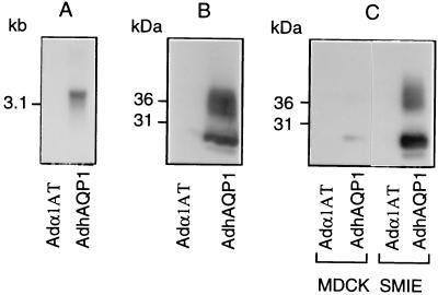
AdhAQP1 was constructed by homologous recombination between pCMVhAQP1 and pJM17 plasmids, and its structure verified by restriction and sequence analyses. The ability of AdhAQP1 to direct the expression of hAQP1 was initially assessed in 293 cells (human kidney) in vitro by both Northern blot and Western blot analyses. Northern blot analysis showed no hAQP1 transcript present in uninfected 293 cells (data not shown) or in 293 cells infected for 24 h with an adenovirus expressing α-1 antitrypsin (Adα1AT; see ref. 15; Fig. 1A). However, in AdhAQP1-infected 293 cells, abundant amounts of a hybridization-positive transcript of ≈3 kb were detected 24 h postinfection (Fig. 1A). This is the length of the transcript expected for hAQP1 (22). Western blot analysis of AdhAQP1-infected 293 cells showed the presence of both the nonglycosylated (≈28 kDa) and glycosylated (≈35–45 kDa) forms of hAQP1 (Fig. 1B) (22, 23). Additionally, we have observed that AdhAQP1 was able to drive hAQP1 expression in other epithelial cell lines, e.g. MDCK (derived from dog kidney) and SMIE (derived from rat submandibular) cells (Fig. 1C).
Figure 1.
hAQP1 expression in 293 cells and other epithelial cells. (A) Northern blot analysis was performed using 10 μg of total RNA, and 293 cells were infected at a MOI of 100 with either Adα1AT or AdhAQP1 and hybridized with a 813-bp radiolabeled probe of hAQP1 as described in Methods. (B) Ten micrograms of crude membranes from 293 cells infected at a MOI of 100 with either Adα1AT or AdhAQP1 were analyzed by Western blot as described in Methods. (C) Ten micrograms of crude membranes from MDCK and SMIE cells were analyzed by Western blot as above. The cells were infected at a MOI of 100 with either Adα1AT or AdhAQP1.
Sorting and Function of hAQP1 in MDCK Cells After AdhAQP1 Infection.
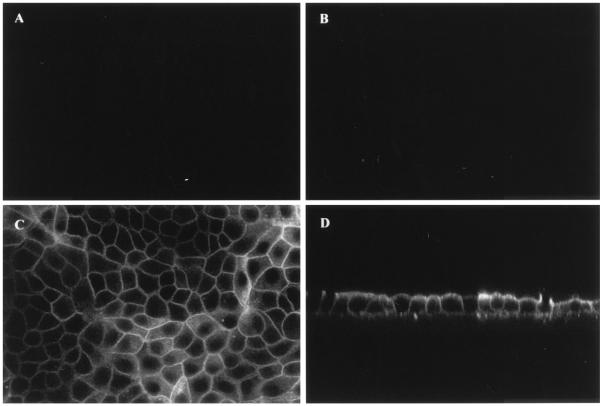
MDCK cells were used to study the sorting and function of the hAQP1 protein after AdhAQP1 infection. These cells are able to form polarized monolayers with distinct apical and basolateral membranes when grown on collagen-coated permeable filters (32). Uninfected MDCK cells (not shown) and cells infected with Adα1AT (Fig. 2A and B) at a multiplicity of infection (MOI) of 100 showed no hAQP1 expression. Conversely, cells infected with AdhAQP1 exhibited hAQP1 immunolabeling at both the apical and basolateral membranes (Fig. 2 C and D), similar to its distribution in vivo for several epithelial and endothelial cells (13, 20, 21).
Figure 2.
Localization of hAQP1 in polarized MDCK cells. Confluent MDCK cells, grown on filters, were infected for 24 h at the basolateral side at a MOI of 100 with either Adα1AT (A and B) or AdhAQP1 (C and D). hAQP1 expression was determined as described in Methods. Micrographs of horizontal (xy; A and C), and vertical (xz; B and D) optical sections through MDCK cells are shown.
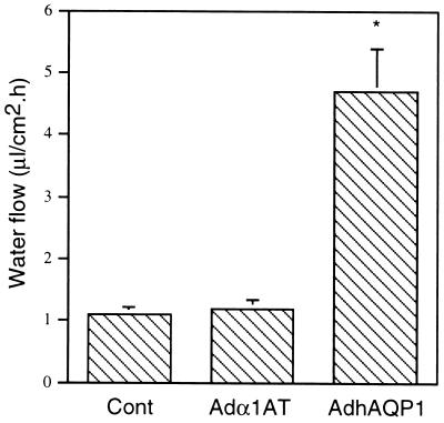
We used MDCK cells as an in vitro model to determine if the AdhAQP1-directed hAQP1 protein expression led to increased water permeability (27, 29). The net movement of fluid across MDCK cells was measured in the presence of a transepithelial (apical > basal) osmotic gradient. After 4 h incubation, the net fluid movement across uninfected cells, or cells infected with Adα1AT, was similar, approximately 1 μl/cm2 per h. However, in AdhAQP1-infected MDCK cells net fluid movement was increased ≈4-fold (Fig. 3), demonstrating that the expressed hAQP1 was functional.
Figure 3.
Net fluid secretion rate of MDCK cells. Net fluid secretion rate of control, Adα1AT-(MOI of 100)- and AdhAQP1-(MOI of 100)-infected MDCK cells was measured as described in Methods. The results are expressed as water flow in microliters of fluid secreted per cm2 per h and are the mean ± SEM of three separate experiments, each performed in triplicate. The results were analyzed using an ANOVA. ∗, P < 0.05 compared with control and Adα1AT-infected cells.
AdhAQP1-Mediated hAQP1 Expression in Vivo in Rat Submandibular Glands.
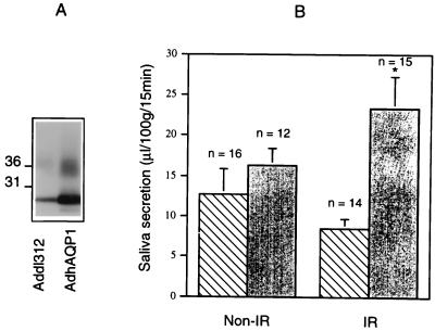
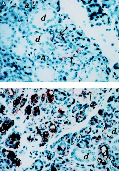
To determine if AdhAQP1 infection could lead to hAQP1 expression in vivo, we administered AdhAQP1 to rat submandibular glands via retrograde ductal instillation. After 24 h we prepared membranes from these glands and from glands either infected with Addl312, or uninfected. Membranes from uninfected rat submandibular glands (data not shown) or glands infected with Addl312 showed evidence of some nonglycosylated and glycosylated forms of AQP1 (Fig. 4A). This represents the endogenous rat AQP1, which is known to be present in blood vessels within the gland (Fig. 5A; see ref. 14). After infection with AdhAQP1, AQP1 levels in membranes from whole glands were increased ≈5-fold as judged from Western blot analyses (Fig. 4A). We also obtained samples of control and AdhAQP1-infected glands for localization of hAQP1 in various gland cell types. In nonirradiated glands infected with Addl312, 2.1 ± 0.5% (mean ± SEM) of the cells were immunopositive for AQP1 (these represent endothelial cells in capillaries and venules; refs. 14, 25). Similarly, Addl312-infected, irradiated (17.5 Gy) glands showed low levels of cellular immunolabeling (1.6 ± 0.3%). AdhAQP1-infected glands showed high AQP1 immunolabeling levels in both acinar and ductal cells (Fig. 5B). In AdhAQP1-infected nonirradiated glands 15.0 ± 3.6% of the cells were immunopositive; a significant increase above that seen in Addl312-infected glands (P < 0.05). In irradiated glands infected with AdhAQP1, the proportion of cells expressing AQP1 was significantly greater (33.6 ± 1.34%; P < 0.05)
Figure 4.
Expression and function of the recombinant hAQP1 in vivo. (A) Ten micrograms of crude membranes from rat submandibular glands, which were cannulated and infected for 24 h with 5 × 10⁹ pfu of either Addl312 or AdhAQP1, were used for Western blot analysis as in Fig. 1. (B) Submandibular glands of sham-irradiated (Non-IR) or irradiated (IR) rats were cannulated and retrograde instilled with 5 × 10⁹ pfu of either Addl312 (hashed bar) or AdhAQP1 (solid bar), and then saliva was collected after 3 days as described in Methods. The results are expressed as saliva secretion in microliters per 100 g of rat body weight per 15 min and are the mean ± SEM. The results were analyzed using an ANOVA. ∗, P < 0.05 between the two groups with different instilled viruses.
Figure 5.
Localization of hAQP1 in rat submandibular glands infected with AdhAQP1. hAQP1 localization was determined by immunohistochemistry (see Methods) on paraffin sections from sham-irradiated rat submandibular glands infected with 5 × 10⁹ pfu of either Addl312 (A) or AdhAQP1 (B). Arrows indicate endothelial cells in capillaries and venules. a, acinar cells; d, ductal cells. Similar results were found with irradiated glands (not shown).
Saliva Secretion from AdhAQP1-Infected Rat Submandibular Glands.
We next determined if the expression of hAQP1 occurring subsequent to AdhAQP1 infection was associated with increased salivary fluid secretion. We first examined salivary secretion from rat submandibular glands that were either sham-irradiated or given a single exposure of 17.5 Gy. Three months postsham or actual irradiation, all animals were infected with either Addl312 or AdhAQP1. As shown in Fig. 4B, when compared with sham-irradiated rats, rats exposed to radiation and administered Addl312 exhibited a modest, ≈30%, but not statistically significant, reduction in saliva production. We next compared the effect of AdhAQP1 infection on salivary secretion in sham-irradiated and irradiated animals. AdhAQP1 had a modest, but not significant, effect on salivary secretion in the sham-irradiated animals. However, in the irradiated rats, AdhAQP1 infection resulted in a higher level of saliva production. Salivary secretion in these animals was ≈3-fold (P < 0.05) that observed from glands of irradiated animals infected with the control virus (Addl312).
Saliva also was collected from submandibular glands of an additional set of rats that were either sham-irradiated or given a single radiation dose of 21 Gy (Table 1), a condition expected to result in greater salivary hypofunction. In this group of animals, infection with either Addl312 or AdhAQP1 was performed at 4 months postirradiation. Saliva secretion from the sham-irradiated and Addl312-infected glands was 36.6 ± 6.8 μl/100 g. Salivary secretion from irradiated glands infected with Addl312 was significantly lower, by 64% (P < 0.05; 13.2 ± 3.7 μl/100 g) than from the sham-irradiated group, consistent with greater salivary hypofunction. AdhAQP1 infection of sham-irradiated glands was without effect on salivary secretion (28.4 ± 8.0 μl/100 g) compared with that seen with Addl312-infected glands (above). However, AdhAQP1-infection of irradiated glands resulted in a ≈2-fold increase in submandibular gland secretion (30.6 ± 3.5 μl/100 g) compared with Addl312-infected glands (above; P < 0.05).
Table 1.
Effect of AdhAQP1 infection on secretion by rat submandibular glands irradiated with 21 Gy
| Experimental group | Saliva secretion after viral infection, μl/100 g body weight per 15 min
|
|
|---|---|---|
| Addl312 | AdhAQP1 | |
| Sham-irradiation | 36.6 ± 6.8a (n = 4) | 28.4 ± 8.0c (n = 6) |
| 21 Gy irradiation | 13.2 ± 3.7a,b,c (n = 6) | 30.6 ± 3.5b (n = 9) |
Animals were either sham-irradiated or their salivary glands were exposed to a single radiation dose of 21 Gy. Four months later, either a control virus (Addl312) or AdhAQP1 was administered via retrograde ductal instillation to the submandibular glands. Three days later saliva was collected from these glands. The numbers in parentheses represent the number of animals in each treatment group. Values are mean ± SEM; values with the same superscript letter are significantly different, P < 0.05, by a Student’s t test.
DISCUSSION
We have constructed a recombinant adenovirus, AdhAQP1, and shown that it mediated hAQP1 expression in membranes of epithelial cell lines from several species (human, dog, rat) in vitro. The recombinant hAQP1 protein exhibited several characteristics typical of native hAQP1 (13, 20, 21): (i) the presence of nonglycosylated and glycosylated forms; (ii) a nonpolarized membrane distribution; and (iii) the ability to mediate the transepithelial movement of fluid. AdhAQP1 also mediated hAQP1 expression in rat submandibular gland membranes in vivo. Both acinar and ductal cell types were targets for the virus. This wide distribution is consistent with our earlier studies using an adenovirus encoding a reporter gene (β-galactosidase) to evaluate infected cell populations (19).
The impetus for this study was to test a novel strategy for increasing fluid secretion in human salivary glands exposed to ionizing radiation. As noted above, in humans, such radiation especially damages the primary fluid secreting acinar cells, whereas the ductal cells are much less affected. For patients this can result in a marked reduction of salivary flow (4, 5). We used rat salivary glands, which are a convenient and often-used model to study secretory irradiation damage, to test this strategy. Although rat salivary glands are considered relatively more radioresistant than primate and human glands (2), at higher cumulative radiation doses and longer postradiation times rat salivary glands show similar morphological changes (loss of acinar cells) and marked secretory dysfunction (33–36). In the present study, three months after exposure to 17.5 Gy, rats exhibited a modest reduction (≈30%) in salivary flow, but no striking morphological changes in their glands. Nonetheless, salivary secretion was markedly increased if the irradiated glands were infected with AdhAQP1 versus the control virus Addl312. When salivary glands were exposed to a higher radiation dose (21 Gy), and salivary secretion measured 4 months postirradiation, salivary flow rates were dramatically lower (64%), and the submandibular gland parenchyma exhibited clear morphological changes (data not shown). These results are consistent with previous observations, reflecting an experimental situation more like that observed in irradiated patients (e.g., refs. 12, 33, 35). Most importantly, administration of AdhAQP1 to these more severely affected rat salivary glands was able to enhance salivary secretion significantly, approaching levels seen with sham-irradiated glands infected with control virus (30.6 ± 3.5 μl/100 g vs. 36.6 ± 6.8 μl/100 g, respectively). Thus, the strategy suggested above for increasing fluid secretion from irradiated human salivary glands was able to increase fluid secretion from irradiated rat salivary glands.
At present the exact mechanism by which this increase in fluid secretion from irradiated rat salivary glands occurs is unknown. The hypothesis described above, i.e. fluid movement in response to a KHCO3-generated osmotic gradient, is speculative. We have, however, determined the [K+] and [Na+] in the saliva collected from irradiated submandibular glands infected with either Addl312 or AdhAQP1. Although this saliva reflects a mixture of fluid output from all parenchymal tissue present, some of which may be more severely affected by radiation than others, interestingly we found that [K+] in saliva from animals administered AdhAQP1 had >40% higher [K+], 99.1 ± 7.4 mM, than the [K+] found in saliva of animals infected with Addl312 (69.6 ± 10.6; P < 0.05). Conversely, no difference was observed in salivary [Na+] between these two groups (14.1 ± 2.9 vs. 15.1 ± 2.7 mM). While these results are generally in keeping with the above hypothesis, considerably more study is necessary to understand the mechanism for AdhAQP1-mediated increases in fluid secretion from irradiated glands. An additional, but unexpected, finding was that we observed that more (≈2-fold) glandular parenchymal cells were transduced by AdhAQP1 in irradiated glands than in sham-irradiated glands. The reason for this difference is not known, but it was a consistent finding in all animals examined and likely contributed to the increased saliva secretion measured.
There are two current, significant, and presently inescapable limitations to fully understanding the results presented here. First, relatively little is known about the physiology of salivary ductal cells, and what is known represents a mixture of findings from different ductal segments (intralobular; main excretory; refs. 6–10). Second, the exact nature of radiation damage to salivary glands (human and rodent) is not clear and, indeed, is considered enigmatic (3, 36–40). Obviously, designing a specific molecular approach to alter ductal cell function requires detailed knowledge of the molecules functioning normally and pathologically, a situation presently impossible to realize. What is clear, however, is that AQP1 gene transfer to both modestly affected and significantly dysfunctional, irradiated rat submandibular glands leads to enhanced salivary secretion from these glands. These findings, obtained with two separately irradiated and treated cohorts of animals, strongly support the possibility of using gene therapy to correct the defects in salivary glands that occur subsequent to therapeutic irradiation. This approach may have the potential to improve salivary function and relieve the considerable morbidity experienced by patients.
Acknowledgments
We thank Dr. J. Mitchell, A. M. DeLuca, and N. Wersto (Radiation Biology and Radiation Oncology Branches, National Cancer Institute) for help with animal irradiation procedures. We are also grateful to Dr. J. Mitchell and Dr. R. J. Turner for helpful comments on this manuscript. This work was supported in part by National Institutes of Health Grants HL33991, HL48268, and EY11239.
ABBREVIATIONS
- AQP
aquaporin
- hAQP1
human aquaporin 1
- MOI
multiplicity of infection
- pfu
plaque-forming units
References
- 1.Silverman S., Jr Clin Geriatric Med. 1992;8:529–541. [PubMed] [Google Scholar]
- 2.Stephens L C, King G K, Peters L J, Ang K K, Schultheiss T E, Jardine J H. Am J Pathol. 1986;124:469–478. [PMC free article] [PubMed] [Google Scholar]
- 3.Vissink A, Kalicharan D, s’Gravenmade E J, Jongbloed W L, Ligeon E E, Nieuwenhuis P, Konings W T. J Oral Pathol Med. 1991;20:449–456. doi: 10.1111/j.1600-0714.1991.tb00437.x. [DOI] [PubMed] [Google Scholar]
- 4.Franzen L, Funegard U, Ericson T, Henriksson R. Eur J Cancer. 1992;28:457–462. doi: 10.1016/s0959-8049(05)80076-0. [DOI] [PubMed] [Google Scholar]
- 5.Johnson J T, Ferretti G A, Nethery W J, Valdez I H, Fox P C, Ng D, Muscoplat C C, Gallagher S C. N Engl J Med. 1993;329:390–395. doi: 10.1056/NEJM199308053290603. [DOI] [PubMed] [Google Scholar]
- 6.Cook D I, Van Lennep E W, Roberts M L, Young J A. In: Physiology of the Gastrointestinal Tract. Johnson L R, editor. New York: Raven; 1994. pp. 1061–1117. [Google Scholar]
- 7.Zhao H, Xu X, Diaz J, Muallem S. J Biol Chem. 1995;270:19599–19605. [PubMed] [Google Scholar]
- 8.Moran A, Davis V H, Turner R J. Am J Physiol. 1995;268:C350–C355. doi: 10.1152/ajpcell.1995.268.2.C350. [DOI] [PubMed] [Google Scholar]
- 9.He X, Tse C-M, Donowitz M, Alper S L, Gabriel S E, Baum B J. Pflügers Arch. 1997;433:260–268. doi: 10.1007/s004240050276. [DOI] [PubMed] [Google Scholar]
- 10.Knauf H, Lubcke R, Kreutz W, Sachs G. Am J Physiol. 1982;242:F132–F139. doi: 10.1152/ajprenal.1982.242.2.F132. [DOI] [PubMed] [Google Scholar]
- 11.Turner R J. Ann NY Acad Sci. 1993;694:24–35. doi: 10.1111/j.1749-6632.1993.tb18339.x. [DOI] [PubMed] [Google Scholar]
- 12.Kashima H K, Kirkham W R, Andrews J R. Am J Roentgenol. 1965;94:271–291. [Google Scholar]
- 13.Agre P, Brown D, Nielsen S. Curr Opin Cell Biol. 1995;7:472–483. doi: 10.1016/0955-0674(95)80003-4. [DOI] [PMC free article] [PubMed] [Google Scholar]
- 14.Li J, Nielsen S, Dai Y, Lazowski K W, Christensen E I, Tabak L A, Baum B J. Pflügers Arch. 1992;428:455–460. doi: 10.1007/BF00374565. [DOI] [PubMed] [Google Scholar]
- 15.Rosenfeld M A, Siegfried W, Yoshimura K, Yoneyama K, Fukuyama M, Stier L E, Paako P K, Gilardi P, Stratford-Perricaudet L P, Perricaudet M, Jallat S, Pavirani A, Lecocq J P, Crystal R G. Science. 1991;252:431–434. doi: 10.1126/science.2017680. [DOI] [PubMed] [Google Scholar]
- 16.Gomez-Foix A M, Coats W S, Baque S, Alam T, Gerard R D, Newgard C B. J Biol Chem. 1992;267:25129–25134. [PubMed] [Google Scholar]
- 17.Mitani K, Graham F L, Caskey T. Hum Gene Ther. 1994;5:941–948. doi: 10.1089/hum.1994.5.8-941. [DOI] [PubMed] [Google Scholar]
- 18.Kozarsky K F, McKinley D R, Austin L L, Raper S E, Stratford-Perricaudet L D, Wilson J. J Biol Chem. 1994;269:13695–13702. [PubMed] [Google Scholar]
- 19.Mastrangeli A, O’Connell B, Aladib W, Fox P C, Baum B J, Crystal R G. Am J Physiol. 1994;266:G1146–G1155. doi: 10.1152/ajpgi.1994.266.6.G1146. [DOI] [PubMed] [Google Scholar]
- 20.Verkman A S, Van Hoek A N, Ma T, Frigeri A, Skach W R, Mitra A, Tamarapoo B K, Farinas J. Am J Physiol. 1996;270:C12–C30. doi: 10.1152/ajpcell.1996.270.1.C12. [DOI] [PubMed] [Google Scholar]
- 21.Knepper M A. Proc Natl Acad Sci USA. 1994;91:6255–6258. doi: 10.1073/pnas.91.14.6255. [DOI] [PMC free article] [PubMed] [Google Scholar]
- 22.Preston G M, Agre P. Proc Natl Acad Sci USA. 1991;88:11110–11114. doi: 10.1073/pnas.88.24.11110. [DOI] [PMC free article] [PubMed] [Google Scholar]
- 23.Denker B M, Smith B J, Kuhajda F P, Agre P. J Biol Chem. 1988;263:15634–15642. [PubMed] [Google Scholar]
- 24.Preston G M, Carroll T P, Guggino W B, Agre P. Science. 1992;256:385–387. doi: 10.1126/science.256.5055.385. [DOI] [PubMed] [Google Scholar]
- 25.Nielsen S, Smith B L, Christensen E I, Agre P. Proc Natl Acad Sci USA. 1993;90:7275–7279. doi: 10.1073/pnas.90.15.7275. [DOI] [PMC free article] [PubMed] [Google Scholar]
- 26.Becker T C, Noel R J, Coats W S, Gomez-Foix A M, Alam T, Gerard R D, Newgard C B. Methods Cell Biol. 1994;43:161–189. doi: 10.1016/s0091-679x(08)60603-2. [DOI] [PubMed] [Google Scholar]
- 27.Delporte C, O’Connell B C, He X, Ambudkar I S, Agre P, Baum B J. J Biol Chem. 1996;271:22070–22075. doi: 10.1074/jbc.271.36.22070. [DOI] [PubMed] [Google Scholar]
- 28.Lazowski K W, Li J, Delporte C, Baum B J. J Cell Physiol. 1995;164:613–619. doi: 10.1002/jcp.1041640320. [DOI] [PubMed] [Google Scholar]
- 29.Neufeld T K, Grant M E, Grantham J J. J Tiss Cult Meth. 1991;13:229–234. [Google Scholar]
- 30.Adesanya M, Redman R S, Baum B J, O’Connell B C. Hum Gene Ther. 1996;7:1085–1093. doi: 10.1089/hum.1996.7.9-1085. [DOI] [PubMed] [Google Scholar]
- 31.Bodner L, Qwarnstrom E, Omnell K-A, Hand A R, Baum B J. Comp Biochem Physiol. 1983;74A:829–831. doi: 10.1016/0300-9629(83)90354-7. [DOI] [PubMed] [Google Scholar]
- 32.Cereijido M, Robbins E S, Dolan W J, Rotunno C A, Sabatini D D. J Cell Biol. 1978;77:853–880. doi: 10.1083/jcb.77.3.853. [DOI] [PMC free article] [PubMed] [Google Scholar]
- 33.Cherry C P, Glucksmann A. Brit J Radiol. 1959;32:596–608. doi: 10.1259/0007-1285-32-381-596. [DOI] [PubMed] [Google Scholar]
- 34.Chomette G, Auriol M, Vaillant J M, Bertrand J Ch, Chenal Ch. Virchows Arch Pathol Anat. 1981;391:291–299. doi: 10.1007/BF00709161. [DOI] [PubMed] [Google Scholar]
- 35.Franzen L, Gustaffson H, Sundstrom S, Karlson M, Littbrand B, Henriksson R. Int J Radiat Biol. 1993;64:93–101. doi: 10.1080/09553009314551141. [DOI] [PubMed] [Google Scholar]
- 36.Nagler R M, Baum B J, Fox P C. Radiat Res. 1993;136:392–396. [PubMed] [Google Scholar]
- 37.Stephens L C, Schultheiss T E, Ang K K, Peters L J. Cancer Bull. 1989;41:106–114. [Google Scholar]
- 38.Mertz P M, Fox P C, Pluta A, Baum B J, Kouslevari E E. Radiat Res. 1992;130:104–112. [PubMed] [Google Scholar]
- 39.Peter B, Van Waarde M A W H, Vissink A, ’s-Gravenmade E, Konings A W T. Radiat Res. 1994;140:257–265. [PubMed] [Google Scholar]
- 40.Nagler, R., Marmary, Y., Fox, P. C., Baum, B. J., Har-El, R. & Chevion, M. (1997) Radiat. Res. in press. [PubMed]


