

Abstract
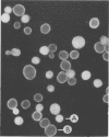
Existing methods for quantitating yeasts in beverages include time-consuming plate counts that detect only living cells and hemacytometer counts that are reliable only at very high concentrations (e.g., 10(6) to 20 X 10(6) cells per ml). The new method described here involves the use of fluorescence microscopy with the fluorescent stain aniline blue to differentiate yeasts (and other fungi) from backgrounds for easy counting and also may be used in conjunction with membrane filtration to concentrate yeasts from liquids before cell enumeration. Recoveries averaged 91.5% for beverages spiked with levels of 500 to 600,000 organisms per ml. The correlation coefficient of count to spike level was 0.996.
Full text
PDF
Images in this article
Selected References
These references are in PubMed. This may not be the complete list of references from this article.
- von Sengbusch P., Hechler J., Müller U. Molecular architecture of fungal cell walls. An approach by use of fluorescent markers. Eur J Cell Biol. 1983 May;30(2):305–312. [PubMed] [Google Scholar]