Abstract
Microvascular function in patients with type 1 diabetes without chronic complications was assessed using skin capillary recruitment during post-occlusive reactive hyperemia (PORH). Structural (maximal) capillary density was evaluated during venous occlusion. The study included 48 consecutive outpatients aged 26.3 ± 10.8 years with type 1 diabetes (duration of 9.5 years) without chronic complications and 34 control subjects. Intravital capillary video-microscopy was used in the dynamic study of skin capillaries in the dorsum of the fingers and toes. Capillary recruitment during PORH (% increase in mean capillary density, MCD) was significantly higher in the controls than the patients in both the fingers (p < 0.001) and toes (p < 0.001). During venous occlusion, MCD increase was also higher in the controls than the patients in both the fingers (p < 0.05) and toes (p < 0.0001). In patients, no difference was found between MCD at baseline and after venous occlusion in the fingers but a decrease was observed in the toes (p < 0.001). It is concluded that skin capillary function is significantly impaired in both fingers and toes of patients with type 1 diabetes without chronic complications. Moreover, capillary density during venous occlusion did not increase in either extremity in the patients, suggesting that their capillaries at rest are already maximally recruited.
Keywords: BMI, mean capillary density, type 1 diabetes, skin capillar, hyperemia, capillary video-microscopy
Introduction
Microvascular endothelial dysfunction is associ ated with the development of diabetic nephropathy and atherosclerosis in type 1 diabetes [1]. The main early alterations of capillary circulation in type 1 diabetes patients include increased capillary flow and permeability associated with basement membrane thickening [2]. Functional microvascular disturbances in the nail fold of the great toe in type 1 diabetic patients, evaluated by capillary blood flow velocity, have been shown to precede late diabetic complications [3, 4] and to be related to poor metabolic control [5]. Nevertheless, the presence of microvascular alterations in uncomplicated type 1 diabetes is still a controversial matter [1, 6-8]. The present study assessed microvascular function using skin capillary recruitment during post-occlusive reactive hyperemia, which is related to endothelium-dependent vasodilatation at the pre-capillary level [9, 10], in the upper and lower extremities of patients with type 1 diabetes without chronic complications under routine clinical care.
Methods
Patients and controls
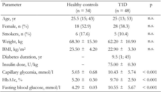
This cross-sectional observational study included 48 outpatients, 58.3% of whom were female, with a mean age of 25 (range; 13 to 53) years, with type 1 diabetes (Table 1). Patients consecutively attended the diabetes clinic at the State University of Rio de Janeiro and had a mean disease duration of 9.5 years, a fasting blood glucose (FBG) level of 10.55 ± 5.67 mmol/l, a HbA1c of 9.7 ± 2.5% and a BMI of 22.9 ± 3.3 kg/m2. Thirty-four control subjects, 52.9% of whom were female with a mean age of 25 (15-43) years, without diabetes (FBG < 5.5 mmol/l) and with an HbA1c of 5.2 ± 0.3% were also studied. Patients and controls were matched for age, gender, smoking habits and BMI. The inclusion criteria were patients with diabetes diagnosed before 30 years of age who had used insulin only since the diagnosis, without symptoms of diabetes decompensation or acute infection. The exclusion criteria were evidence of clinical cardiovascular and microvascular disease (albumin excretion rate < 20 μg/min in two sets of three consecutive overnight urine collections, peripheral neuropathy and retinopathy), Raynaud's syndrome and connective tissue diseases. In female patients and controls, all measurements were performed during the follicular phase of the menstrual cycle. The study was approved by the local ethics committee and patients and controls or their parents gave written informed consent.
Table 1. Baseline characteristics of study subjects.

Data are mean ± SD or median. Data in parantheses (minimum; maximum). T1D: type 1 diabetes patients. BMI: body mass index. n.s.: not significant.
Intravital video capillaroscopy and microvascular parameters
Intravital video-microscopy was carried out in the morning using a standardized and well-validated technique [9-12] in a temperature-controlled room (21-24°C). After measurement of capillary glycemia (Accu-Check Active, Roche, Germany), all subjects were given breakfast and diabetic patients were recommended to take their usual morning insulin dose (see Table 1). All the patients were using two shots of insulin NPH by day; regular insulin was also prescribed according to glycemia before meals. The investigations were carried out approximately 60 min after breakfast and 90 min after injection of insulin. Subjects were seated and the dorsal skin of the middle phalanx on the left hand and of the proximal phalanx of the big toe on the left foot was examined. Capillary density (CD) was defined as the total number of spontaneously perfused capillaries per mm2 of skin (functional CD). Percentage capillary recruitment was assessed by post-occlusive reactive hyperemia (PORH) after stopping arterial blood flow to the forearm and hand or to the foot by inflating a sphygmomanometer cuff over 3 min to 200 mmHg. Ten min after PORH, maximization of skin capillaries was obtained with 2 min of venous occlusion inflating the cuff to 60 mmHg (upper limb) or 90 mmHg (lower limb).
Statistical Analysis
Data were analyzed using SPSS 13.0. Comparisons of independent variables with normal distributions (capillary glycemia, HbA1c, fasting blood glucose, weight, BMI) were made by Student's t test. Comparisons of independent variables without normal distributions (age, comparison of skin capillaroscopy data between diabetic patients and controls) were made by the Mann-Whitney U test. Comparisons of microvascular variables with time in diabetic patients and controls were made by Wilcoxon signed rank test. Comparisons of dichotomous variables (gender, smoking habits and microvascular variables according to glycemic control) were made by χ2 tests. We considered p-values < 0.05 to be statistically significant.
Results
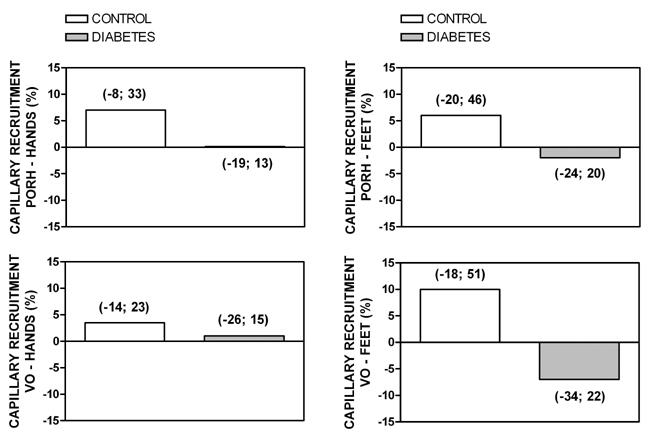
Mean capillary density (MCD) of the fingers at baseline did not differ between controls and patients (Table 2). In contrast, baseline MCD of the toes was lower in controls than in patients (83.5 ± 18.9 and 95.6 ± 25.4 capillaries/mm2 respectively, p < 0.01). Capillary recruitment during PORH (% increase in MCD, Figure 1) was significantly higher in controls than in patients in both the fingers (p < 0.001) and the toes (p < 0.001). During venous occlusion, MCD increase was also higher in controls than in patients in both the fingers (p < 0.05) and the toes (p < 0.0001). In the patient group, no difference was observed between MCD at baseline and during PORH, either in the fingers or the toes. In contrast, in the controls, MCD during PORH was significantly higher compared with baseline values in both the fingers (p < 0.001) and the toes (p < 0.001). In the patients, no difference was found between MCD at baseline and after venous occlusion in the fingers, but a decrease was noted in the toes (p < 0.001). In the controls, we observed a higher MCD after venous occlusion in comparison to baseline in both the fingers (p < 0.05) and the toes (p < 0.005).
Table 2. Skin capillaroscopy data of study subjects.

Data are MCD in capillaries/mm2 ± SD. MCD: mean capillary density. T1D: type 1 diabetes patients. PORH: post-occlusive reactive hyperemia. VO: venous occlusion. * p < 0.05, ** p < 0.01, *** p < 0.001 vs. baseline.
Figure 1.

Skin capillary recruitment measured during post-occlusive reactive hyperemia (PORH) or venous occlusion (VO) in the hands and feet of control subjects and patients with type 1 diabetes, using intravital video-microscopy. The percentage change in capillary density (capillary recruitment) was calculated using absolute change in capillary density during PORH and VO divided by basal capillary density (x 100). Values in parantheses represent minimum and maximum. Capillary recruitment during PORH in the fingers and toes: diabetics vs. controls, p < 0.001.
In our group of patients, there were no differences between patients with HbA1c < 7% (n = 8, 17%) and HbA1c < 7% (n = 40, 83%) concerning all the microvascular parameters studied.
Discussion
The occurrence of macro and/or microvascular endothelial dysfunction in patients with type 1 diabetes without chronic complications is not well established [13]. Notwithstanding, microvascular dysfunction may progress in silence for many years and antedate the development of vascular complications such as retinal and renal microangiopathy. The present study of patients without chronic complications demonstrated that skin capillary function, measured as post-ischemia capillary recruitment, is significantly impaired in both the fingers and toes of patients with type 1 diabetes, as compared to matched control subjects. Moreover, capillary density during venous occlusion, which is considered to represent structural capillary density [12], did not increase in either extremity in patients with type 1 diabetes, thus suggesting that their capillaries at rest are already maximally recruited. These results corroborate the hypothesis that early type 1 diabetes is characterized by increased microvascular pressure and flow and the loss of vasodilatory reserve and autoregulatory capacity [2].
In our study, basal capillary density in the feet of patients was higher than in control subjects, in contrast to the reduced nail fold capillary density described in patients with type 1 diabetes with retinopathy [14], thus suggesting that capillary rarefaction is present only in patients presenting with late complications of the disease. Jörneskog and colleagues [5] reported on significant differences in capillary blood flow between patients with good or bad metabolic control. In our study, the small number of patients with HbA1c < 7% precluded adequate conclusions concerning the correlations of microvascular alterations with glycemic control. Thus, a study of skin capillary recruitment in a subset of well-controlled type 1 patients in our population is warranted.
In conclusion, this study demonstrates early changes in skin capillary function in patients with type 1 diabetes without chronic complications. The causal and temporal relationship of capillary dysfunction with the development of chronic complications should be addressed in prospective studies designed for therapeutic intervention.
Acknowledgments
This investigation was supported by grants from FAPERJ (Fundação de Amparo à Pesquisa, Rio de Janeiro, Brazil), CNPq (Conselho Nacional de Desenvolvimento Tecnológico, grant #471014/2004-4) and FIOCRUZ (Oswaldo Cruz Foundation).
References
- 1.Stehouwer CD, Lambert J, Donker AJ, van Hinsbergh VW. Endothelial dysfunction and pathogenesis of diabetic angiopathy. Cardiovasc Res. 1997;34:55–68. doi: 10.1016/s0008-6363(96)00272-6. [DOI] [PubMed] [Google Scholar]
- 2.Tooke JE. Microvascular function in human diabetes. A physiological perspective. Diabetes. 1995;44:721–726. doi: 10.2337/diab.44.7.721. [DOI] [PubMed] [Google Scholar]
- 3.Jorneskog G, Brismar K, Fagrell B. Skin capillary circulation severely impaired in toes of patients with IDDM, with and without late diabetic complications. Diabetologia. 1995;38:474–480. doi: 10.1007/BF00410286. [DOI] [PubMed] [Google Scholar]
- 4.Jorneskog G, Fagrell B. Discrepancy in skin capillary circulation between fingers and toes in patients with type 1 diabetes. Int J Microcirc Clin Exp. 1996;16:313–319. doi: 10.1159/000179191. [DOI] [PubMed] [Google Scholar]
- 5.Jorneskog G, Brismar K, Fagrell B. Pronounced skin capillary ischemia in the feet of diabetic patients with bad metabolic control. Diabetologia. 1998;41:410–415. doi: 10.1007/s001250050923. [DOI] [PubMed] [Google Scholar]
- 6.Ladeia AM, Ladeia-Frota C, Pinho L, Stefanelli E, Adan L. Endothelial dysfunction is correlated with microalbuminuria in children with short-duration type 1 diabetes. Diabetes Care. 2005;28:2048–2050. doi: 10.2337/diacare.28.8.2048. [DOI] [PubMed] [Google Scholar]
- 7.Khan F, Elhadd TA, Greene SA, Belch JJ. Impaired skin microvascular function in children, adolescents, and young adults with type 1 diabetes. Diabetes Care. 2000;23:215–220. doi: 10.2337/diacare.23.2.215. [DOI] [PubMed] [Google Scholar]
- 8.Pichler G, Urlesberger B, Jirak P, Zotter H, Reiterer E, Muller W, Borkenstein M. Reduced forearm blood flow in children and adolescents with type 1 diabetes (measured by near-infrared spectroscopy) Diabetes Care. 2004;27(8):1942–1946. doi: 10.2337/diacare.27.8.1942. [DOI] [PubMed] [Google Scholar]
- 9.Serne EH, Gans RO, ter Maaten JC, ter Wee PM, Donker AJ, Stehouwer CD. Capillary recruitment is impaired in essential hypertension and relates to insulin's metabolic and vascular actions. Cardiovasc Res. 2001;49:161–168. doi: 10.1016/s0008-6363(00)00198-x. [DOI] [PubMed] [Google Scholar]
- 10.Serne EH, Stehouwer CD, ter Maaten JC, ter Wee PM, Rauwerda JA, Donker AJ, Gans RO. Microvascular function relates to insulin sensitivity and blood pressure in normal subjects. Circulation. 1999;99:896–902. doi: 10.1161/01.cir.99.7.896. [DOI] [PubMed] [Google Scholar]
- 11.Debbabi H, Uzan L, Mourad JJ, Safar M, Levy BI, Tibirica E. Increased skin capillary density in treated essential hypertensive patients. Am J Hypertens. 2006;19:477–483. doi: 10.1016/j.amjhyper.2005.10.021. [DOI] [PubMed] [Google Scholar]
- 12.Antonios TF, Rattray FE, Singer DR, Markandu ND, Mortimer PS, MacGregor GA. Maximization of skin capillaries during intravital video-microscopy in essential hypertension: comparison between venous congestion, reactive hyperaemia and core heat load tests. Clin Sci (Lond) 1999;97:523–528. [PubMed] [Google Scholar]
- 13.Schalkwijk CG, Stehouwer CD. Vascular complications in diabetes mellitus: the role of endothelial dysfunction. Clin Sci (Lond) 2005;109:143–159. doi: 10.1042/CS20050025. [DOI] [PubMed] [Google Scholar]
- 14.Gasser P, Berger W. Nailfold videomicroscopy and local cold test in type I diabetics. Angiology. 1992;43:395–400. doi: 10.1177/000331979204300504. [DOI] [PubMed] [Google Scholar]


