

Abstract
The growth and development of the human skeleton requires an adequate supply of many different nutritional factors. Classical nutrient deficiencies are associated with stunting (e.g. energy, protein, Zn), rickets (e.g. vitamin D) and other bone abnormalities (e.g. Cu, Zn, vitamin C). In recent years there has been interest in the role nutrition may play in bone growth at intakes above those required to prevent classical deficiencies, particularly in relation to optimising peak bone mass and minimising osteoporosis risk. There is evidence to suggest that peak bone mass and later fracture risk are influenced by the pattern of growth in childhood and by nutritional exposures in utero, in infancy and during childhood and adolescence. Of the individual nutrients, particular attention has been paid to Ca, vitamin D, protein and P. There has also been interest in several food groups, particularly dairy products, fruit and vegetables and foods contributing to acid–base balance. However, it is not possible at the present time to define dietary reference values using bone health as a criterion, and the question of what type of diet constitutes the best support for optimal bone growth and development remains open. Prudent recommendations (Department of Health, 1998; World Health Organization/Food and Agriculture Organization, 2003) are the same as those for adults, i.e. to consume a Ca intake close to the reference nutrient intake, optimise vitamin D status through adequate summer sunshine exposure (and diet supplementation where appropriate), be physically active, have a body weight in the healthy range, restrict salt intake and consume plenty of fruit and vegetables.
Keywords: Bone growth and development, Bone health, Nutritional factors, Dietary and lifestyle recommendations, Bone measurements in children
Many nutrients are essential for the growth and development of the skeleton. Bone tissue consists of crystals of hydroxyapatite ((Ca)10(PO4)6(OH)2) and other ions embedded in fibrils of collagen and a ground substance of glycoproteins and proteoglycans. Bone formation therefore requires adequate supplies of energy, amino acids and the main bone-forming minerals (Ca, P, Mg, Zn) and of other ions (e.g. Cu, Mn, carbonate, citrate) and vitamins (e.g. vitamins C, D, K) that are involved in crystal and collagen formation, cartilage and bone metabolism and/or Ca and phosphate homeostasis.
Development of the human skeleton begins in early embryonic life by the differentiation of cells into chondrocytes (cartilage-forming cells). Mesenchymal cells proliferate and differentiate into pre-osteoblasts and osteoblasts (bone-forming cells). The notochord is evident within 2 weeks and limb buds emerge by the second month of gestation. Expansion of bone mass occurs through intramembranous ossification (apposition) on the periosteal (outer) and endosteal (inner) bone surfaces and, at the ends of the long bones, by endochondral ossification within epiphyseal growth plates (layers of hyaline cartilage). Growth in the length of the long bones occurs in the growth plates through the chondrocytic production of cartilage, which then matures, calcifies and is subsequently resorbed and replaced by bone. Endochondral ossification ceases during puberty and the growth plates fuse (fully ossify) but intramembranous bone growth continues slowly throughout life. Thus, growth in stature ceases at maturity, but the outer skeletal envelope continues to expand by periosteal apposition during adulthood while the endosteal surfaces expand and contract depending on the stage of life (e.g. pregnancy, lactation, menopause). Skeletal growth and development are influenced by genetic variation, and polymorphisms in several genes have been identified that are associated with inter-individual variability in bone-related measures and hormone levels, such as in the vitamin D receptor, LDL receptor-related protein 5, insulin-like growth factor (IGF) 1 and growth hormone genes.
The velocity of skeletal growth is high immediately after birth, slows rapidly and increases again later in infancy. At this stage longitudinal bone growth is more rapid in the appendicular (arms and legs) than axial skeleton (trunk), and remains so until puberty. With the secretion of the sex hormones at puberty, axial bone growth accelerates while that of the long bones decelerates until the epiphyses fuse and linear growth ceases.
Gender difference in body size are evident from birth those in bone mass are relatively small before puberty. Bone mass accumulation increases sharply during puberty. In females the accretion rate in the lumbar spine and femoral neck increases about 4-fold before menarche, then slows such that bone mass changes little or even decreases thereafter. In males bone mass accretion increases approximately 6-fold during puberty with a slower but still marked accretion at many skeletal sites thereafter. In addition, there are gender differences in the cortical porosity of bone between adolescent boys and girls that may reflect greater intracortical bone remodelling in boys at this time. As a result of these differences males have a larger bone size and greater cortical thickness after puberty than females, but there is little difference in volumetric density.
More detailed accounts of the biology of human skeletal growth and development, and of the influences of genetic variability, can be found elsewhere (for example, see Tanner, 1962; Widdowson & Dickerson, 1964; Marcus et al. 1996; Seeman & Hopper, 1997; Prentice, 2001; Bilezikian et al. 2002; Glorieux et al. 2003; Davies et al. 2005; American Society for Bone and Mineral Research, 2006; Cooper et al. 2006; Ralston, 2007). In the present review the aim is to consider the influence of nutrition on bone growth and the extent to which the diets of children and adolescents impact on their current and future bone health. To this end, the effects of the classical nutritional deficiencies on skeletal health and the possible influences of diet composition and nutrient supply above that required to prevent classical deficiency on bone growth and fracture risk will be discussed separately.
Bone health
In the context of public health, the term ‘bone health’ largely refers to the quality of being at low risk of three common skeletal disorders: (a) stunting; (b) rickets and osteomalacia; (c) osteoporosis and fragility fractures.
Stunting represents chronic growth retardation and is defined as a height- or length-for-age that is <2 sd below the mean for reference children (de Onis & Blossner, 2003). Both skeletal growth and somatic growth are affected. Stunting is associated with increased mortality, delayed mental development and poor educational attainment in childhood. It can also set-up problems for later life, such as an increased risk of obstructed labour, which is a major cause of maternal and perinatal mortality in developing countries.
Rickets and osteomalacia refer to abnormalities of the skeleton in which mineral:bone collagen is low (Pettifor, 2003). Rickets is a failure of calcification in the growth plates of the long bones, producing characteristic bone deformities. It therefore occurs only in children, before the growth plates fuse. Osteomalacia is a failure of mineralisation on the trabecular and cortical surfaces of all bones, and can occur in both children and adults. Generally, children who present with rickets also have histological features of osteomalacia.
Osteoporosis describes a lack of bone within the skeletal envelope where the bone tissue that is present has a normal composition (Marcus et al. 1996). In post-menopausal women and in old age, when there is net loss of bone tissue, this condition is associated with deterioration of the skeletal trabecular architecture and thinning and increased porosity of the bone cortices. In growing individuals, when there is net accretion of bone tissue, osteoporosis generally refers to a low bone mass for achieved size with high porosity. In both cases osteoporosis is associated with an increased risk of fragility fracture, i.e. fracture caused with minimal trauma, such as a fall from a standing position. In children such fractures are most common in the distal forearm, and in older individuals at the wrist, spine and hip, but osteoporotic fractures can occur anywhere in the skeleton.
Classical nutritional deficiencies in children
An inadequate dietary supply from whatever cause may result in negative effects on growth and development. Severe protein–energy malnutrition and chronic under-nutrition lead to linear growth retardation (de Onis & Blossner, 2003). Energy for basal metabolism, physical activity and growth comes from the macronutrients, i.e. proteins, fats and carbohydrates. The sources of the different macronutrients are not important in relation to their use as metabolic fuels, but they are in the context of the other nutrients they provide (if any) and their composition (e.g. amino acid and fatty acid profiles, simple v. complex carbohydrates). Inadequate intakes and/or poor absorption of the bone-forming minerals, especially Ca and Zn, may also contribute to linear growth retardation (Prentice & Bates, 1994). Other micronutrients (vitamins, minerals and trace elements) are required for organ and tissue function and integrity, for metabolic processes and/or for uptake and metabolism of other nutrients (Gibney et al. 2002). With the exception of vitamin D, which can be synthesised in the skin by the action of sunlight at specific wavelengths, micronutrients cannot be made or inter-converted in the body and have to be supplied by the diet. There are well-characterised classical deficiency diseases that are caused by an inadequate supply of specific micronutrients, for example: night blindness, xerophthalmia and keratinization of skin (vitamin A); beri-beri (thiamin); scurvy (vitamin C); rickets and osteomalacia (vitamin D); impaired blood clotting and haemorrhagic disease (vitamin K); anaemia (Fe, folate). Such single-nutrient deficiencies in children tend to be associated with impaired growth and hence affect normal skeletal growth and development. Some deficiencies also affect cartilage and bone production directly, and cause characteristic skeletal abnormalities (e.g. Cu, vitamins C and D).
At the present time, the prevalence of nutrition-related deficiency disorders among children in many parts of the world is high. For example, it is estimated that globally 30% of children <5 years of age (>150 million children) are stunted, with the highest prevalence (>40%) in south Asian and sub-Saharan African countries (World Health Organization, 2006). High prevalence rates of rickets are also reported in many countries, even in tropical countries where sunlight with the appropriate wavelengths is abundant all year round (Table 1; Pettifor, 2003). Vitamin D deficiency is the most common underlying cause, often associated with restricted sunshine exposure at the required wavelengths because of latitude, cultural dress habits, lifestyle or atmospheric pollution (Pettifor, 2003). A very low Ca or P intake may also predispose to rickets even in the absence of frank vitamin D deficiency (Pettifor, 2003).
Table 1.
Prevalence of rickets*
| Country | Year | % |
|---|---|---|
| Asia, Middle East and Africa | ||
| Mongolia | 1998 | 70 |
| Tibet | 1994 | 66 |
| Ethiopia | 1997 | 42 |
| Yemen | 1987 | 27 |
| Turkey | 1994 | 10 |
| Nigeria | 1998 | 9 |
| Europe | ||
| The Netherlands: Macrobiotics | 1990 | 55 |
| UK: Manchester, minorities† | 2002 | 1·6 |
Data are the prevalence of clinical or radiological rickets in children not suffering from other diseases (Underwood & Margetts, 1987; Dagnelie et al. 1990; Ashraf & Mughal, 2002; Pettifor, 2003; Fraser, 2004).
Ethnic minorities, 77% were of south-east Asian origin (predominantly from Pakistan).
Thankfully, overt nutritional deficiencies in the UK, the Republic of Ireland and other parts of Western Europe have largely been consigned to history, although an increase in the prevalence of rickets has been recorded in recent years, and is a particular problem among some ethnic and cultural minority groups (Dagnelie et al. 1990; Shaw & Pal, 2002; Allgrove, 2004; Shenoy et al. 2005). However, the extent to which populations are at risk of subclinical or marginal deficiencies is difficult to gauge.
Assessment of nutritional adequacy
An assessment of dietary adequacy to prevent classical nutritional deficiencies can be made by comparing nutrient intakes to dietary reference values (DRV). In doing so, however, it needs to be borne in mind that considerable uncertainties surround the setting of DRV. The nutrient intake required to prevent or treat deficiency disease is usually only one of the criteria used to develop DRV, and other factors are considered including the intake required to maintain balance (the difference between nutrient intake and excretion), the extent of enzyme saturation or tissue concentration, or a given level of a validated biological marker. Bioavailability (encompassing absorption, metabolism, tissue distribution, excretion, chemical form of the nutrient) and the age, physiological stage and nutritional status of the individual also have to be considered. For children and adolescents the additional nutritional demands imposed by growth and development have to be factored in. However, in many instances a lack of experimental data means that it is not possible to determine DRV for children and adolescents specifically, and they are often extrapolated from adult or neonatal data.
There are wide variations in DRV between countries, often by as much as 2–3-fold for each nutrient (Prentice et al. 2004). This disparity reflects differences in the methods, philosophies and assumptions used, plus the fact that the values selected are generally tailored for the diet composition of the population for which the references are formulated. These variations are particularly acute at certain ages in childhood because of differences in the definition of boundaries between different stages of life, in the data used to denote average or desirable growth and in the methods selected for interpolation and extrapolation. All these issues and a comparison of methodological approaches have been addressed in depth, specifically in relation to children and adolescents, in a recent review of current DRV from twenty-nine European counties plus USA and Canada and WHO (Prentice et al. 2004).
Furthermore, assessments of dietary intakes and nutritional status in children and adolescents are difficult to compare between populations because of the use of different assessment techniques and food composition databases, as well as different DRV (Lambert et al. 2004). Few countries have intake data that are nationally representative; the UK's National Diet and Nutrition Survey (Gregory et al. 1995, 2000) and Ireland's National Children's Food Survey (Irish Universities Nutrition Alliance, 2006) are notable exceptions.
Using National Diet and Nutrition Survey data to consider children and adolescents in the UK (Gregory et al. 1995, 2000), it is clear that the intakes of macronutrients and many of the micronutrients do not give rise to concern in relation to frank nutritional deficiencies (Table 2 gives examples of protein, thiamin, vitamin C). However, for some nutrients average intakes are at or below the reference nutrient intake (which represents the intake of a nutrient considered sufficient to meet the needs of 97·5% of the population) and a sizeable proportion of individuals have intakes below the lower reference nutrient intake (which represents the intake considered unlikely to meet the needs of all but 2·5% of the population). Examples of such nutrients include Ca (Fig. 1), vitamin A, Fe, Zn and Mg (Gregory et al. 1995, 2000). While this finding cannot be taken to signify clinical or subclinical deficiency for any of these nutrients, and caution must be exercised about placing too much weight on such assessments because of the many uncertainties surrounding DRV and estimates of dietary intake, such data do give rise to concerns that a marked proportion of children in the UK may have intakes of some micronutrients that are marginal for optimal growth and development.
Table 2.
Mean protein, thiamin and vitamin C intakes of a representative sample of UK children expressed as a percentage of the reference nutrient intake*
| Age (years) | Protein | Thiamin | Vitamin C | |
|---|---|---|---|---|
| 1·5–4·5 | All | 244 | 156 | 172 |
| 4–6 | M | 249 | 183 | 247 |
| F | 226 | 167 | 230 | |
| 7–10 | M | 194 | 205 | 257 |
| F | 181 | 184 | 263 | |
| 11–14 | M | 152 | 190 | 224 |
| F | 128 | 202 | 210 | |
| 15–18 | M | 139 | 175 | 216 |
| F | 121 | 176 | 203 |
M, male; F, female.
Data from the National Diet and Nutrition Survey (Gregory et al. 1995, 2000). No child who participated in the survey had a thiamin or vitamin C intake less than the lower reference nutrient intake; no lower reference nutrient intake is set for protein.
Fig. 1.
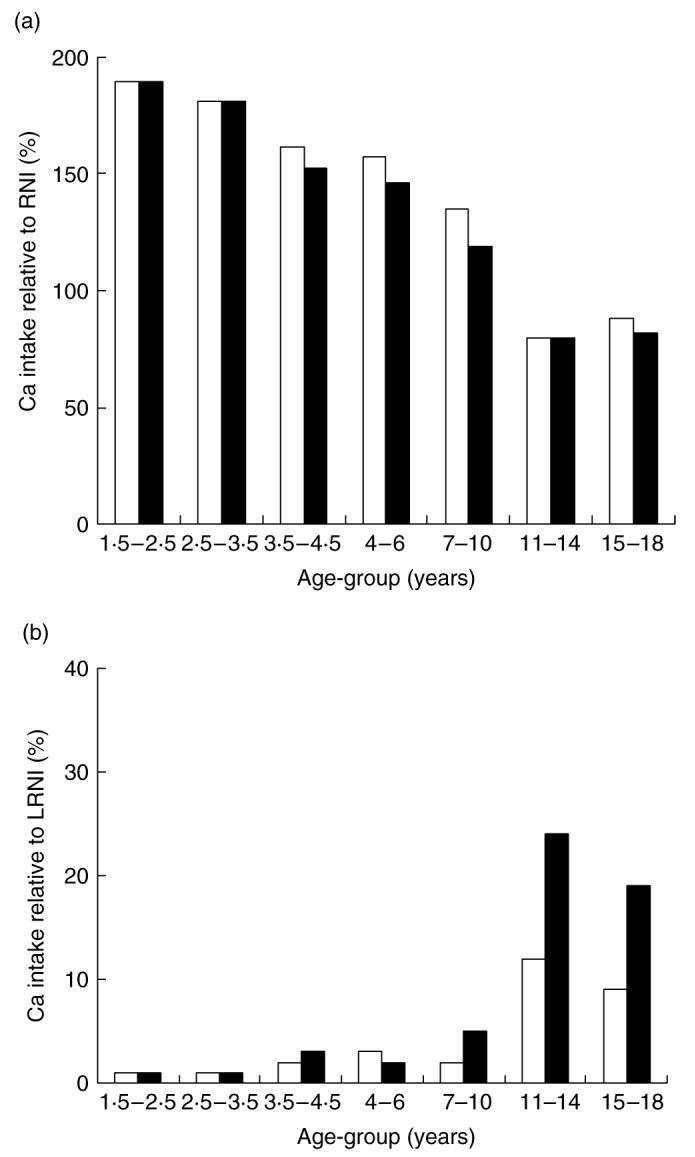
Calcium intake of a representative sample of UK children (□, boys; ■, girls) by age-group expressed relative to (a) the reference nutrient intake (RNI) and (b) the lower RNI (LRNI). Data are from the National Diet and Nutrition Survey (Gregory et al. 1995, 2000).
Adequacy of some nutrients can also be assessed by measuring biochemical markers of nutritional status or function. Of particular value is 25-hydroxyvitamin D as a marker of vitamin D status, because vitamin D is obtained from endogenous synthesis as well as from the diet and thus status cannot be assessed from intake. Patients with rickets and osteomalacia caused by vitamin D deficiency have plasma concentrations of 25-hydroxyvitamin D that range from undetectable to about 20 nmol/l (Arnaud et al. 1976). For this reason a threshold value of 25 nmol/l has been used conventionally to denote the lower limit of vitamin D adequacy (Department of Health, 1998; Willett, 2005). Applying this cut-off to National Diet and Nutrition Survey data (Fig. 2), it would appear that young children in the general UK population have reasonable vitamin D status but that the prevalence of low status increases to 10–15% among teenagers. However, in a survey of 2-year-old British Asian children (Fig. 2; Lawson & Thomas, 1999) the prevalence of low vitamin D status has been found to be 25%, suggesting that there is real cause for concern about the risk of vitamin D deficiency among some ethnic minorities in the UK.
Fig. 2.
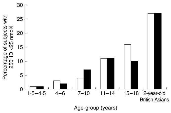
The vitamin D status of a representative sample of UK children (□, boys; ■, girls) by age-group expressed as the percentage with a plasma 25-hydroxyvitamin D (25OHD) concentration of <25 nmol/l (Department of Health, 1998). Data are for all seasons combined and are taken from the National Diet and Nutrition Survey (Gregory et al. 1995, 2000) and the Survey of Asian Children Living in England (Lawson & Thomas, 1999).
Optimal health
In addition to the avoidance of deficiency, concerns are voiced that the diets of children and adolescents in the UK and the Republic of Ireland may not be the best for their current or long-term health because the intakes or status of some nutrients may be lower (e.g. Ca, vitamin D, Zn, Fe, folate) or higher (e.g. Na) than may be optimal for growth or physiological function (Gregory et al. 2000). There are also anxieties expressed that the main sources of some nutrients may not be the most appropriate (e.g. vitamin K from oils rather than vegetables; Prynne et al. 2004), that food group recommendations are not being met (e.g. 5-a-day fruit and vegetable intake; Gregory et al. 2000) and that recent or anticipated changes in eating and lifestyle habits may compromise nutritional adequacy further (e.g. displacement of milk by less-nutrient-dense drinks, use of sunscreens). These concerns are coupled with fears that the DRV, food-based guidelines or status marker thresholds may have been set too conservatively and may therefore give false reassurance about nutrient adequacy and disease risk. A current example is the possibility that optimal health may require a vitamin D status >25 nmol/l, the threshold for 25-hydroxyvitamin D set on the basis of avoiding clinical deficiency. However, there is little consensus about what that higher threshold level should be. Cut-offs ranging from 25 nmol/l to >100 nmol/l have been proposed, largely based on the inverse relationship between 25-hydroxyvitamin D and parathyroid hormone (PTH) because a high PTH is a risk factor for osteoporosis in older adults (Willett, 2005). Most recently, a threshold level of ≥ 75 nmol/l has been suggested, based on multiple health outcomes in older adults (Bischoff-Ferrari et al. 2006). The extent to which such models are relevant to children and adolescents is not known, not least because physiological increases in PTH occur during periods of rapid growth such as puberty. However, with a threshold of >50 nmol/l, over one-third of 4–18-year-olds in the UK would be regarded as having suboptimal vitamin D status, and this value would rise to about 70% using the ≥ 75 nmol/l cut-off (Gregory et al. 2000). Clearly, the choice of threshold substantially alters the impression of vitamin D adequacy among children in the UK.
Fragility fractures
In Western countries concerns about children's diets in relation to bone health tend to centre on how best to optimise the development of strong healthy bones that have a low risk of fragility fracture during childhood and in later life.
Fractures are common in children, particularly at the wrist (Khosla et al. 2003; Ferrari et al. 2006). The incidence in both girls and boys peaks at puberty when distal forearm fracture incidence reaches a level similar to that seen in post-menopausal women (Heaney et al. 2000; Khosla et al. 2003). This peak may be related to the fact that the gain in bone mineral content (BMC) during puberty temporarily lags behind that of height and hence skeletal size, reducing bone strength. Although many of these fractures may be attributable to risk-taking in adventurous individuals, children who have experienced one fracture tend to be at increased risk of repeated fractures (Goulding et al. 2005; Ferrari et al. 2006) and to have lower bone mineral density (BMD) and accretion than their peers (Goulding et al. 2001; Ferrari et al. 2006; Manias et al. 2006). These findings suggests that there may be an underlying tendency in some children that may be related in part to diet or other environmental factors.
Fragility fractures in older adults are a major public health problem. Recent data from the National Osteoporosis Society (2006) indicate that osteoporosis affects over three million individuals in the UK and that one in two women and one in five men of >50 years of age can expect to fracture a bone during the rest of their lifetime. As a result of the high prevalence of osteoporosis in Caucasian populations, these data are magnified many times when considering the fracture burden across Europe, North America and Australasia. Although osteoporosis is currently regarded as a problem of Caucasian women, it is predicted that the number of hip fractures occurring annually throughout the world, currently over 2 × 10⁶, will rise steeply over the next 40–50 years because of the changing demographics, with Asia expected to see the greatest increases (Cooper et al. 1992; Prentice, 2004).
Assessment of bone health
Fracture incidence is a difficult end point to use as an outcome in studies attempting to define optimal diets for children and adolescents. BMC and BMD are good predictors of fracture risk in both children and older adults, and several techniques have been developed to provide estimates of bone mineral status in otherwise healthy individuals (Prentice, 1995, 2004; Prentice et al. 2003). The most commonly used technique is dual-energy X-ray absorptiometry (DXA), which has largely replaced its predecessor, dual-photon absorptiometry (which used radioisotopes rather than an X-ray source). DXA quantifies fat, lean and bone mineral masses and is the only widely-used technique that can measure regions in both the axial and appendicular skeleton. Software has been developed for use on some DXA systems to measure angles and dimensions within specific regions of the skeleton (especially the hip and spine). Single X-ray absorptiometry is a similar technique that is suitable for scanning regions in the appendicular skeleton, but it does not produce information about body composition. Peripheral quantitative computed tomography and radiogrammetry are X-ray techniques that also can only be applied to sites in the appendicular skeleton, but they provide additional information on bone shape and dimensions as well as mineral content. Classical anthropometry also provides information on external skeletal dimensions and geometry. Quantitative bone ultrasound is a technique that does not use X-rays and gives a measure of bone status in the appendicular skeleton based on the attenuation of ultrasound.
Most of these techniques provide measures of areal bone density (g/cm2; DXA) or volumetric bone density (g/cm3; peripheral quantitative computed tomography), BMC and areal (DXA) or cross-sectional (peripheral quantitative computed tomography) bone area for the skeletal regions scanned. The measures are not equivalent across the different techniques and comparisons cannot be made between them. Even comparing measurements using the same technique but on instruments developed by different manufacturers can be a problem because of the use of different calibration methods and materials. For example, DXA measurements can differ by 10–20% between instruments made by different manufacturers. Furthermore, these measurements are precise (1–2%) but not accurate. The precision of measurements depends on the region of the skeleton being scanned. With DXA precision is generally good for well-defined anatomical regions (e.g. spine and whole body), but relatively poor (>3%) for regions in the hip and forearm where there are few well-defined landmarks to aid localisation of the region of interest.
Most bone-densitometry techniques have been designed and evaluated for adults. Such measurements are particularly challenging in growing children, not least because bones are continually changing in size, shape and mass, the regions of interest are often difficult to define and bone edge detection can be a problem in small individuals (Fewtrell, 2003; Fulkerson et al. 2004; Petit et al. 2005; Prentice et al. 2005). Manufacturers have introduced paediatric and infant software to minimise technical problems related to the small size and the low tissue attenuation of children (Laskey & Prentice, 1999; Fulkerson et al. 2004), but because the software is appropriate only for children within specific age- or weight- ranges, the necessity of moving from one scan mode to another as a child grows can introduce problems in monitoring changes in bone measures over time.
There are some inherent difficulties with these bone-scanning techniques that limit their usefulness, both practically and interpretatively, for understanding the relationships between diet and resilience to fracture (Prentice, 2004). DXA data expressed as areal BMD are obtained by dividing BMC by bone area. It is, therefore, not a true density but an integration of X-ray attenuation across a two-dimensional projection of the scanned bone region. Such data can be heavily influenced by bone and body size (Prentice et al. 1994). Correction for bone and body size is critical when comparing populations with different anthropometric characteristics, when assessing individuals with impaired growth and when judging the influence of factors, such as dietary intake, that are themselves related to body size. Size-confounding can be a particularly difficult problem in studies of growing children, and different methods have been proposed to adjust for this size dependence, each having advantages and disadvantages (Molgaard et al. 1997; Fewtrell, 2003; Klein et al. 2005). Arguably, BMC and bone area separately, rather than areal BMD, are the more appropriate measures for charting influences on bone mineral accretion and skeletal development in childhood (Prentice, 2004).
Bone remodelling transients can also complicate interpretation of bone densitometry, especially in intervention studies (Prentice, 1997, 2004; Heaney, 2001). Bone remodelling (turnover) is the continual process by which the skeleton is renewed through the resorption (breakdown) of existing bone and the formation of new bone. Since these processes occur in sequence, remodelling gives rise to a temporary deficit of bone tissue, known as the remodelling space. Any intervention that alters the balance between resorption and formation produces a temporary phase lag while the system readjusts to the new remodelling rate, known as the bone remodelling transient. This readjustment process can lead to an increase or decrease in the remodelling space depending on whether resorption is temporarily increased or decreased relative to bone formation respectively. As a result of the precision of bone scanning techniques, particularly DXA, and the fact that these instruments provide a static measure of the skeleton at one specific time, alterations in the bone remodelling space can be detected as a change in BMC and BMD. Children, with their rapid bone growth and reshaping, have larger remodelling spaces than adults and a shorter remodelling period (several weeks compared with 6–18 months for older adults), making it more likely that bone remodelling transients will be detected.
Information about bone remodelling can be obtained through measuring biochemical markers in blood or urine (Szulc et al. 2000; Levine, 2003; Prentice et al. 2003), which include markers of osteoblast activity (e.g. osteocalcin, bone alkaline phosphatase), osteoclast activity (e.g. tartrate-resistant acid phosphatase), various collagen propeptides (e.g. amino-terminal and carboxy-terminal propeptides of type 1 procollagen) and breakdown products (e.g. hydroxyproline, deoxypyridinoline, N- and C-terminal telopeptides of collagen). Further insight into mechanisms can be obtained by measuring biochemical markers of bone metabolism, Ca homeostasis and growth modulators, such as PTH, vitamin D metabolites, cortisol, growth hormone and IGF-1 and -2 (Juul et al. 1994; Glorieux et al. 2003; American Society for Bone and Mineral Research, 2006). Levels of many of these markers are high in the first 2 years of life, decrease in childhood, increase again during puberty and decrease thereafter to the lower levels in adulthood. Thus, the concentrations or outputs of these markers are highly dependent on age, skeletal and sexual maturity, growth velocity and mineral accrual and can be difficult to interpret. In addition, the levels and pattern of change differ between boys and girls, and between ethnic groups. Further sources of variability include circadian periodicity, season, diet, exercise, kidney function and, in older girls, the phase of the menstrual cycle and the use of oral contraceptives. For technical reasons, there are also substantial differences between currently-available bone marker assays that severely limit any comparison or pooling of data between laboratories, and there are few reference values available for children. Furthermore, the patterns of change in these markers more closely match that of height than mineral accumulation; for example, the increase in bone formation markers precedes that of whole-body mineral accretion. Thus, for all these reasons, the concentrations or outputs of circulating and urinary markers are not directly translatable into amounts of bone formed or lost and thus the net bone balance (Szulc et al. 2000). Thus, bone markers can only be used in combination with measures of bone mineral to evaluate the relationship with diet or the net outcome of an intervention and to shed light on the mechanisms of action (Szulc et al. 2000; Prentice et al. 2003).
Bone strength is determined by many factors over and above bone mineral mass and BMD (Prentice et al. 2003; Prentice, 2004). These factors include bone size, bone geometry, distribution of bone tissue within the bone envelope and bone turnover, all of which change during childhood. On purely engineering principles, two bones with an identical BMC but different size will have different strengths under bending; the larger bone will have greater strength despite its lower areal BMD, while an increase in bone length will be detrimental to strength unless counteracted by growth in width (Seeman, 2002; Leonard et al. 2004). Furthermore, relationships between bone fragility and biochemistry such as bone remodelling markers and PTH established in elderly adults at risk of bone loss and osteoporosis may not apply to growing children (Prentice, 2006). The aspects of skeletal growth that need to be optimised in order to maximise bone strength and decrease fracture risk are not yet understood. For example, optimisation might be best achieved by one or more of the following: promoting bone and body size; increasing the amount of mineral within the bone envelope; altering the distribution of mineral within the skeleton; changing skeletal architecture and geometry; improving bone quality by altering bone turnover or microfracture repair mechanisms; influencing the tempo of growth and skeletal maturation (Prentice et al. 2005; Prentice, 2006). Many of these aspects are encapsulated within the measurement of BMC and BMD by the techniques currently available. Thus, it is important to recognise that, at present, only blunt tools are available for the assessment of bone health in children and adolescents and this factor needs to be borne in mind when interpreting studies into the influence of diet and nutrition on bone growth and development.
Maternal diet and bone health of the child
Maternal nutrition may have long-lasting effects on the growth and development of the child, because fetal life is a critical period for organogenesis and the development of metabolic systems, including the skeleton (Davies et al. 2005; Cooper et al. 2006; Langley-Evans, 2006). In studies relating growth in fetal and early life to later skeletal outcomes, birth weight and postnatal weight have been shown to be predictors of adult bone mass and skeletal size (Cooper et al. 1995, 1997; Dennison et al. 2005) and shortness at birth and slow childhood growth predict adult hip fracture (Cooper et al. 2001). The effects of early growth restriction may be particularly evident if a child undergoes an acceleration of growth beyond the normal rate for age, such as during catch-up growth after periods of nutritional deprivation. A greater risk of hip fracture in late adulthood has been demonstrated in individuals who were short at birth but were normal height by 7 years of age (Cooper et al. 2001). The influence of events occurring in fetal and early life on later health may be mediated by programming of the hormonal axes that persists into old age. For the skeleton, there is evidence of programming of the growth hormone–IGF-1, hypothalamo–pituitary–adrenal and PTH–vitamin D axes (Gluckman & Pinal, 2003; Tobias & Cooper, 2004; Tobias et al. 2005; Kapoor et al. 2006). Early nutrition and environment may also programme skeletal growth through interactions with underlying genotype (Jones & Dwyer, 2000; Davies et al. 2005). The interpretation of studies investigating the influence of early life on later bone growth is complicated because of the difficulty in distinguishing between the influence of pre- and postnatal exposures from those of current body weight and size, since anthropometric measures track through childhood and because bone and body size at any age are important determinants of bone mineral mass (Cole, 2004).
Maternal vitamin D status and intake of nutrients such as protein, fat, Ca, P, Mg, K and folate during pregnancy have been shown to predict height, BMC, bone area and areal BMD, and hence fracture risk, in prepubertal children (Jones et al. 2000; Tobias et al. 2005; Ganpule et al. 2006; Javaid et al. 2006). As bone mass tracks across puberty (Ferrari et al. 2006), it is also likely that maternal nutrition in pregnancy influences skeletal development of the offspring peri- and post-pubertally. To date there have been few intervention studies designed to investigate the influence of maternal diet on child bone growth and development. Two exceptions are Ca and vitamin D. Ca supplementation of pregnant Gambian and Indian mothers with a very low Ca intake was not found to be associated with marked increases in weight, length, BMC or areal BMD of the child after birth or during the first year of life (Raman et al. 1978; Jarjou et al. 2006). This outcome contrasts with that of an intervention study in the USA in which an effect of Ca supplementation on BMC was observed at 2 d postpartum in infants whose mothers had a low baseline Ca intake (Koo et al. 1999). Vitamin D supplementation of pregnant British Asian mothers at risk of poor vitamin D status was reported to result in a trend towards increased birth weight and in higher weight and length at 1 year (Brooke et al. 1980, 1981). Similarly, vitamin D supplementation of Swiss infants was found to be associated with higher prepubertal bone mass (Zamora et al. 1999).
Children's diets and bone health
The questions of most concern to parents and policy makers are what should the diets of children be to optimise their bone growth and development, and what aspects of modern-day Western diets are unfavourable to optimal bone health. As discussed earlier, there are concerns that the intakes of key nutrients by children are too low or too high, and that there are interactions between dietary components that render some food patterns more or less desirable. Many of the nutrients or dietary components that have received attention are those that are involved directly in bone metabolism or affect growth through actions on growth modulators, although mostly they are those that have actions, either positive or negative, on Ca absorption and excretion. These are of potential interest because 99% of the body's Ca is in the skeleton and therefore an effect on Ca retention is synonymous with an effect on bone mineral mass (Prentice, 1997; Prentice et al. 2003). Some of these dietary components have actions that enhance Ca absorption (e.g. Ca, vitamin D, sugars) or decrease excretion (e.g. B, alkali producers) and therefore have the potential to promote bone mineral accretion. Others have the potential to act in the opposite direction by inhibiting Ca absorption (e.g. fat, phytates, oxalates) or promoting urinary Ca excretion (e.g. Na, caffeine, animal protein, acid producers). However, given the complex nature of human diets, the extent to which any of these components or food patterns in isolation influences long-term Ca retention or fracture risk is uncertain (Prentice, 1997, 2004; Department of Health, 1998). The effects of some components may be offset by others (Heaney, 2004) while some may act synergistically (Bonjour, 2003).
In addition, other environmental and lifestyle factors, especially physical activity and, in older children, alcohol consumption, smoking and use of oral contraceptives, may interact with diet and nutritional status to influence bone growth and development (Bonjour, 2003). Furthermore, the effects of diet on children's bones may depend on their stage of maturity. For example, the skeleton appears to be more responsive to Ca, protein or exercise before the onset of puberty (Bonjour, 2003; Davies et al. 2005). In addition as mentioned earlier, BMC and areal BMD are influenced by overall body size and weight, and a low body weight is associated with greater fracture risk in children as well as in adults (Seeman, 1998; Prentice, 2004). Whether this increased risk is an effect of weight per se or is related to the different components of body composition (fat and/or muscle) and whether the effects differ by life-stage remain to be firmly established. At present, the evidence suggests that at the same weight a higher proportion of lean tissue compared with fat tissue (lean:fat) is associated with higher BMC and areal BMD in children and adolescents and adult men whereas a higher fat:lean is associated with a higher areal BMD in older women (Prentice, 2004). Accumulating evidence also suggests that a high body weight and obesity are risk factors for fracture in childhood (Goulding et al. 2001; Jones et al. 2004; Manias et al. 2006) and have been associated with low BMC, areal BMD and/or size for their weight in some studies (Goulding et al. 2001; Skaggs et al. 2001; Manias et al. 2006), but not in all (Leonard et al. 2004; Petit et al. 2005). Gains in total body weight and in fat, lean and bone mineral masses do not occur synchronously during growth (Seeman, 1998). In children body weight is more closely correlated with bone size than with BMC. Consequently, changes in skeletal strength may not match the increases in weight and body fat, especially when growth is rapid in early puberty, and this disparity may result in transient bone fragility (Seeman, 1998; Seeman et al. 2000; Skaggs et al. 2001). This mismatch may be particularly accentuated in heavy and obese children and in adolescents with low body weight as a result of anorexia nervosa, and may account for the greater fracture incidence in these groups (Goulding et al. 2000, 2002; Seeman et al. 2000).
Several comprehensive reviews are available that discuss the complexities of the evidence in relation to children's diets and bone health (Institute of Medicine Food and Nutrition Board, 1997; Department of Health, 1998; Bonjour, 2003; Prentice, 2004, 2006). The following section is a brief summary highlighting selected key nutrients (Ca, protein, P) and dietary patterns (milk and milk products, acid–base balance, fruit and vegetable consumption) that are at the forefront of current attention.
Key nutrients and dietary patterns
There continues to be considerable debate about whether Ca intakes at or close to current reference values are sufficient to optimise bone growth and development and to minimise current and later fracture risk (Prentice, 2004, 2006). Intervention studies using Ca from supplements or food sources have often demonstrated increases in BMC and areal BMD of about 1–5% in children and adolescents, including studies from the authors' group in the UK and The Gambia (Prentice, 1997, 2006; Department of Health, 1998; Dibba et al. 2000; Stear et al. 2003; Prentice et al. 2005). However, a recent Cochrane systematic review of nineteen trials in children (Winzenberg et al. 2006) has concluded that, taken overall, this effect is small. In this meta-analysis no evidence was found of effect modification by baseline Ca intake, gender, ethnicity, physical activity or pubertal stage. The lack of a relationship between the magnitude of the effect of Ca intervention and baseline Ca intake makes it difficult to use the results of intervention studies to identify an optimal Ca intake for promoting bone health or to consider the effect of the additional Ca on bone as recovery from an underlying dietary insufficiency (Prentice et al. 2005; Prentice, 2006). In the Cambridge Bone Studies (Stear et al. 2003; Prentice et al. 2005; Prentice, 2006), in which the diets of 294 teenage girls and boys aged 16–18 years were supplemented with 1000 mg Ca as Ca(CO3)2/d or a matching placebo for 12–15 months, different effects of the supplement were observed in girls and boys. In the girls BMC and areal BMD at several skeletal sites were found to be increased with little or no effect on bone area or overall skeletal size. However, in the boys the consumption of additional Ca was found to be associated not only with a higher BMC but also with an increase in skeletal size including stature. Consequently, the effect on BMC or areal BMD after size adjustment was not significant. In both cases an increase in IGF-1 was observed (Ginty et al. 2004). These findings raise questions about what is ultimately optimal for bone health, a greater bone density, as in the girls, or a greater bone size, as in the boys, and about whether the effect is transitory, as a result of changes in bone remodelling, or long-lasting. Follow-up studies of this cohort are in progress to try and answer some of these questions.
The observation of increased bone growth in the boys taking part in the Cambridge Bone Studies is the first time in a developed world context that supplementation with a Ca salt, in this case CaCO3, has been associated with an increase in stature and skeletal size (Prentice & Bates, 1994; Department of Health, 1998). In contrast, milk has long been known to be anabolic to the skeleton, probably via the promotion of IGF-1 (Cadogan et al. 1997; Department of Health, 1998; Ginty, 2003). The effect is unlikely to be entirely related to Ca because milk is rich in protein and other growth-promoting components. Milk intake in Danish children has been associated with height and serum IGF-1 concentration at 2.5 years, a relationship not seen with meat or vegetable protein (Hoppe et al. 2004b). Similarly, stimulation of IGF-1 by milk has been observed in 8-year-old Danish boys (Hoppe et al. 2004a). Furthermore, a low milk intake is a risk factor for childhood fractures (Goulding et al. 2004; Manias et al. 2006). However, studies investigating the relationship between milk and dairy product consumption and bone mineral measures in children and adolescents have not shown consistent results (Lanou et al. 2005).
Positive associations between total protein intake, BMC and bone size have been reported in children and adolescents. However, these results are difficult to interpret because protein intakes are driven partly by the requirements for growth (Ginty, 2003; Massey & Whiting, 2003) and because the level of protein intake may influence the tempo of growth and hence the shape and structure of the skeleton (Prentice, 2004). In addition, the effect of protein on bone may be biphasic, with potentially detrimental effects at high intakes because of its calciuric effect (Ginty, 2003; Massey & Whiting, 2003). However, the greater urinary Ca excretion associated with a high protein intake may be offset by higher Ca absorption or reduced urinary Ca excretion associated with other components in the protein source (such as Ca in milk, P in meat) and the overall acid–base balance of the diet (Ginty, 2003; Heaney, 2004; Prentice, 2004).
Acid–base balance refers to the potential of dietary components to contribute to the acid and alkali load of the body (Remer & Manz, 1995; Ginty, 2003; New, 2003; Prynne et al. 2004). When protons are produced during metabolism they are neutralised by the buffering action of anions generated from food or liberated from bone (Buclin et al. 2001). Since increases in bone resorption lead to greater urinary Ca excretion, a high renal acid load could potentially be associated with increased bone loss in older adults or reduced bone gain in children (Barzel, 1995; New et al. 2004). The main contributors to acid load are foods rich in S amino acids, P or chloride (e.g. in meat, grains, nuts, dairy products), while major contributors to alkali load are the K and Mg salts of organic acids (e.g. in fruit and vegetables). The acidity or alkalinity of the food before consumption does not predict its contribution to acid–base balance after metabolism (e.g. citric acid contributes to alkali load). The extent to which the potential renal acid load of the diet contributes to low bone gain or fracture risk in children and adolescence is not yet known. This aspect is currently under investigation in the Cambridge teenage cohort. Preliminary data would suggest that there are relationships between size-adjusted BMC and intake of fruit and vegetables (Prynne et al. 2006) but not with potential renal acid load (Ginty et al. 2005).
Carbonated drinks have been the focus of some concern because of the perception that such drinks contribute to acid load. In fact, carbonated drinks in which citric acid is the acidulant (e.g. lemonade) are net alkali producing, while the acidogenic potential of P in those in which phosphoric acid is the acidulant (e.g. colas) is relatively modest compared with milk and is similar, for example, to orange juice (Remer & Manz, 1995; Fitzpatrick & Heaney, 2003). Associations have been noted between high consumption of carbonated drinks and low areal BMD and risk of fracture in adolescents (Whiting et al. 2001; McGartland et al. 2003; Ma & Jones, 2004; Manias et al. 2006). A low milk intake and a high-carbonated drink consumption appear to be independent risk factors (Manias et al. 2006). It is considered likely that the associations with high-carbonated drink consumption may be partly attributed to confounding environmental factors, the displacement of milk from the diet and/or an effect of caffeine rather than to differences in acid–base balance (Heaney & Rafferty, 2001; Whiting et al. 2001; Fitzpatrick & Heaney, 2003; Ma & Jones, 2004).
P has actions that may affect Ca retention at high intakes, in addition to its contribution to acid load, such as the stimulation of PTH, luminal inhibition of Ca absorption and reduction in urinary Ca output (Calvo & Carpenter, 2003; Heaney, 2004). Similarly, fruit and vegetables, in addition to their contribution to an alkaline environment, may promote bone health in children and adolescents through components such as vitamin K (Bugel, 2003; Kalkwarf et al. 2004; Cashman, 2005) and phytooestrogens (Branca, 2003). Fruit and vegetable consumption has been associated in several studies, including the Cambridge Bone Studies, with greater bone mineral status in children and adolescents (New, 2003; Lanham-New, 2006; Prynne et al. 2006). Despite these findings, cross-sectional and longitudinal population-based studies do not suggest that there is a difference in areal BMD between vegetarians and omnivores, other than that which may be accounted for by differences in weight and lifestyle (New, 2004; Prentice, 2004).
Some interesting paradoxes arise when considering the composition of an optimal diet for bone health on the basis of diet composition (Prynne et al. 2004). For example, the Cambridge Bone Studies the greatest contributions to acid load in the teenage boys and girls were found to be from cheese and yoghurt, foods that were the main contributors to Ca intake, while the greatest contribution to alkali load was from potatoes and beverages. A low net acid excretion was observed not only in those with a high intake of fruit and vegetables and little meat, but also in those with a diet of chips, baked beans, crisps, chocolate and lager (Prynne et al. 2004). Thus, a low acid load is not necessarily associated with what is currently regarded as a healthy diet. These and other studies (Neville et al. 2002; Heaney, 2004; Storey et al. 2004; Lanou et al. 2005) demonstrate the importance of considering food groups and the whole diet in assessing the impact of diet on bone health, and may partly explain the lack of consistent relationships between the intakes of single nutrients and bone mineral acquisition in children and adolescents.
Policy perspective
Considering all the uncertainties detailed earlier, at the present time it is not possible to redefine DRV for children and adolescents using bone health as a criterion. Thus, the question of what constitutes a diet that will best support optimal bone growth and development remains open. However, the weight of evidence would suggest that the prudent recommendations for adults defined by the UK Committee on the Medical Aspects of Food Policy and Nutrition (Department of Health, 1998) and more recently by a joint WHO/FAO Consultation (World Health Organization, 2003) are equally applicable to children. The recommendations are to consume a Ca intake close to the reference nutrient intake, optimise vitamin D status through adequate summer sunshine exposure (and diet supplementation where appropriate), be physically active, have a weight in the healthy range, restrict salt intake and consume plenty of fruit and vegetables. Until further evidence becomes available these recommendations provide a valuable yardstick in selecting diets that are compatible with the promotion of good bone health in children and adolescents.
Abbreviations
- BMC
bone mineral content
- BMD
bone mineral density
- DRV
dietary reference values
- DXA
dual-energy X-ray absorptiometry
- IGF
insulin-like growth factor
- PTH
parathyroid hormone
References
- Allgrove J. Is nutritional rickets returning? Archives of Disease in Childhood. 2004;89:699–701. doi: 10.1136/adc.2003.036780. [DOI] [PMC free article] [PubMed] [Google Scholar]
- American Society for Bone and Mineral Research . Primer of the Metabolic Bone Diseases and Disorders of Mineral Metabolism. 6th ed. Washington, DC: The American Society for Bone and Mineral Research; 2006. [Google Scholar]
- Arnaud SB, Stickler GB, Haworth JC. Serum 25-hydroxyvitamin D in infantile rickets. Pediatrics. 1976;57:221–225. [PubMed] [Google Scholar]
- Ashraf S, Mughal MZ. The prevalence of rickets among non-Caucasian children. Archives of Disease in Childhood. 2002;87:263–264. doi: 10.1136/adc.87.3.263-a. [DOI] [PMC free article] [PubMed] [Google Scholar]
- Barzel US. The skeleton as an ion exchange system: implications for the role of acid-base imbalance in the genesis of osteoporosis. Journal of Bone and Mineral Research. 1995;10:1431–1436. doi: 10.1002/jbmr.5650101002. [DOI] [PubMed] [Google Scholar]
- Bilezikian JP, Raisz LG, Rodan GA, editors. Principles of Bone Biology. vol. 1 and 2. San Diego, CA: Academic Press; 2002. [Google Scholar]
- Bischoff-Ferrari HA, Giovannuci E, Willett WC, Dietrich T, Dawson-Hughes B. Estimation of optimal serum concentrations of 25-hydroxyvitamin D for multiple health outcomes. American Journal of Clinical Nutrition. 2006;84:18–28. doi: 10.1093/ajcn/84.1.18. [DOI] [PubMed] [Google Scholar]
- Bonjour J-P, Ammann P, Chevalley T, Ferrari S, Rizzoli R. Nutritional aspects of bone growth: an overview. In: New SA, Bonjour J-P, editors. Nutritional Aspects of Bone Health. Cambridge: The Royal Society of Chemistry; 2003. pp. 111–128. [Google Scholar]
- Branca F. Dietary phyto-oestrogens and bone health. Proceedings of the Nutrition Society. 2003;62:877–887. doi: 10.1079/PNS2003309. [DOI] [PubMed] [Google Scholar]
- Brooke OG, Brown IRF, Bone CDM, Carter ND, Cleeve HJW, Maxwell JD, Robinson VP, Winder SM. Vitamin D supplements in pregnant Asian women: Effects on Ca status and fetal growth. British Medical Journal. 1980;280:751–754. doi: 10.1136/bmj.280.6216.751. [DOI] [PMC free article] [PubMed] [Google Scholar]
- Brooke OG, Butters F, Wood C. Intrauterine vitamin D nutrition and postnatal growth in Asian infants. British Medical Journal. 1981;283:1024. doi: 10.1136/bmj.283.6298.1024. [DOI] [PMC free article] [PubMed] [Google Scholar]
- Buclin T, Cosma M, Appenzeller M, Jacquet AF, Decosterd LA, Biollaz J, Buckhardt P. Diet acids and alkalis influence Ca retention in bone. Osteoporosis International. 2001;12:493–499. doi: 10.1007/s001980170095. [DOI] [PubMed] [Google Scholar]
- Bugel S. Vitamin K and bone health. Proceedings of the Nutrition Society. 2003;62:839–843. doi: 10.1079/PNS2003305. [DOI] [PubMed] [Google Scholar]
- Cadogan J, Eastell R, Jones N, Barker ME. Milk intake and bone mineral acquisition in adolescent girls: randomised, controlled intervention trial. British Medical Journal. 1997;315:1255–1260. doi: 10.1136/bmj.315.7118.1255. [DOI] [PMC free article] [PubMed] [Google Scholar]
- Calvo M, Carpenter T. The influence of phosphorus on the skeleton. In: New SA, Bonjour J-P, editors. Nutritional Aspects of Bone Health. Cambridge: The Royal Society of Chemistry; 2003. pp. 229–265. [Google Scholar]
- Cashman KD. Vitamin K may be an important determinant of childhood bone health. Nutrition Reviews. 2005;63:284–289. doi: 10.1111/j.1753-4887.2005.tb00142.x. [DOI] [PubMed] [Google Scholar]
- Cole TJ. Modeling postnatal exposures and their interactions with birth size. Journal of Nutrition. 2004;134:201–204. doi: 10.1093/jn/134.1.201. [DOI] [PubMed] [Google Scholar]
- Cooper C, Campion G, Melton LJ. Hip fractures in the elderly: a world-wide projection. Osteoporosis International. 1992;2:285–289. doi: 10.1007/BF01623184. [DOI] [PubMed] [Google Scholar]
- Cooper C, Cawley M, Bhalla A, Egger P, Ring F, Morton L, Barker D. Childhood growth, physical activity, and peak bone mass in women. Journal of Bone and Mineral Research. 1995;10:940–947. doi: 10.1002/jbmr.5650100615. [DOI] [PubMed] [Google Scholar]
- Cooper C, Eriksson JG, Forsen T, Osmond C, Tuomilehto J, Barker DJ. Maternal height, childhood growth and risk of hip fracture in later life: a longitudinal study. Osteoporosis International. 2001;12:623–629. doi: 10.1007/s001980170061. [DOI] [PubMed] [Google Scholar]
- Cooper C, Fall C, Egger P, Hobbs R, Eastell R, Barker D. Growth in infancy and bone mass in later life. Annals of the Rheumatic Diseases. 1997;56:17–21. doi: 10.1136/ard.56.1.17. [DOI] [PMC free article] [PubMed] [Google Scholar]
- Cooper C, Westlake S, Harvey N, Javaid K, Dennison E, Hanson M. Review: developmental origins of osteoporotic fracture. Osteoporosis International. 2006;17:337–347. doi: 10.1007/s00198-005-2039-5. [DOI] [PubMed] [Google Scholar]
- Dagnelie PC, Vergote FJ, van Staveren WA, van den Berg H, Dingian PG, Hautvast JG. High prevalence of rickets in infants on macrobiotic diets. American Journal of Clinical Nutrition. 1990;51:202–208. doi: 10.1093/ajcn/51.2.202. [DOI] [PubMed] [Google Scholar]
- Davies JH, Evans BA, Gregory JW. Bone mass acquisition in healthy children. Archives of Disease in Childhood. 2005;90:373–378. doi: 10.1136/adc.2004.053553. [DOI] [PMC free article] [PubMed] [Google Scholar]
- de Onis M, Blossner M. The World Health Organization global database on child growth and malnutrition: methodology and applications. International Journal of Epidemiology. 2003;32:518–526. doi: 10.1093/ije/dyg099. [DOI] [PubMed] [Google Scholar]
- Dennison EM, Syddall HE, Sayer AA, Gilbody HJ, Cooper C. Birth weight and weight at 1 year are independent determinants of bone mass in the seventh decade: the Hertfordshire cohort study. Pediatric Research. 2005;57:582–586. doi: 10.1203/01.PDR.0000155754.67821.CA. [DOI] [PubMed] [Google Scholar]
- Department of Health . Nutrition and Bone Health: With Particular Reference to Ca and Vitamin D. London: The Stationery Office; 1998. (Report on Health and Social Subjects no. 49). [Google Scholar]
- Dibba B, Prentice A, Ceesay M, Stirling DM, Cole TJ, Poskitt EME. Effect of Ca supplementation on bone mineral accretion in Gambian children accustomed to a low Ca diet. American Journal of Clinical Nutrition. 2000;71:544–549. doi: 10.1093/ajcn/71.2.544. [DOI] [PubMed] [Google Scholar]
- Ferrari SL, Chevalley T, Bonjour JP, Rizzoli R. Childhood fractures are associated with decreased bone mass gain during puberty: an early marker of persistent bone fragility? Journal of Bone and Mineral Research. 2006;21:501–507. doi: 10.1359/jbmr.051215. [DOI] [PubMed] [Google Scholar]
- Fewtrell MS. Bone densitometry in children assessed by dual x ray absorptiometry: uses and pitfalls. Archives of Disease in Childhood. 2003;88:795–798. doi: 10.1136/adc.88.9.795. [DOI] [PMC free article] [PubMed] [Google Scholar]
- Fitzpatrick L, Heaney RP. Got soda? Journal of Bone and Mineral Research. 2003;18:1570–1572. doi: 10.1359/jbmr.2003.18.9.1570. [DOI] [PubMed] [Google Scholar]
- Fraser DR. Vitamin D-deficiency in Asia. Journal of Steroid Biochemistry and Molecular Biology. 2004;89–90:491–495. doi: 10.1016/j.jsbmb.2004.03.057. [DOI] [PubMed] [Google Scholar]
- Fulkerson JA, Himes JH, French SA, Jensen S, Petit MA, Stewart C, Story M, Ensrud K, Fillhouer S, Jacobsen K. Bone outcomes and technical measurement issues of bone health among children and adolescents: considerations for nutrition and physical activity intervention trials. Osteoporosis International. 2004;15:929–941. doi: 10.1007/s00198-004-1685-3. [DOI] [PubMed] [Google Scholar]
- Ganpule A, Yajnik CS, Fall CHD, Rao S, Fisher DJ, Kanade A, et al. Bone mass in Indian children; relationships to maternal nutritional status and diet during pregnancy; the Pune Maternal Nutrition Study. Journal of Clinical Endocrinology and Metabolism. 2006;91:2994–3001. doi: 10.1210/jc.2005-2431. [DOI] [PubMed] [Google Scholar]
- Gibney MJ, Vorster HH, Kok FJ, editors. Introduction to Human Nutrition. Oxford: Blackwell Publishing; 2002. [Google Scholar]
- Ginty F. Dietary protein and bone health. Proceedings of the Nutrition Society. 2003;62:867–876. doi: 10.1079/PNS2003307. [DOI] [PubMed] [Google Scholar]
- Ginty F, Prentice A, Laidlaw A, McKenna L, Jones SC, Stear SJ, Cole TJ. Calcium carbonate supplementation is associated with higher plasma IGF-1 in 16–18 year old boys and girls. In: Burckhardt P, Dawson-Hughes B, Heaney RP, editors. Nutritional Aspects of Osteoporosis. 2nd ed. San Diego, CA: Elsevier Science; 2004. pp. 45–57. [Google Scholar]
- Ginty F, Prynne CJ, Muniz-Terrera G, Mishra GD, Prentice A, O'Connell MA. No evidence for a negative assocation between bone mineral status and indirect estimates of renal net acid excretion in adolescents. Proceedings of the Nutrition Society. 2005;64:80A. [Google Scholar]
- Glorieux F, Jueppner H, Pettifor JM. Pediatric Bone – Biology and Diseases. San Diego, CA: Elsevier Science; 2003. [Google Scholar]
- Gluckman PD, Pinal CS. Regulation of fetal growth by the somatotrophic axis. Journal of Nutrition. 2003;133:1741S–1746S. doi: 10.1093/jn/133.5.1741S. [DOI] [PubMed] [Google Scholar]
- Goulding A, Jones IE, Taylor RW, Williams SM, Manning PJ. Bone mineral density and body composition in boys with distal forearm fractures: a dual energy x-ray absorptiometry study. Journal of Paediatrics. 2001;139:509–515. doi: 10.1067/mpd.2001.116297. [DOI] [PubMed] [Google Scholar]
- Goulding A, Jones IE, Williams SM, Grant AM, Taylor RW, Manning PJ, Langley J. First fracture is associated with increased risk of new fractures during growth. Journal of Pediatrics. 2005;146:286–288. doi: 10.1016/j.jpeds.2004.09.029. [DOI] [PubMed] [Google Scholar]
- Goulding A, Rockell JE, Black RE, Grant AM, Jones IE, Williams SM. Children who avoid drinking cow's milk are at increased risk for prepubertal bone fractures. Journal of the American Dietetic Association. 2004;104:250–253. doi: 10.1016/j.jada.2003.11.008. [DOI] [PubMed] [Google Scholar]
- Goulding A, Taylor RW, Jones IE, McAuley KA, Manning PJ, Williams SM. Overweight and obese children have low bone mass and area for their weight. International Journal of Obesity. 2000;24:627–632. doi: 10.1038/sj.ijo.0801207. [DOI] [PubMed] [Google Scholar]
- Goulding A, Taylor RW, Jones IE, Manning PJ, Williams SM. Spinal overload: a concern for obese children and adolescents? Osteoporosis International. 2002;13:835–840. doi: 10.1007/s001980200116. [DOI] [PubMed] [Google Scholar]
- Gregory J, Lowe S, Bates C, Prentice A, Jackson L, Smithers G, Wenlock R, Farron M. National Diet and Nutrition Survey: Young People Aged 4 to 18 Years. vol. 1: Report of the Diet and Nutrition Survey. London: The Stationery Office; 2000. [Google Scholar]
- Gregory JR, Collins DL, Davies PSW, Hughes JM, Clarke PC. National Diet and Nutrition Survey: Children Aged 1.5 to 4.5 Years. London: HM Stationery Office; 1995. [Google Scholar]
- Heaney RP. The bone remodeling transient: interpreting interventions involving bone-related nutrients. Nutrition Reviews. 2001;59:327–334. doi: 10.1111/j.1753-4887.2001.tb06957.x. [DOI] [PubMed] [Google Scholar]
- Heaney RP. Nutrients, interactions and foods: the importance of source. In: Burckhardt P, Dawson-Hughes B, Heaney RP, editors. Nutritional Aspects of Osteoporosis. 2nd ed. San Diego, CA: Elsevier Science; 2004. pp. 61–78. [Google Scholar]
- Heaney RP, Abrams S, Dawson-Hughes B, Looker A, Marcus R, Matkovic V, Weaver C. Peak bone mass. Osteoporosis International. 2000;11:985–1009. doi: 10.1007/s001980070020. [DOI] [PubMed] [Google Scholar]
- Heaney RP, Rafferty K. Carbonated beverages and urinary Ca excretion. American Journal of Clinical Nutrition. 2001;74:343–347. doi: 10.1093/ajcn/74.3.343. [DOI] [PubMed] [Google Scholar]
- Hoppe C, Mølgaard C, Michaelsen KF. High intakes of skimmed milk, but not meat, increase serum IGF-1 and IGFBP-3 in eight year old boys. European Journal of Clinical Nutrition. 2004a;58:1211–1216. doi: 10.1038/sj.ejcn.1601948. [DOI] [PubMed] [Google Scholar]
- Hoppe C, Udam TR, Lauritzen L, Mølgaard C, Juul A, Michaelsen KF. Animal protein intake, serum insulin-like growth factor I, and growth in healthy 2·5-y-old Danish children. American Journal of Clinical Nutrition. 2004b:447–452. doi: 10.1093/ajcn/80.2.447. [DOI] [PubMed] [Google Scholar]
- Institute of Medicine Food and Nutrition Board . Dietary Reference Intakes for Calcium, Phosphorus, Magnesium, Vitamin D, and Fluoride. Washington, DC: National Academy Press; 1997. [PubMed] [Google Scholar]
- Irish Universities Nutrition Alliance National Children's Food Survey. 2006. http://www.iuna.net/childrens_survey/
- Jarjou LMA, Prentice A, Sawo Y, Laskey MA, Bennett J, Goldberg GR, Cole TJ. Randomized, placebo-controlled Ca supplementation study of pregnant Gambian women: effects on breast-milk Ca concentration and infant birth weight, growth and bone mineral accretion in the first year of life. American Journal of Clinical Nutrition. 2006;83:657–666. doi: 10.1093/ajcn.83.3.657. [DOI] [PubMed] [Google Scholar]
- Javaid MK, Crozier SR, Harvey NC, Gale CR, Dennison EM, Boucher BJ, Arden NK, Godfrey KM, Cooper C. Maternal vitamin D status during pregnancy and childhood bone mass at age 9 years: a longitudinal study. Lancet. 2006;367:36–43. doi: 10.1016/S0140-6736(06)67922-1. [DOI] [PubMed] [Google Scholar]
- Jones G, Dwyer T. Birth weight, birth length, and bone density in prepubertal children: evidence for an association that may be mediated by genetic factors. Calcified Tissues International. 2000;67:304–308. doi: 10.1007/s002230001148. [DOI] [PubMed] [Google Scholar]
- Jones G, Riley MD, Dwyer T. Maternal diet during pregnancy is associated with bone mineral density in children: a longitudinal study. European Journal of Clinical Nutrition. 2000;54:749–756. doi: 10.1038/sj.ejcn.1601082. [DOI] [PubMed] [Google Scholar]
- Jones IE, Williams SM, Goulding A. Associations of birth weight and length, childhood size, and smoking with bone fractures during growth: evidence from a birth cohort study. American Journal of Epidemiology. 2004;159:343–350. doi: 10.1093/aje/kwh052. [DOI] [PubMed] [Google Scholar]
- Juul A, Bang P, Hertel NT, Main K, Dalgaard P, Jorgensen K, Muller J, Hall K, Skakkebaek NE. Serum insulin-like growth factor-I in 1030 healthy children, adolescents, and adults: relation to age, sex, stage of puberty, testicular size, and body mass index. Journal of Clinical Endocrinology and Metabolism. 1994;78:744–752. doi: 10.1210/jcem.78.3.8126152. [DOI] [PubMed] [Google Scholar]
- Kalkwarf HJ, Khoury JC, Bean J, Elliot JG. Vitamin K, bone turnover, and bone mass in girls. American Journal of Clinical Nutrition. 2004;80:1075–1080. doi: 10.1093/ajcn/80.4.1075. [DOI] [PubMed] [Google Scholar]
- Kapoor A, Dunn E, Kostaki A, Andrews MH, Matthews SG. Fetal programming of hypothalamo-pituitary-adrenal function: prenatal stress and glucocorticoids. Journal of Physiology. 2006;572:31–44. doi: 10.1113/jphysiol.2006.105254. [DOI] [PMC free article] [PubMed] [Google Scholar]
- Khosla S, Melton LJ, III, Dekutoski MB, Achenbach SJ, Oberg AL, Riggs BL. Incidence of childhood distal forearm fractures over 30 years. A population-based study. Journal of the American Medical Association. 2003;290:1479–1485. doi: 10.1001/jama.290.11.1479. [DOI] [PubMed] [Google Scholar]
- Klein GL, Fitzpatrick LA, Langman CB, Beck TJ, Carpenter TO, Gilsanz V, Holm IA, Leonard MB, Specker BL. The state of pediatric bone: summary of the ASBMR Pediatric Bone Initiative. Journal of Bone and Mineral Research. 2005;20:2075–2081. doi: 10.1359/JBMR.050901. [DOI] [PubMed] [Google Scholar]
- Koo WW, Walters JC, Esterlitz J, Levine RJ, Bush AJ, Sibai B. Maternal Ca supplementation and fetal bone mineralisation. Obstetrics and Gynecology. 1999;94:577–582. doi: 10.1016/s0029-7844(99)00371-3. [DOI] [PubMed] [Google Scholar]
- Lambert J, Agostoni C, Elmadfa I, Hulshof K, Krause E, Livingstone B, Socha P, Pannemans D, Samartin S. Dietary intake and nutritional status of children and adolescents in Europe. British Journal of Nutrition. 2004;92(Suppl. 2):S147–S211. doi: 10.1079/bjn20041160. [DOI] [PubMed] [Google Scholar]
- Langley-Evans SC. Developmental programming of health and disease. Proceedings of the Nutrition Society. 2006;65:97–105. doi: 10.1079/pns2005478. [DOI] [PMC free article] [PubMed] [Google Scholar]
- Lanham-New SA. Fruit and vegetables: the unexpected natural answer to the question of osteoporosis prevention? American Journal of Clinical Nutrition. 2006;83:1254–1255. doi: 10.1093/ajcn/83.6.1254. [DOI] [PubMed] [Google Scholar]
- Lanou AJ, Berkow SE, Barnard ND. Calcium, dairy products, and bone health in children and young adults: a reevaluation of the evidence. Pediatrics. 2005;115:736–743. doi: 10.1542/peds.2004-0548. [DOI] [PubMed] [Google Scholar]
- Laskey MA, Prentice A. Comparison of adult and paediatric spine and whole body software for the Lunar dual energy X-ray absorptiometer. British Journal of Radiology. 1999;72:967–976. doi: 10.1259/bjr.72.862.10673948. [DOI] [PubMed] [Google Scholar]
- Lawson M, Thomas M. Vitamin D concentrations in Asian children aged 2 years living in England: population survey. British Medical Journal. 1999;318:28–29. doi: 10.1136/bmj.318.7175.28. [DOI] [PMC free article] [PubMed] [Google Scholar]
- Leonard MB, Shults J, Elliott DM, Stallings VA, Zemel BS. Interpretation of whole body dual energy X-ray absorptiometry measures in children: comparison with peripheral quantitative computed tomography. Bone. 2004;34:1044–1052. doi: 10.1016/j.bone.2003.12.003. [DOI] [PubMed] [Google Scholar]
- Levine RA. Biochemical markers of bone metabolism: application to understanding bone remodeling in children and adolescents. Journal of Pediatric Endocrinology and Metabolism. 2003;16(Suppl. 3):661–672. [PubMed] [Google Scholar]
- McGartland C, Robson PJ, Murray L, Cran G, Savage MJ, Watkins D, Rooney M, Boreham C. Carbonated soft drink consumption and bone mineral density in adolescence: The Northern Ireland Young Hearts Project. Journal of Bone and Mineral Research. 2003;18:1563–1569. doi: 10.1359/jbmr.2003.18.9.1563. [DOI] [PubMed] [Google Scholar]
- Ma D, Jones G. Soft drink and milk consumption, physical activity, bone mass and upper limb fractures in children: a population-based case-control study. Calcified Tissue International. 2004;75:286–291. doi: 10.1007/s00223-004-0274-y. [DOI] [PubMed] [Google Scholar]
- Manias K, McCabe D, Bishop N. Fractures and recurrent fractures in children; varying effects of environmental factors as well as bone size and mass. Bone. 2006;39:652–657. doi: 10.1016/j.bone.2006.03.018. [DOI] [PubMed] [Google Scholar]
- Marcus R, Feldman D, Kelsey J. Osteoporosis. San Diego, CA: Academic Press; 1996. [Google Scholar]
- Massey LK, Whiting SJ. Excess dietary protein and bone health. In: New SA, Bonjour J-P, editors. Nutritional Aspects of Bone Health. Cambridge: The Royal Society of Chemistry; 2003. pp. 213–228. [Google Scholar]
- Mølgaard C, Thomsen BL, Prentice A, Cole TJ, Michaelsen KF. Whole body bone mineral content in healthy children and adolescents. Archives of Diseases in Childhood. 1997;76:9–15. doi: 10.1136/adc.76.1.9. [DOI] [PMC free article] [PubMed] [Google Scholar]
- National Osteoporosis Society All about osteoporosis. 2006. http://www.nos.org.uk/about.htm.
- Neville CE, Robson PJ, Murray LJ, Strain JJ, Twisk J, Gallagher AM, McGuinness M, Cran GW, Ralston SH, Boreham CA. The effect of nutrient intake on bone mineral status in young adults: the Northern Ireland Young Hearts project. Calcified Tissue International. 2002;70:89–98. doi: 10.1007/s00223-001-1023-0. [DOI] [PubMed] [Google Scholar]
- New SA. Intake of fruit and vegetables: implications for bone health. Proceedings of the Nutrition Society. 2003;62:889–899. doi: 10.1079/PNS2003310. [DOI] [PubMed] [Google Scholar]
- New SA. Do vegetarians have a normal bone mass? Osteoporosis International. 2004;15:679–688. doi: 10.1007/s00198-004-1647-9. [DOI] [PubMed] [Google Scholar]
- New SA, MacDonald HM, Campbell MK, Martin JC, Garton MJ, Robins SP, Reid DM. Lower estimates of net endogenous non-carbonic acid production are positively associated with indexes of bone health in premenopausal and perimenopausal women. American Journal of Clinical Nutrition. 2004;79:131–138. doi: 10.1093/ajcn/79.1.131. [DOI] [PubMed] [Google Scholar]
- Petit MA, Beck TJ, Kontulainen SA. Examining the developing bone: What do we measure and how do we do it? Journal of Musculoskeletal and Neuronal Interactions. 2005;5:213–224. [PubMed] [Google Scholar]
- Pettifor JM. Nutritional rickets. In: Glorieux F, Jueppner H, Pettifor JM, editors. Pediatric Bone – Biology and Diseases. San Diego, CA: Elsevier Science; 2003. pp. 541–566. [Google Scholar]
- Prentice A. Application of dual-energy X-ray absorptiometry and related techniques to the measurement of bone and body composition. In: Davies PSW, Cole TJ, editors. Body Composition Techniques in Health and Disease. Cambridge: Cambridge University Press; 1995. pp. 1–13. [Google Scholar]
- Prentice A. Is nutrition important in osteoporosis? Proceedings of the Nutrition Society. 1997;56:357–367. doi: 10.1079/pns19970038. [DOI] [PubMed] [Google Scholar]
- Prentice A. The relative contribution of diet and genotype to bone development. Proceedings of the Nutrition Society. 2001;60:1–8. doi: 10.1079/pns200072. [DOI] [PubMed] [Google Scholar]
- Prentice A. Diet, nutrition and osteoporosis. Public Health Nutrition. 2004;7:237–254. doi: 10.1079/phn2003590. [DOI] [PubMed] [Google Scholar]
- Prentice A. Studies of Gambian and UK children and adolescents: insights into Ca requirements and adaptation to a low Ca intake. Proceedings of the 6th International Symposium on Nutritional Aspects of Osteoporosis; Lausanne, Switzerland. 2006. In the Press. [Google Scholar]
- Prentice A, Bates CJ. Adequacy of dietary mineral supply for human bone growth and mineralisation. European Journal of Clinical Nutrition. 1994;48(Suppl. 1):S161–S176. [PubMed] [Google Scholar]
- Prentice A, Bonjour J-P, Branca F, Cooper C, Flynn A, Garabedian M, Muller D, Pannemans D, Weber P. PASSCLAIM – Bone Health and Osteoporosis. European Journal of Nutrition. 2003;42(Suppl. 1):I/28–I/49. doi: 10.1007/s00394-003-1103-1. [DOI] [PubMed] [Google Scholar]
- Prentice A, Branca F, Decsi T, Michaelsen KF, Fletcher R, Guesry P, Manz F, Vidailhet M, Pannemans D, Samartin S. Energy and nutrient dietary reference values for children in Europe: methodological approaches and current nutritional recommendations. British Journal of Nutrition. 2004;92(Suppl. 2):S83–S146. doi: 10.1079/bjn20041159. [DOI] [PubMed] [Google Scholar]
- Prentice A, Ginty F, Stear SJ, Jones SC, Laskey MA, Cole TJ. Calcium supplementation increases stature and bone mineral mass of 16- to 18-year-old boys. Journal of Clinical Endocrinology and Metabolism. 2005;90:3153–3161. doi: 10.1210/jc.2004-2114. [DOI] [PubMed] [Google Scholar]
- Prentice A, Parsons TJ, Cole TJ. Uncritical use of bone mineral density in absorptiometry may lead to size-related artifacts in the identification of bone mineral determinants. American Journal of Clinical Nutrition. 1994;60:837–842. doi: 10.1093/ajcn/60.6.837. [DOI] [PubMed] [Google Scholar]
- Prynne CJ, Ginty F, Paul AA, Bolton-Smith C, Stear SJ, Jones SC, Prentice A. Dietary acid-base balance and intake of bone-related nutrients in Cambridge teenagers. European Journal of Clinical Nutrition. 2004;58:1462–1471. doi: 10.1038/sj.ejcn.1602006. [DOI] [PubMed] [Google Scholar]
- Prynne CJ, Mishra G, O'Connell MA, Muniz G, Laskey MA, Yan L, Prentice A. Fruit and vegetable intakes and bone mineral status: a cross-sectional study in 5 age and sex cohorts. American Journal of Clinical Nutrition. 2006;83:1420–1428. doi: 10.1093/ajcn/83.6.1420. [DOI] [PubMed] [Google Scholar]
- Ralston SH. Genetics of osteoporosis. Proceedings of the Nutrition Society. 2007 doi: 10.1017/S002966510700540X. In the Press. [DOI] [PubMed] [Google Scholar]
- Raman L, Rajalakshmi K, Krishnamachari KAVR, Sastry KG. Effect of Ca supplementation on undernourished mothers during pregnancy on the bone density of the neonates. American Journal of Clinical Nutrition. 1978;21:466–469. doi: 10.1093/ajcn/31.3.466. [DOI] [PubMed] [Google Scholar]
- Remer T, Manz F. Potential renal acid load of foods and its influence on urine pH. Journal of the American Dietetic Association. 1995;95:791–797. doi: 10.1016/S0002-8223(95)00219-7. [DOI] [PubMed] [Google Scholar]
- Seeman E. Growth in bone mass and size – are racial and gender differences in bone mineral density more apparent than real? Journal of Clinical Endocrinology and Metabolism. 1998;83:1414–1419. doi: 10.1210/jcem.83.5.4844. [DOI] [PubMed] [Google Scholar]
- Seeman E. Pathogenesis of bone fragility in women and men. Lancet. 2002;359:1841–1850. doi: 10.1016/S0140-6736(02)08706-8. [DOI] [PubMed] [Google Scholar]
- Seeman E, Hopper J. Genetic and environmental components of the population variance in bone density. Osteoporosis International. 1997;7(Suppl. 3):S10–S16. doi: 10.1007/BF03194336. [DOI] [PubMed] [Google Scholar]
- Seeman E, Karlsson MK, Duan Y. On exposure to anorexia nervosa, the temporal variation in axial and appendicular skeletal development predisposes to site-specific deficits in bone size and density: a cross-sectional study. Journal of Bone and Mineral Research. 2000;15:2259–2265. doi: 10.1359/jbmr.2000.15.11.2259. [DOI] [PubMed] [Google Scholar]
- Shaw NJ, Pal BR. Vitamin D deficiency in UK Asian families: activating a new concern. Archives of Disease in Childhood. 2002;86:147–149. doi: 10.1136/adc.86.3.147. [DOI] [PMC free article] [PubMed] [Google Scholar]
- Shenoy SD, Swift P, Cody D, Iqbal J. Maternal vitamin D deficiency, refractory neonatal hypocalcaemia, and nutritional rickets. Archives of Disease in Childhood. 2005;90:437–438. doi: 10.1136/adc.2004.065268. [DOI] [PMC free article] [PubMed] [Google Scholar]
- Skaggs DL, Loro ML, Pitukcheewanont P, Tolo V, Gilsanz V. Increased body weight and decreased radial cross-sectional dimensions in girls with forearm fractures. Journal of Bone and Mineral Research. 2001;16:1337–1342. doi: 10.1359/jbmr.2001.16.7.1337. [DOI] [PubMed] [Google Scholar]
- Stear SJ, Prentice A, Jones SC, Cole TJ. Effect of a Ca and exercise intervention on the bone mineral status of 16–18 y-old adolescent girls. American Journal of Clinical Nutrition. 2003;77:985–992. doi: 10.1093/ajcn/77.4.985. [DOI] [PubMed] [Google Scholar]
- Storey ML, Forshee RA, Anderson PA. Associations of adequate intake of Ca with diet, beverage consumption, and demographic characteristics among children and adolescents. Journal of the American College of Nutrition. 2004;23:18–33. doi: 10.1080/07315724.2004.10719339. [DOI] [PubMed] [Google Scholar]
- Szulc P, Seeman E, Delmas PD. Biochemical measurements of bone turnover in children and adolescents. Osteoporosis International. 2000;11:281–294. doi: 10.1007/s001980070116. [DOI] [PubMed] [Google Scholar]
- Tanner JM. Growth at Adolescence. 2nd ed. Oxford: Blackwell Scientific; 1962. [Google Scholar]
- Tobias JH, Cooper C. PTH/PTHrP activity and the programming of skeletal development in utero. Journal of Bone and Mineral Research. 2004;19:177–182. doi: 10.1359/JBMR.0301235. [DOI] [PubMed] [Google Scholar]
- Tobias JH, Steer CD, Emmett PM, Tonkin RJ, Cooper C, Ness AR. Bone mass in childhood is related to maternal diet in pregnancy. Osteoporosis International. 2005;16:1731–1741. doi: 10.1007/s00198-005-1912-6. [DOI] [PubMed] [Google Scholar]
- Underwood P, Margetts B. High levels of childhood rickets in rural North Yemen. Social Science and Medicine. 1987;24:37–41. doi: 10.1016/0277-9536(87)90136-5. [DOI] [PubMed] [Google Scholar]
- Whiting SJ, Healey A, Psiuk S, Mirwald R, Kowalski K, Bailey DA. Relationship between carbonated and other low nutrient dense beverages and bone mineral content of adolescents. Nutrition Research. 2001;21:1107–1115. [Google Scholar]
- Widdowson EM, Dickerson JWT. Chemical composition of the body. In: Comar CL, Bronner F, editors. Mineral Metabolism. New York: Academic Press; 1964. pp. 1–247. [Google Scholar]
- Willett AM. Vitamin D status and its relationship with parathyroid hormone and bone mineral status in older adolescents. Proceedings of the Nutrition Society. 2005;64:193–203. doi: 10.1079/pns2005420. [DOI] [PubMed] [Google Scholar]
- Winzenberg TM, Shaw K, Fryer J, Jones G. Calcium supplementation for improving bone mineral density in children. Chichester, West Sussex: John Wiley & Sons Ltd.; 2006. (Cochrane Database of Systematic Reviews 2006, issue 3.). [DOI] [PMC free article] [PubMed] [Google Scholar]
- World Health Organization WHO global database on child growth and malnutrition. 2006. http://www.who.int/nutgrowthdb/en/ [DOI] [PubMed]
- World Health Organization/Food and Agriculture Organization . Diet, Nutrition and the Prevention of Chronic Disease. Geneva: WHO; 2003. [Google Scholar]
- Zamora SA, Rizzoli R, Belli DC, Slosman DO, Bonjour JP. Vitamin D supplementation during infancy is associated with higher bone mineral mass in prepubertal girls. Journal of Clinical Endocrinology and Metabolism. 1999;84:4541–4544. doi: 10.1210/jcem.84.12.6183. [DOI] [PubMed] [Google Scholar]