Abstract
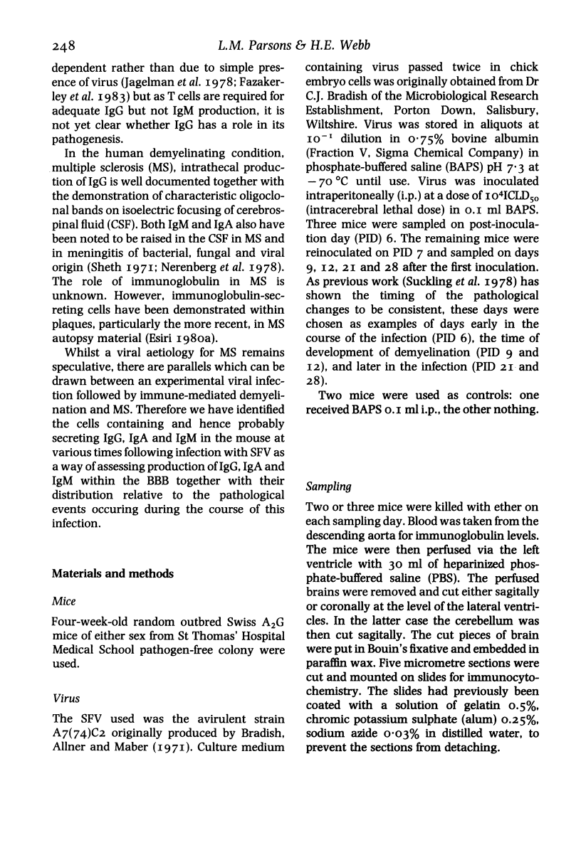
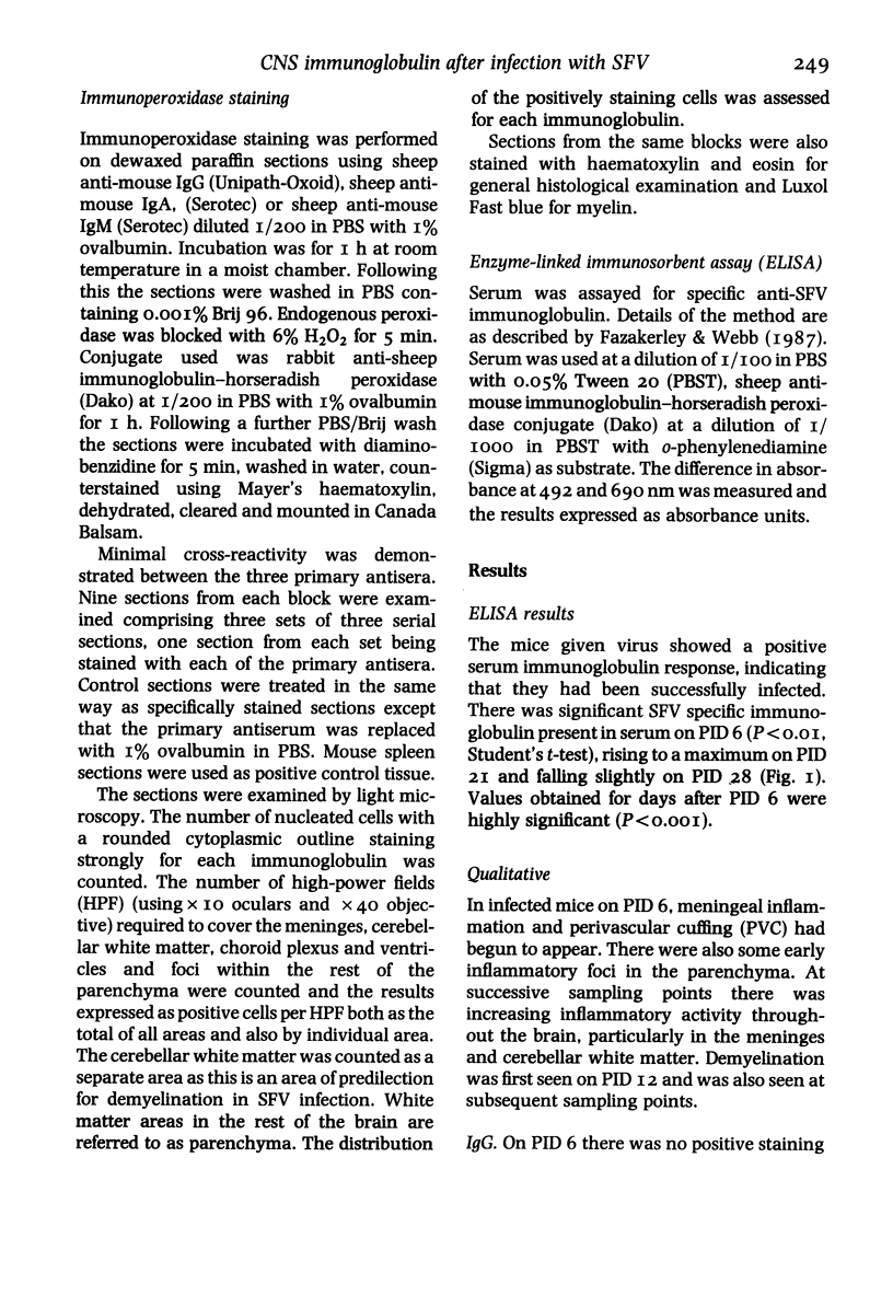
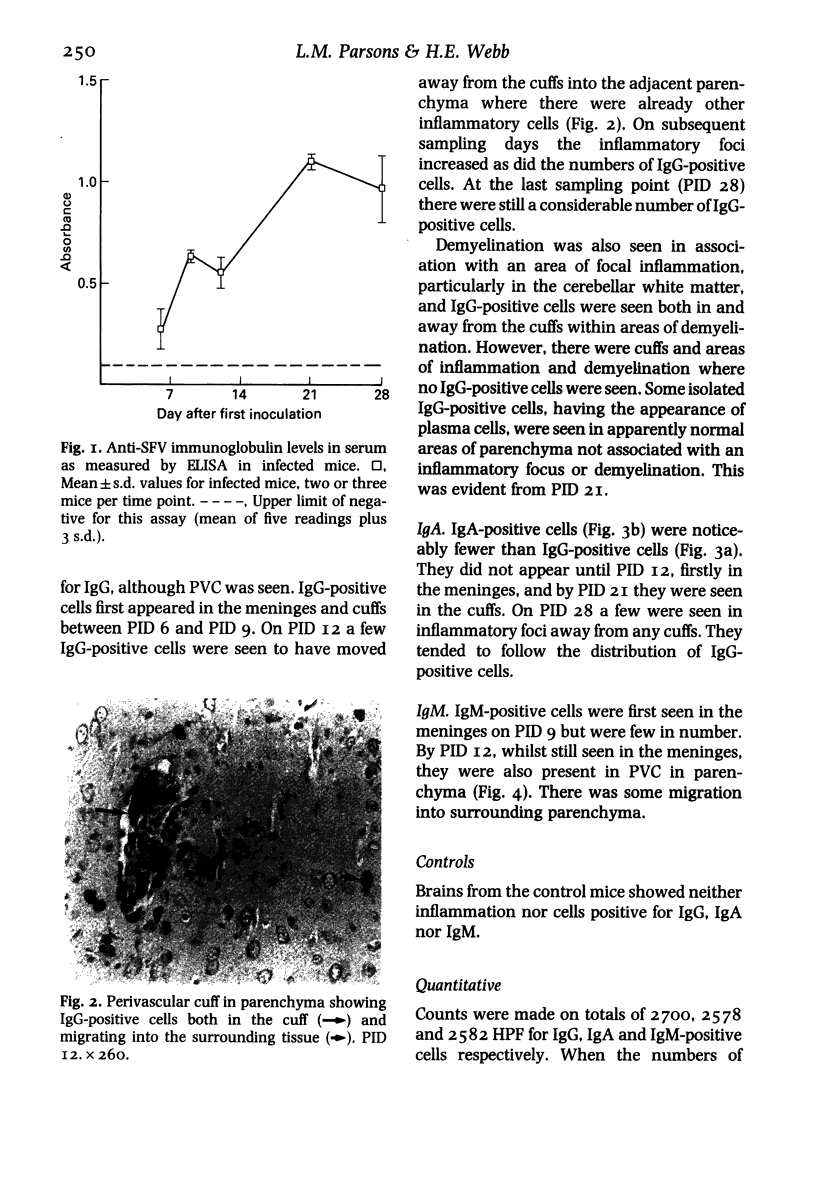
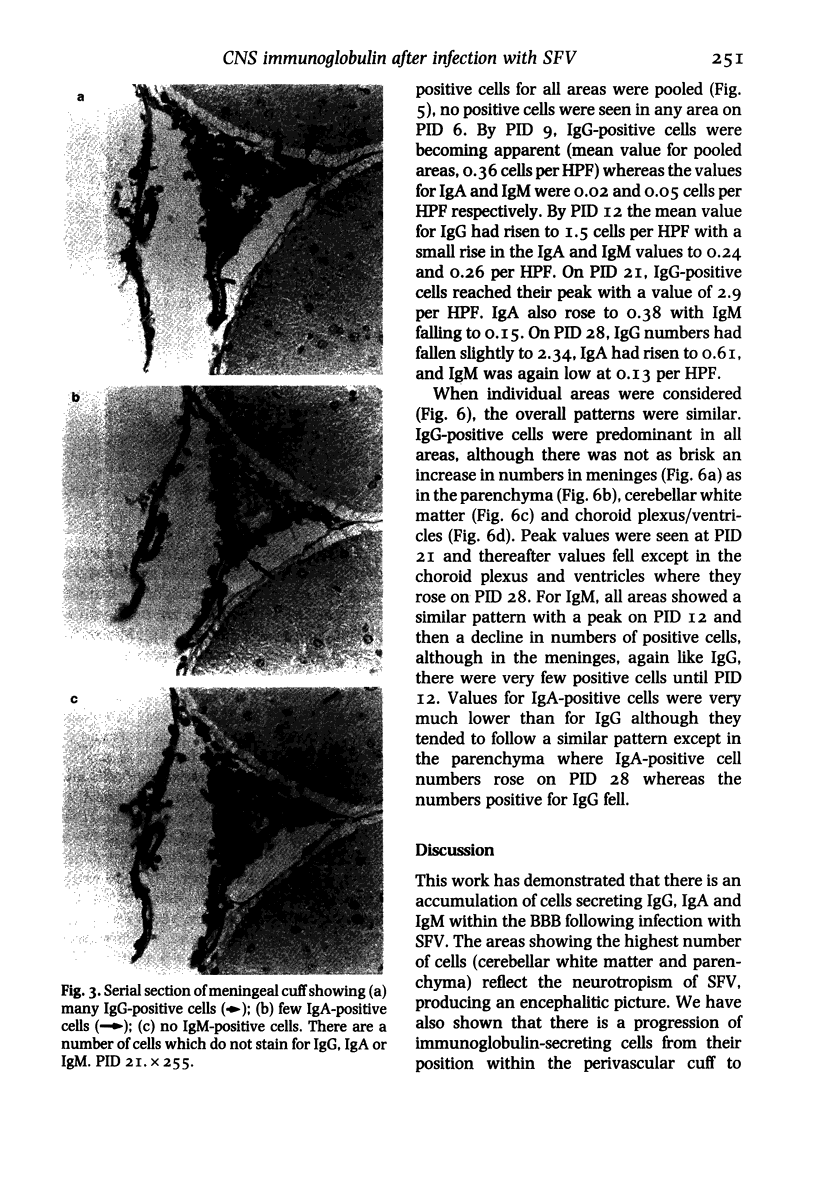
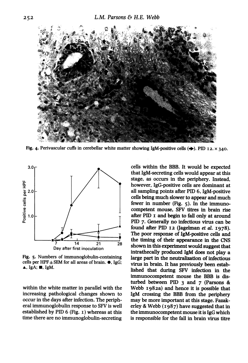
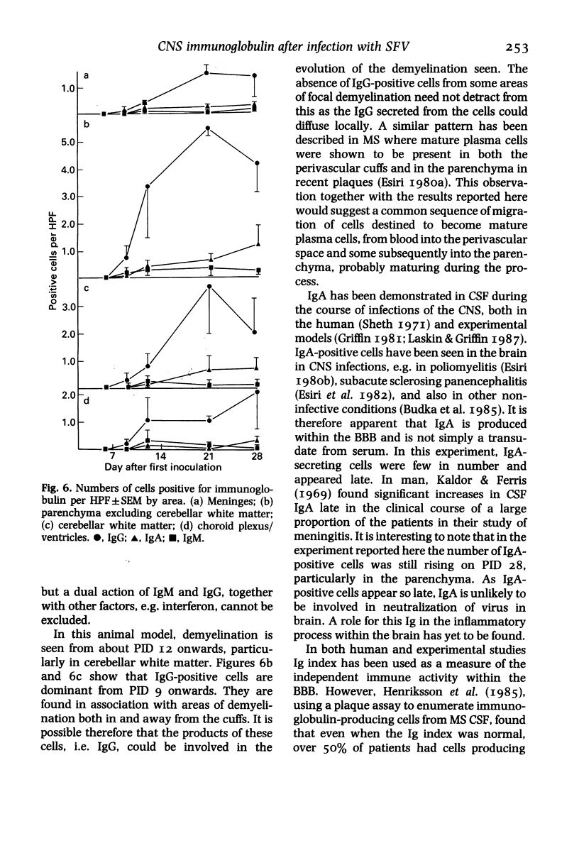
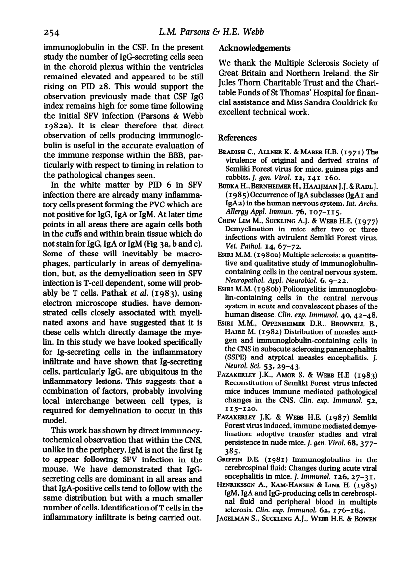
Cells within the central nervous system were identified as containing immunoglobulin G, A and M using immunocytochemistry in mice previously infected with Semliki Forest virus, a togavirus causing primary immune-mediated demyelination. Cells positive for these immunoglobulins were counted in cerebellar white matter, parenchyma, meninges and choroid plexus/ventricles. No positively staining cells were seen on day 6 after infection although other inflammatory cells were present at this time and virus-specific immunoglobulin was found in serum. Cells positive for IgG appeared in all areas by day 9 and remained dominant in numbers throughout. IgM-secreting cells appeared in small numbers in the parenchyma first on day 9 and subsequently in other areas, their numbers rising to a maximum on day 12 in all areas and falling thereafter. The number of IgA-secreting cells was small. They appeared by PID 12 and continued to rise on successive sampling days. Initially IgG-positive cells were seen in the perivascular cuffs but by day 12 a few had moved away from the cuffs into the adjacent parenchyma. IgG-positive cells were seen both in and away from cuffs within areas of demyelination. IgM and IgA-positive cells tended to follow the distribution of IgG-positive cells, but in fewer numbers.
Full text
PDF
Images in this article
Selected References
These references are in PubMed. This may not be the complete list of references from this article.
- Bradish C. J., Allner K., Maber H. B. The virulence of original and derived strains of Semliki forest virus for mice, guinea-pigs and rabbits. J Gen Virol. 1971 Aug;12(2):141–160. doi: 10.1099/0022-1317-12-2-141. [DOI] [PubMed] [Google Scholar]
- Budka H., Bernheimer H., Haaijman J. J., Radl J. Occurrence of IgA subclasses (IgA1 and IgA2) in the human nervous system. Correlation with disease. Int Arch Allergy Appl Immunol. 1985;76(2):107–115. doi: 10.1159/000233675. [DOI] [PubMed] [Google Scholar]
- Fazakerley J. K., Amor S., Webb H. E. Reconstitution of Semliki forest virus infected mice, induces immune mediated pathological changes in the CNS. Clin Exp Immunol. 1983 Apr;52(1):115–120. [PMC free article] [PubMed] [Google Scholar]
- Fazakerley J. K., Webb H. E. Semliki Forest virus-induced, immune-mediated demyelination: adoptive transfer studies and viral persistence in nude mice. J Gen Virol. 1987 Feb;68(Pt 2):377–385. doi: 10.1099/0022-1317-68-2-377. [DOI] [PubMed] [Google Scholar]
- Kaldor J., Ferris A. A. Immunoglobin levels in cerebro-spinal fluid in viral and bacterial meningitis. Med J Aust. 1969 Dec 13;2(24):1206–1209. doi: 10.5694/j.1326-5377.1969.tb103336.x. [DOI] [PubMed] [Google Scholar]
- Laskin O. L., Griffin D. E. Changes in cerebrospinal fluid cells, IgG and IgA during herpes simplex virus encephalitis in rabbits. J Neuroimmunol. 1987 Apr;14(3):283–292. doi: 10.1016/0165-5728(87)90015-4. [DOI] [PubMed] [Google Scholar]
- Nerenberg S. T., Prasad R., Rothman M. E. Cerebrospinal fluid IgG, IgA, IgM, IgD, and IgE levels in central nervous system disorders. Neurology. 1978 Oct;28(10):988–990. doi: 10.1212/wnl.28.10.988. [DOI] [PubMed] [Google Scholar]
- Parsons L. M., Webb H. E. Virus titres and persistently raised white cell counts in cerebrospinal fluid in mice after peripheral infection with demyelinating Semliki Forest virus. Neuropathol Appl Neurobiol. 1982 Sep-Oct;8(5):395–401. doi: 10.1111/j.1365-2990.1982.tb00307.x. [DOI] [PubMed] [Google Scholar]
- Sheth N. K. The possible significance of IgA in abnormal cerebrospinal fluid. J Clin Pathol. 1971 May;24(4):363–365. doi: 10.1136/jcp.24.4.363. [DOI] [PMC free article] [PubMed] [Google Scholar]


