

Abstract
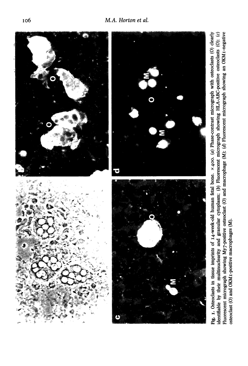
A method for the isolation of osteoclasts from human fetal long bones in sufficient numbers for phenotypic studies has been devised. Using this technique we have studied the expression of cell surface antigens characteristic of mononuclear phagocytes and other haemopoietic cell types on fetal osteoclasts and compared their phenotype with mononuclear cells in the same preparations. We found that osteoclasts failed to express DrW(Ia) and 24 of 26 antigens (in 6 of 7 antigenic clusters) found on mononuclear phagocytes of the same developmental stage. This implies that osteoclasts represent the maturational end-stage of a cell lineage separate from that of conventional blood cells, and of mononuclear phagocytes in particular.
Full text
PDF

Images in this article
Selected References
These references are in PubMed. This may not be the complete list of references from this article.
- Chambers T. J., Magnus C. J. Calcitonin alters behaviour of isolated osteoclasts. J Pathol. 1982 Jan;136(1):27–39. doi: 10.1002/path.1711360104. [DOI] [PubMed] [Google Scholar]
- Chambers T. J., Revell P. A., Fuller K., Athanasou N. A. Resorption of bone by isolated rabbit osteoclasts. J Cell Sci. 1984 Mar;66:383–399. doi: 10.1242/jcs.66.1.383. [DOI] [PubMed] [Google Scholar]
- Loutit J. F., Peters J., Marshall M. J. Colony forming units and haematopoietic stem cells in osteoclastopoiesis. Metab Bone Dis Relat Res. 1981;3(2):131–133. doi: 10.1016/0221-8747(81)90031-x. [DOI] [PubMed] [Google Scholar]