Abstract
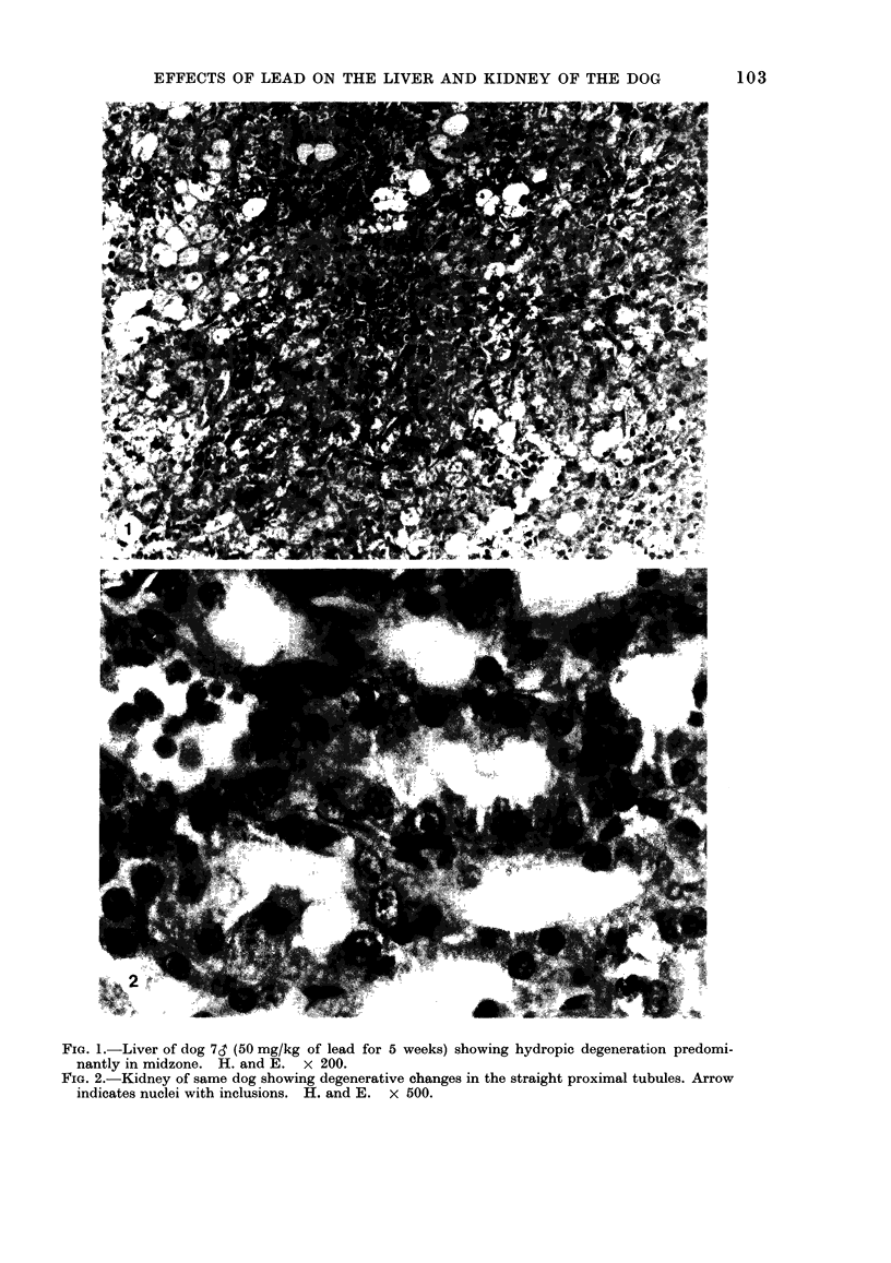
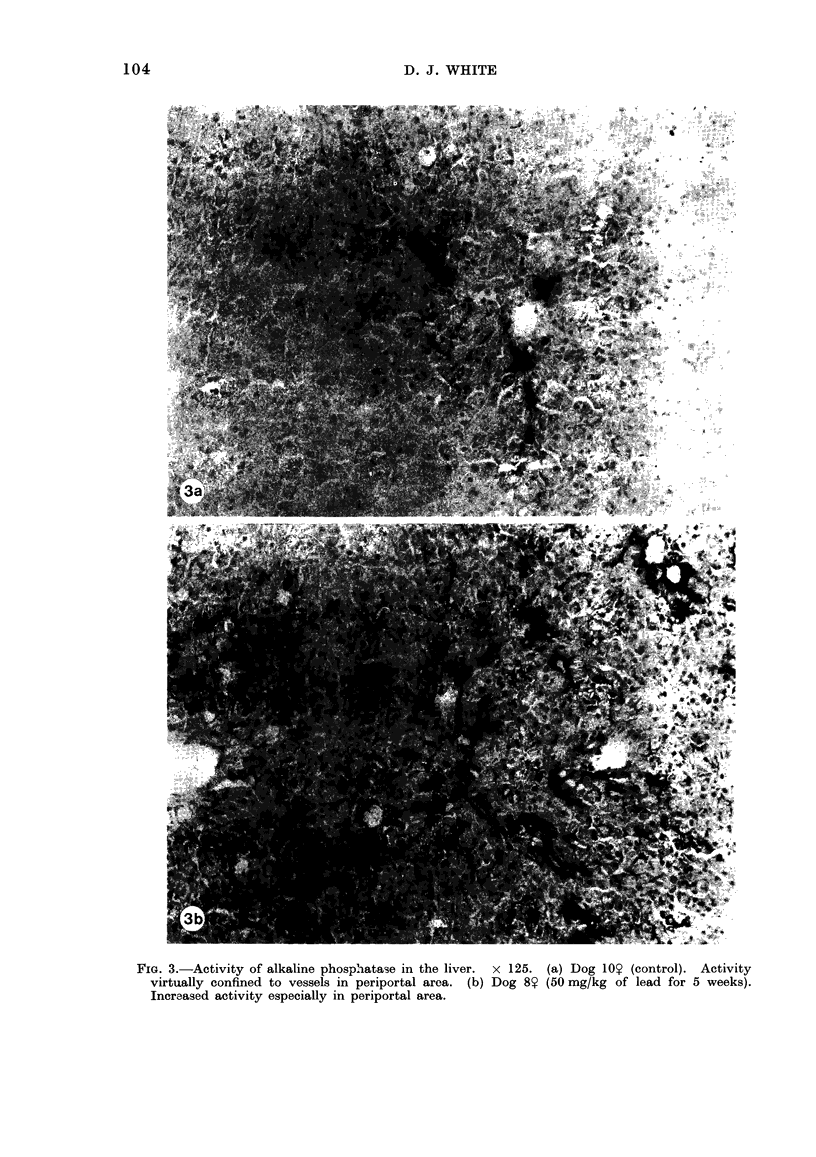
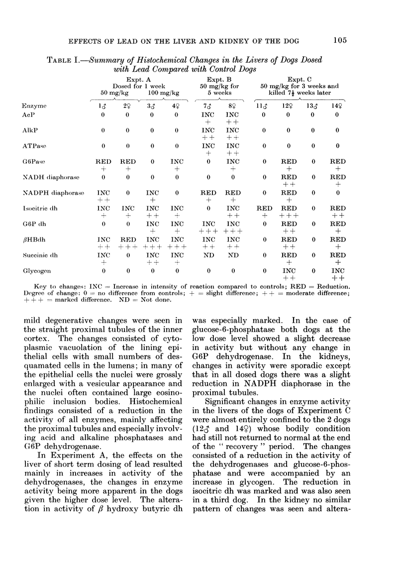
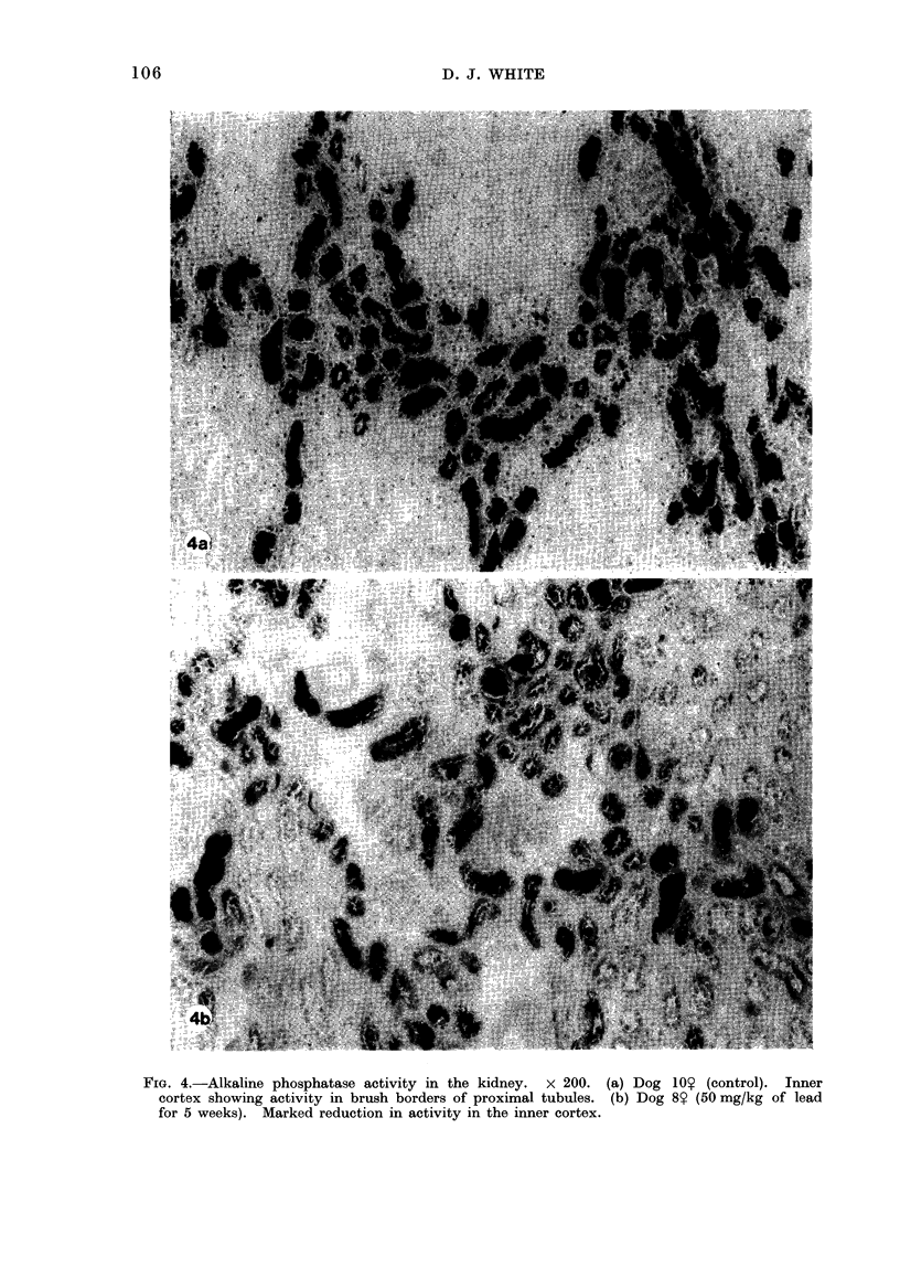
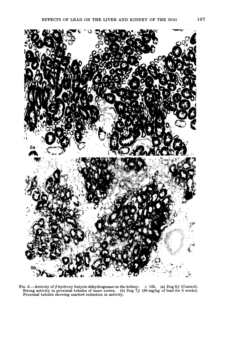
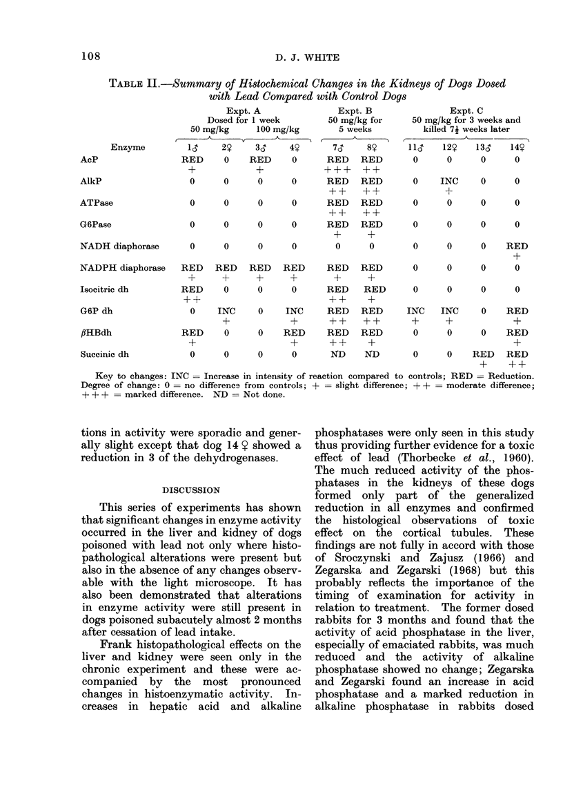
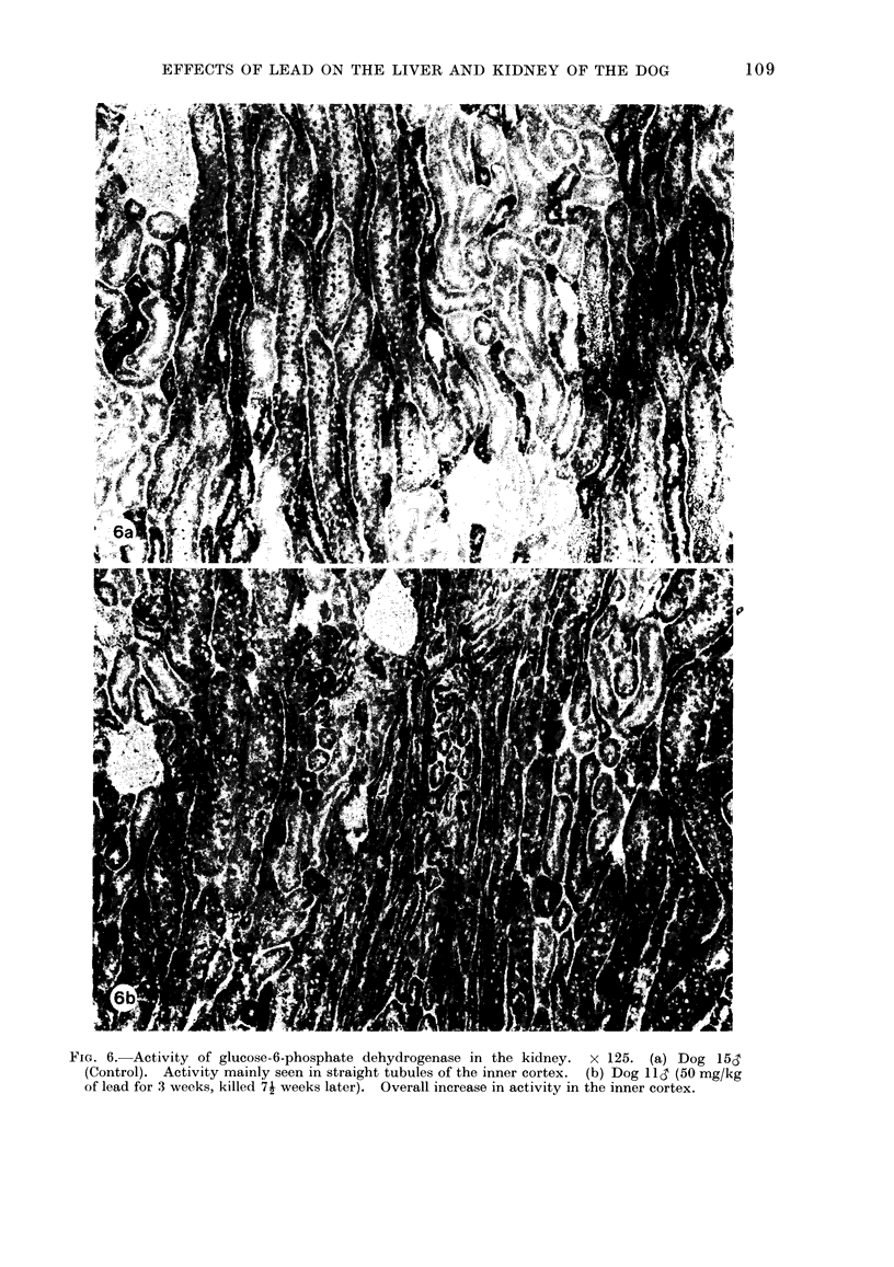
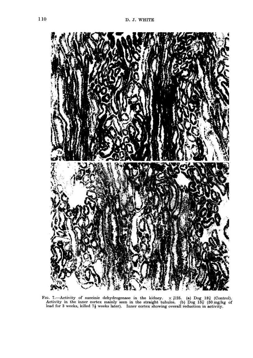
In a series of 3 experiments, beagle dogs were dosed orally with lead carbonate and the histochemical and histological changes in the liver and kidney assessed. Dosing at 50 mg/kg per day for 5 weeks resulted in well documented histological changes in the kidney and hydropic degeneration in the liver; significant alterations in the activity of the majority of enzymes studied were also seen in both organs. In dogs dosed for one week at 50 or 100 mg/kg no histological changes were seen and histochemical alterations were mainly confined to the dehydrogenases and NADPH diaphorase. A third group of dogs were dosed for 3 weeks; during a subsequent recovery period of almost 2 months the mild clinical effects produced by lead during the dosing period were quickly reversible except in 2 dogs. At the end of the recovery period histochemical alterations were evident in both organs of these 2 dogs principally shown by a reduction in the dehydrogenases of the liver. The findings are interpreted as an effect by lead on a range of cellular enzymes particularly those involved in energy production, these effects being still demonstrable after an extended recovery period.
Full text
PDF
Images in this article
Selected References
These references are in PubMed. This may not be the complete list of references from this article.
- Alvares A. P., Leigh S., Cohn J., Kappas A. Lead and methyl mercury: effects of acute exposure on cytochrome P-450 and the mixed function oxidase system in the liver. J Exp Med. 1972 Jun 1;135(6):1406–1409. doi: 10.1084/jem.135.6.1406. [DOI] [PMC free article] [PubMed] [Google Scholar]
- Barltrop D., Barrett A. J., Dingle J. T. Subcellular distribution of lead in the rat. J Lab Clin Med. 1971 May;77(5):705–712. [PubMed] [Google Scholar]
- Cardona E., Lessler M. A., Brierley G. P. Mitochondrial oxidative phosphorylation: interaction of lead and inorganic phosphate. Proc Soc Exp Biol Med. 1971 Jan;136(1):300–304. doi: 10.3181/00379727-136-35252. [DOI] [PubMed] [Google Scholar]
- Castellino N., Aloj S. Intracellular distribution of lead in the liver and kidney of the rat. Br J Ind Med. 1969 Apr;26(2):139–143. doi: 10.1136/oem.26.2.139. [DOI] [PMC free article] [PubMed] [Google Scholar]
- Goyer R. A. The renal tubule in lead poisoning. I. mMitochondrial swelling and aminoacidura. Lab Invest. 1968 Jul;19(1):71–77. [PubMed] [Google Scholar]
- JONES W. A., COHEN R. B. The effect of a murine hepatitis virus on the liver. An anatomic and histochemical study. Am J Pathol. 1962 Sep;41:329–347. [PMC free article] [PubMed] [Google Scholar]
- Sroczyński J., Zajusz K. Histoenzymatic reactions in experimental lead poisoning in rabbits. Arch Immunol Ther Exp (Warsz) 1966;14(3):391–403. [PubMed] [Google Scholar]
- Vallee B. L., Ulmer D. D. Biochemical effects of mercury, cadmium, and lead. Annu Rev Biochem. 1972;41(10):91–128. doi: 10.1146/annurev.bi.41.070172.000515. [DOI] [PubMed] [Google Scholar]
- Zegarska Z., Zegarski W. The behavior of some enzymes in the liver in the course of subacute experimental lead poisoning. Acta Med Pol. 1968;9(1):119–127. [PubMed] [Google Scholar]


