

Abstract
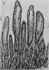
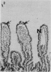
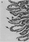
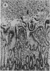
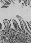
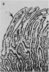
The rate of development of postmortem artefacts was investigated in the mucosa of the small intestine of a calf dually infected with an enteropathogenic strain of Esch. coli and rotavirus, and an uninfected control calf. Samples were removed under general anaesthesia from the proximal, middle and distal small intestine and compared with those taken from adjacent sites 1-30 min after severing the major blood vessels of the neck. In the challenged calf, changes occurred in the villous mucosa by 3 min after severance, whilst in the control calf good fixation was obtained until 10 min after severance.
Full text
PDF
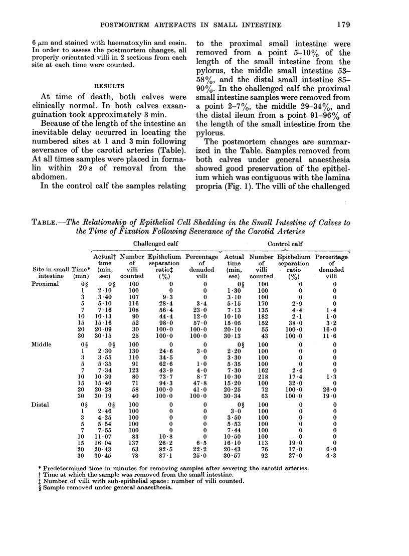

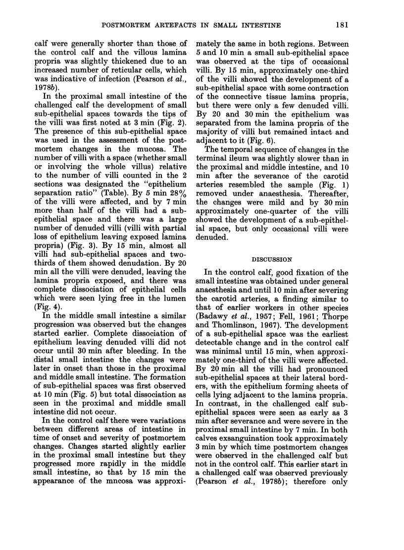
Images in this article
Selected References
These references are in PubMed. This may not be the complete list of references from this article.
- Cross R. F., Kohler E. M. Autolytic changes in the digestive system of germfree, escherichia coli monocontaminated, and conventional baby pigs. Can J Comp Med. 1969 Apr;33(2):108–112. [PMC free article] [PubMed] [Google Scholar]
- Doughri A. M., Storz J. Light and ultrastructural pathologic changes in intestinal coronavirus infection of newborn calves. Zentralbl Veterinarmed B. 1977;24(5):367–385. doi: 10.1111/j.1439-0450.1977.tb01011.x. [DOI] [PMC free article] [PubMed] [Google Scholar]
- FELL B. F. Cell shedding in the epithelium of the intestinal mucosa: fact and artefact. J Pathol Bacteriol. 1961 Jan;81:251–254. doi: 10.1002/path.1700810130. [DOI] [PubMed] [Google Scholar]
- McNulty M. S., Allan G. M., McFerran J. B. Isolation of a cytopathic calf rotavirus. Res Vet Sci. 1976 Jul;21(1):114–115. [PubMed] [Google Scholar]