Abstract
Pointed, rod-shaped bacteria colonizing the cuticular surface of the hindgut of the terrestrial isopod crustacean Porcellio scaber (Crustacea: Isopoda) were investigated by comparative 16S rRNA gene sequence analysis and electron microscopy. The results of phylogenetic analysis, and the absence of a cell wall, affiliated these bacteria with the class Mollicutes, within which they represent a novel and deeply branched lineage, sharing less than 82.6% sequence similarity to known Mollicutes. The lineage has been positioned as a sister group to the clade comprising the Spiroplasma group, the Mycoplasma pneumoniae group, and the Mycoplasma hominis group. The specific signature sequence was identified and used as a probe in in situ hybridization, which confirmed that the retrieved sequences originate from the attached rod-shaped bacteria from the hindgut of P. scaber and made it possible to detect these bacteria in their natural environment. Scanning and transmission electron microscopy revealed a spherically shaped structure at the tapered end of the rod-shaped bacteria, enabling their specific and exclusive attachment to the tip of the cuticular spines on the inner surface of the gut. Specific adaptation to the gut environment, as well as phylogenetic positioning, indicate the long-term association and probable coevolution of the bacteria and the host. Taking into account their pointed, rod-shaped morphology and their phylogenetic position, the name “Candidatus Bacilloplasma” has been proposed for this new lineage of bacteria specifically associated with the gut surface of P. scaber.
The common woodlouse Porcellio scaber is a widely spread species of the terrestrial isopod crustaceans (Crustacea, Isopoda, Oniscidea) living in temperate climates. Like other terrestrial isopods, P. scaber is a herbivorous scavenger, feeding predominantly on decayed plant material and thus contributing to nutrient and energy cycling in terrestrial ecosystems (43). Due to its ecological importance, easy handling, breeding capability under laboratory conditions, and tolerance to polluted environments as well as the considerable body of knowledge about its biology, this crustacean is commonly used as a test organism in terrestrial ecotoxicological and ecophysiological studies (12, 17).
The tripartite digestive system of P. scaber consists of a short foregut comprising an esophagus and stomach, a midgut consisting of two pairs of blind-ended tubular digestive glands, and a long, tube-like hindgut. The last of these comprises two functionally different parts, an anterior chamber and a papillate region with a rectum (17). The foregut of terrestrial isopods is generally poorly inhabited by microorganisms. On the other hand, a high microbial density is found in the hindgut, particularly in the papillate region, where favorable conditions (44) allow the multiplication of those microorganisms that have survived the digestion in the anterior part of the digestive tract of P. scaber (24, 26) and those of other terrestrial isopods like Oniscus asellus (15) and Trachelipus rathkii (35).
During previous observations of the digestive tracts of several isopod species (11) and various populations of P. scaber, rod-shaped bacteria associated with the gut cuticle were frequently observed. These bacteria are morphologically distinct and differ from other bacteria observed in the P. scaber digestive system (references 15, 25, 39, and 40; summarized in reference 26).
In the present study, the phylogenetic affiliation of rod-shaped bacteria attached to the hindgut cuticle structures of the terrestrial isopod P. scaber was investigated by 16S rRNA gene analysis. The morphological features of these bacteria were investigated by electron microscopy, and their location and distribution on the gut surface were determined by hybridization with a specific fluorescence-labeled oligonucleotide probe. The plausible role of the attached bacteria in the host and the routes of recolonization of the gut surface after molting are also discussed.
MATERIALS AND METHODS
Animals.
The animals were collected in their natural environments from locations near Ljubljana, Cerknica, Postojna, Idrija, and Radenci in Slovenia and near Vienna in Austria. They were kept at 20°C in glass tanks filled with soil under conditions of high humidity and a 16-h/8-h day/night cycle. Leaf litter from the unpolluted collection site near Ljubljana was provided as food. Healthy, adult animals of both sexes were used in the experiment.
Microscopy.
The hindguts were extracted from the animals by use of fine-tipped forceps. They were then opened and rinsed several times with sterile phosphate-buffered saline (PBS) (pH 7.4) (130 mM NaCl, 3 mM NaH2PO4, 7 mM Na2HPO4) in order to remove the gut content (24). For light microscopy, the hindguts were spread on microscope slides and examined. For transmission electron microscopy, the hindguts were fixed in 3.5% glutaraldehyde in a 0.1 M phosphate buffer (pH 7.2) for 2 h, washed in the phosphate buffer, and postfixed in 1% OsO4. After additional washing, the gut tissues were dehydrated in a graded alcohol series and embedded in Spurr's medium. Ultrathin sections were stained with uranyl acetate and Reynold's lead citrate and examined with a Philips CM 100 transmission electron microscope operating at 80 kV. For scanning electron microscopy, the removed and rinsed guts were fixed in 1% paraformaldehyde and 0.4% glutaraldehyde in a 0.1 M sodium cacodylate buffer solution (pH = 7.2) for 2 hours at 4°C and postfixed by modified ligand binding of osmium with a thiocarbohydrazide-binding technique [9]). Fixed tissues were critical-point dried, mounted on aluminum stubs, sputter coated with gold, and examined with a Jeol 840A scanning electron microscope.
DNA extraction, PCR amplification, and cloning.
The hindguts were extracted from five animals with sterile fine-tipped forceps. The papillate regions were removed from the hindguts, opened with sterile needles, and washed in PBS in order to remove the gut content. The cuticle surface of the gut was carefully separated from the gut wall and washed vigorously in PBS. Isolated cuticles with attached bacteria were crushed in 1 ml of PBS by use of a Teflon homogenizer. Genomic DNA was extracted from the gut by a previously described method (24). Segments of 16S rRNA genes were amplified with a combination of evolutionary conserved bacterial primers fD1 (41), a universal primer corresponding to the sites 522 to 536 (14), and the reverse primer 1392r (28) in PCR. The sample DNA (1 μl) was amplified in a reaction mixture that consisted of 1U reaction buffer (GIBCO BRL), 2.5 mM MgCl2, 0.2 mM of each deoxynucleoside triphosphate, 6 pmol of each primer, and 1 U of Taq polymerase (GIBCO BRL) in a final volume of 20 μl. The PCR was performed at 95°C for 5 min followed by 25 cycles of 40 s at 95°C, 30 s at 65°C, and 80 s at 72°C, with a final 10-min extension at 72°C. The amplification products were separated by gel electrophoresis, and amplified 16S rRNA genes of the expected size (i.e., approximately 1,400 bp) were purified with a QIA gel extraction kit (QIAGEN). Purified amplicons were ligated into a p-BAD TOPO vector and cloned into Escherichia coli TOP10 recipient cells by use of a p-BAD TOPO TA cloning kit (Invitrogen) according to the manufacturer's instructions.
ARDRA and sequencing.
Plasmid DNA was isolated from the recombinant cells by the “mini-prep method” (37). Selected cloned amplicons were separately digested with DdeI, HaeIII, and HhaI restriction endonucleases. Sequences of the representative amplicons from established amplified 16S rRNA gene restriction analysis (ARDRA) groups were sequenced at our request by Mycrosynth GmbH (Baglach, Switzerland) with the above-described primers.
Phylogenetic analysis.
Retrieved ribosomal sequences were subjected to the Check-Chimera program on the RDP website (6) or to the Bellerophon program on the Greengenes website (10) for the elimination of plausible chimeric sequences and compared with the sequences deposited in nucleotide sequence databases by use of BLAST and fasta algorithms in order to find the most related sequences in publicly available databases. Sequence data were aligned using the NAST algorithm on the Greengenes website (10) and checked manually. A data set of 89 unambiguously aligned 16S rRNA sequences, with approximately 1,250 nucleotide positions, was used for the construction of phylogenetic trees. Maximum likelihood trees were constructed using the PHYML program, version 2.4.4 (16). The most appropriate model of nucleotide substitution, the general time-reversible model, with gamma distributed rate heterogeneity and a significant proportion of invariable sites, was selected by testing alternative models of evolution by the Modelgenerator program (23) and used in the PHYML phylogenetic interface. Neighbor-joining trees were constructed based on the Kimura two-parameter model of sequence evolution using phylogenetic analysis algorithms implemented in the PHYLIP package (13). Maximum-parsimony trees were constructed using PAUP* program version 4.0b10 (D. Swofford, Sinauer Associates, Sunderland, MA). Parsimony searches included heuristic searches using random sequence addition with 100 replicates and a tree-bisection-reconnection branch-swapping algorithm. Nodal support was estimated using bootstrap analysis in a set of 1,000 replications under the appropriate model. A Bayesian inference of phylogeny was generated with MrBayes software package 3.1.2 (36), using the general time-reversible model of evolution. The analysis was run in duplicate with four chains for 106 generations with a sampling frequency of 100 generations; of the 106 total generations, the initial 250,000 were discarded as burn-in.
In situ hybridization.
A group-specific oligonucleotide probe was designed to specifically target the 16S rRNA of the clones obtained in this study, using a Probe-check program (6). The universal probe EUB338 (1) was used as a positive control, and the species-specific Prevotella brevis probe GA33 (3) was used as a negative control to exclude nonspecific probe binding. All probes were synthesized at our request at MWG-BIOTECH AG (Ebersberg, Germany) and 5′ labeled with Cy3 cyanine dye.
The hindguts were removed, opened, and washed as described above. The cuticle surface of the gut with the attached bacteria was carefully separated from the gut wall in order to reduce background fluorescence and spread on adhesive slides (MJ Research, MA). Tissues were fixed in 4% paraformaldehyde for 2 h and dehydrated by immersing the slides in 50%, 80%, and 96% ethanol for 3 min each. The samples were hybridized as previously described (25), with 4 h of incubation at 46°C and a 20-min wash at 48°C. After being washed, the slides were rinsed with distilled water, air dried, and examined. The examination was performed by use of an Axio Imager.Z1 microscope (Zeiss, Germany) upgraded with an ApoTome system (Zeiss, Germany) for the optimization of fluorescence microscopy.
Nucleotide sequence accession numbers.
The 16S rRNA sequences have been deposited in the GenBank nucleotide sequence database under accession numbers DQ485973 to DQ485977.
RESULTS
Morphology and distribution.
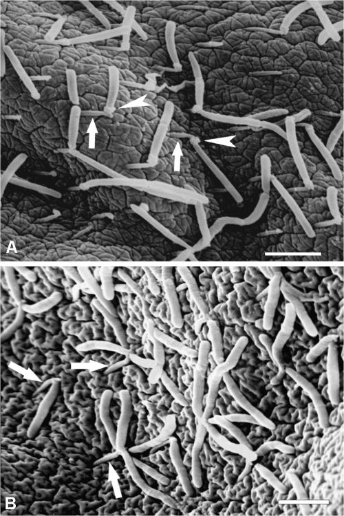
Microscopic examination of the inner hindgut surface revealed evenly distributed spine-like cuticular protrusions facing backwards and the presence of several bacterial morphotypes associated with the gut wall, among which rod-shaped bacteria were very strongly predominant. The latter were observed by light and electron microscopy in the hindguts of 75% of 345 examined animals from all collection sites. The densities of the attached rod-shaped bacteria varied among the animals, ranging from mat-like microcolonies with a density of 50 or more cells per 100 μm2 (not shown) to patches of more-evenly distributed bacteria (Fig. 1A) with a density of approximately 20 cells per 100 μm2. Detailed microscopic observation revealed that each rod-shaped bacterium was attached to the tip of a cuticular spine protruding from the gut wall (Fig. 1A). Commonly, a single bacterium was found attached to a spine, where the total number of observed bacteria was low. When the density of the attached bacteria was higher, several bacteria were observed as attached to a single spine, thus forming a rosette-like structure (Fig. 1B).
FIG. 1.
Scanning electron micrographs of rod-shaped bacteria associated with the hindgut cuticle of P. scaber showing (A) a colonized gut surface and the connection between the rod-shaped bacteria and the tips of cuticular spines (arrows) via spherical structures (arrowheads) and (B) a group of rod-shaped bacteria attached to a single cuticular spine (arrows) forming a rosette-like structure. Scale bars = 2 μm.
Most of the observed rod-shaped bacteria were 2.5 ± 1 μm long and 200 ± 50 nm wide (Fig. 2A), although morphotypes of up to 6 μm long were observed as well. The attached bacteria were slightly pointed at the end attached to the cuticular spine and rounded at the distal end (Fig. 2A). Ultrastructural examination of the attached bacteria showed a single trilaminar plasma membrane and the absence of a cell wall (Fig. 2B). Further observations of the site of bacterial attachment to the gut wall revealed a spherical structure between the tip of the cuticular spine and the tapered end of the attached bacterium (Fig. 1B and 2C). Electron micrographs of the cross-sections of the spherical structure showed that this structure consists of electron-dense filaments extending radially from the attached end of bacterial cell outwards (Fig. 2C), indicating that such spherical structures are parts of, or produced by, the attached bacteria. The bacterial origin of the spherical attachment structure was also indicated by the longitudinal section through the cuticular spine (Fig. 2D), revealing the absence of any channels connecting the underlying epithelia and the tip of the spine, which would enable the secretion of the spherical structures through the cuticle.
FIG. 2.
Transmission electron micrographs of a rod-shaped bacterium associated with the hindgut wall of P. scaber showing (A) a longitudinal section across the attached bacterium, (B) a detail from the attached bacterium revealing a single trilaminar plasma membrane (arrowhead), (C) the ultrastructure of the spherical structure (arrowhead) between the cuticular spine (asterisk) and the attached bacterium (arrow), and (D) the anatomy of the gut cuticle (EC, epithelial cell; C, cuticle; CS, cuticular spine). Scale bars = 500 nm (panels A and D), 100 nm (panel B), and 200 nm (panel C).
Cloning, sequencing, and phylogenetic analysis.
In order to determine the phylogenetic affiliation of the rod-shaped bacteria attached to the hindgut, DNA was extracted from the hindgut sample of P. scaber. The cuticle was previously peeled from the underlying epithelium and washed in PBS several times in order to remove unattached bacteria and reduce the amount of the host's DNA in the samples. Following amplification and cloning of 16S rRNA genes, randomly selected clones containing bacterial rRNA gene inserts were separately digested with the DdeI, HaeIII, and HhaI endonucleases. The obtained restriction patterns differed only slightly (results not shown). ARDRA resulted in two groups consisting of 14 and 7 clones, respectively. Three clones (named P5, P6, and P14) from the first ARDRA group and two clones (named P7 and P10) from the second ARDRA group were selected for sequencing.
Pairwise alignments between the obtained sequences revealed similarity between the sequences from the first ARDRA group ranging between 93.3 and 96%, whereas two sequences from the second ARDRA group shared 98% similarity. Levels of similarity between sequences from both ARDRA groups and other sequences in the databases were below 82.6%, with the highest sequence identity to known taxa from the class Mollicutes. Partial sequences from uncultured gut bacteria of P. scaber, retrieved in our previous study (24), and the clone C13, belonging to uncultured bacterium from the gut of the fish Gillichthys mirabilis (DQ340200), provided exceptions, sharing 86.4% to 99.5% similarities to the sequences obtained in the present study.
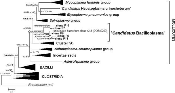
In order to determine the phylogenetic position of the sequences from both ARDRA groups, representative 16S rRNA sequences from major Mollicutes groups, including the sequences from group incertae sedis 8, which is positioned within the class Mollicutes according to the RDP nomenclature hierarchy (6), were included in the phylogenetic analysis. Sequences of intestinal Mollicutes from two additional clades, i.e., the recently described “Candidatus Hepatoplasma crinochetorum” (39) from P. scaber and a group of uncultured Mollicutes known as “unknown cluster A” from a vertebrate digestive tract (8, 29), were also included in the analysis. Despite high similarity to the sequences from both ARDRA groups, the partial sequences from our previous study (24) were not included in the phylogenetic analysis because of their insufficient lengths. Due to the low similarity to other known sequences, selected 16S rRNA sequences from all major Firmicutes clades were used as out-groups.
Phylogenetic trees based on full or nearly full 16S rRNA sequences and constructed with parsimony, neighbor-joining, and Bayesian treeing methods revealed topologies the same as those of the condensed maximum-likelihood tree presented in Fig. 3 and a maximum-likelihood tree constructed by complete selection of the sequences (see Fig. S1 in the supplemental material). Both trees show that the clones from both ARDRA groups from the gut of P. scaber and clone C13 from the gut of G. mirabilis form a distinct and deeply branched phylogenetic lineage within Mollicutes. The lineage branches at the base of the main phylogenetic clades within Mollicutes, where it is positioned as a sister group to the clade comprising the Spiroplasma group, the Mycoplasma pneumoniae group, “Candidatus Hepatoplasma crinochetorum,” and the Mycoplasma hominis group. Despite the low bootstrap values supporting some of the branching points of the major Mollicutes clades and the novel lineage (Fig. 3), the topology of the branches between major clades remain unchanged regardless of the treeing method used, which supports the affiliation of the novel lineage with the Mollicutes. Within this novel lineage, the sequences are gathered in two clades corresponding to both ARDRA groups, and the branching points are supported by bootstrap values above 94%. The first clade consists of the clones P5, P6, and P14 from the first ARDRA group and the clone C13 from G. mirabilis. The second clade consists of clones P7 and P10 from the second ARDRA group.
FIG. 3.
Condensed phylogenetic tree showing the relationships of the 16S rRNA gene sequences of the clones P5, P6, P7, P10, and P14, obtained from bacteria attached to the gut wall of P. scaber, to the main phylogenetic clades of Mollicutes. The tree is based on maximum-likelihood analysis of the 1,250-nucleotide region of 16S rRNA. Numbers at the branching points indicate bootstrap support values above 50%, as calculated in percentages by maximum-likelihood/maximum parsimony/neighbor-joining/posterior Bayesian probabilities. The tree was rooted with E. coli, and 32 selected species belonging to the main phylogenetic clades of Firmicutes were used as out-groups. The scale bar represents 10% estimated sequence divergence per nucleotide position.
In situ hybridization.
To confirm that the sequences obtained from the selected clones truly originated from the rod-shaped bacteria attached to the gut cuticle of P. scaber, a specific oligonucleotide probe named S-G-Bpl-0089-a-A-23 [5′-CGTTCGCCACTAAC(G/T)G(A/C)AAAT(T/C)C −3′] (E. coli positions 89 to 111) was designed to be complementary to the specific 16S rRNA gene sequence region of the selected clones and applied to whole-cell hybridization of the hindgut tissues. Despite three degenerative nucleotide sites, the designed oligonucleotide probe differs by at least 2 nucleotides from any one of the 16S rRNA gene sequences of other bacteria in the Ribosomal Database Project database, including all Mollicutes (Table 1).
TABLE 1.
Alignment of the S-G-Bpl-0089-a-A-23 oligonucleotide probe sequence against the closest target sequences of 16S rRNA genes and representative sequences of major Mollicutes taxa
| Sequence origin | Taxonomic affiliationa | GenBank accession no. | Sequencec |
|---|---|---|---|
| S-G-Bpl-0089-a-A-23 (E. coli positions 89-111) | 5′-CGTTCGCCACTAACKGMAAATYC-3′ | ||
| UCb clones from the gut of isopod crustacean Porcellio scaber | Mollicutes/“Candidatus Bacilloplasma” | DQ485973-DQ485977 | 3′-GCAAGCGGTGATTGMCKTTTARG-5′ |
| UC clone from the guts of fish Gillichthys mirabilis | Mollicutes/“Candidatus Bacilloplasma” | DQ340200 | 3′-....................T..-5′ |
| UC clone from the gut of scarabid larva Pachnoda ephippiata | Mollicutes/incertae sedis 8 | AJ576412 | 3′-.............CG........-5′ |
| UC clone from the termite gut | Mollicutes/incertae sedis 8 | AB231068 | 3′-.............CG........-5′ |
| UC clone from cattle feces | Firmicutes/unclassified Clostridiales | AB107541 | 3′-.............C......C..-5′ |
| UC clone from hypersaline wastewater | Firmicutes/unclassified Clostridiales | AM157472 | 3′-....A........C.........-5′ |
| UC clones from the hindgut of shrimp Neotrypaea californiensis | Unclassified Firmicutes | AF434118-AF434128 | 3′-............CT.........-5′ |
| Spiroplasma gladiatoris | Mollicutes/Entomoplasmatales | M24475 | 3′-............CC......C..-5′ |
| UC clone from the gut of scarabid larva Pachnoda ephippiata | Mollicutes/incertae sedis 8 | AJ629069 | 3′-.............CG.......G-5′ |
| UC clones from the pig gastrointestinal tract | Mollicutes/incertae sedis 8 | AF371518 | 3′-...............A...AG.T-5′ |
| Erysipelothrix rhusiopathiae | Mollicutes/incertae sedis 8 | M23728 | 3′-.............C.A.G..CCT-5′ |
| Entomoplasma melaleucae | Mollicutes/Entomoplasmatales | AY345990 | 3′-...........C.C.ACG..C.T-5′ |
| Mycoplasma pneumoniae | Mollicutes/Mycoplasmatales | M29061 | 3′-........A....T.A.AA.G.T-5′ |
| Acholeplasma laidlawii | Mollicutes/Acholeplasmatales | U14905 | 3′-...........CCT.ACG..C.T-5′ |
| Mesoplasma syrphidae | Mollicutes/Entomoplasmatales | AY231458 | 3′-...........CCC.ACG..C.T-5′ |
| Ureaplasma urealyticum | Mollicutes/Mycoplasmatales | M23935 | 3′-.....T.......CGGA...TCC-5′ |
| Anaeroplasma varium | Mollicutes/Anaeroplasmatales | M23934 | 3′-.........ACCAT...A.GGTA-5′ |
| “Candidatus Hepatoplasma crinochetorum” | Unclassified Firmicutes | AY500249, AY500250 | 3′-...........G.T.AAGAATTA-5′ |
| Mycoplasma hominis | Mollicutes/Mycoplasmatales | M24473 | 3′-.T......C...CA.TAACG.TT-5′ |
| Asteroleplasma anaerobium | Mollicutes/Anaeroplasmatales | M22351 | 3′-...T.......CCCGA.GC.TCT-5′ |
According to RDP nomenclature hierarchy (6).
UC, uncultured bacterium.
R, A or G; Y, T or C; M, A or C; K, G or T.
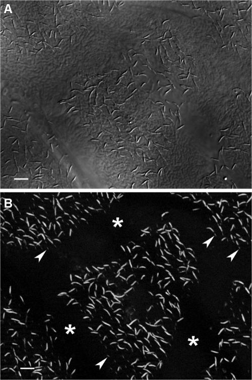
Fluorescence microscopy revealed a strong signal for all the observed rod-shaped cells (Fig. 4A and B) attached to cuticular spines after hybridization with the bacterial probe EUB338 (not shown) and the group-specific probe S-G-Bpl-0089-a-A-23 (Fig. 4B). After hybridization with nonspecific probe GA33, a fluorescent signal was not observed for the attached bacteria (not shown). An additional negative control was used in order to test the specificity of designed probe. Cells from a fresh culture of Mycoplasma gallisepticum strain Rlow gave no hybridization signal under the same hybridization conditions (not shown). The results confirmed the presence of the targeted sequences in the rod-shaped bacteria attached to the cuticular spines (Fig. 4B). To elucidate the distribution of the attached bacteria in the gut of P. scaber, hybridization with the specific probe S-G-Bpl-0089-a-A-23 was performed on the gut surfaces of at least three animals from the Austrian population and from each Slovenian population. Upon in situ hybridization, fluorescently labeled bacterial cells with a distinctive rod morphology were observed on the gut surfaces of all of the animals. The rod-shaped bacteria were always found in the papillate region of the hindgut and only occasionally in the rectum or in the anterior chamber of the hindgut. The distribution of the hybridized rod-shaped bacteria was patchy, and their densities varied among individual animals from sporadic bacteria to mat-like microcolonies. In the latter case, distinctive lines without attached bacteria were observed on the gut surface (Fig. 4B). More-detailed observations revealed that the bacterium-free zones on the cuticle correspond to the regions directly above the junctions of epithelial cells, where the cuticular spines are missing.
FIG. 4.
Specific detection of rod-shaped bacteria associated with the gut wall of P. scaber by in situ hybridization. (A) Gut wall of P. scaber viewed by differential interference contrast. (B) Gut wall shown by fluorescence microscopy after hybridization with the specific probe S-G-Bpl-0089-a-A-23 showing rod-shaped bacteria associated with the cuticular spines (arrows) and patches of bacteria separated by bacterium-free areas (asterisks). Scale bars = 5 μm.
DISCUSSION
As for other terrestrial invertebrates, the digestive tracts of isopods, especially the hindguts, are colonized by diverse bacterial microbiota (24, 43). The majority of the known gut microbiota of terrestrial isopods are represented by ubiquitous microorganisms from a wider environment (summarized in reference 26), whereas the data on resident or truly autochthonous gut microbiota are less clear (22, 35). Moreover, in earlier studies even the absence of resident microbiota in the guts of terrestrial isopods, mainly due to the straight tube-like anatomy of the gut, the short retention time of the food in the digestive tract, and the frequent removal of the gut surface during the molting cycle, has been suggested (19, 20). Nevertheless, our observations strongly indicate the presence of bacteria adapted to persist in the digestive tracts of terrestrial isopods for longer periods.
Morphological observations of the rod-shaped bacteria attached to the cuticular lining of the hindgut of P. scaber revealed their close and specific association with the gut surface. Detailed observations revealed specific and exclusive attachment of these bacteria at the tip of cuticular spines, suggesting a certain advantage of this attachment site over other surfaces in the gut. Since the chemical structure of the tip does not differ from that of other gut surfaces (31) and the tip of the spine is not a site of secretion from the underlying cells, it is possible that the tip of the spine is advantageous for the bacteria simply as the most exposed site on the gut surface. The difference in the oxic and chemical conditions between the gut surface and the lumen of the gut (44) might also have an important role in favoring the tip of the cuticular spines as an attachment site. However, the observed attachment of several rod-shaped bacteria to a single spine, forming a rosette-like structure, strongly suggests that competition between these bacteria for the attachment site at the tip of the spine exists, indicating the advantage of the tip of the spine as an attachment site over other gut surfaces for these rod-shaped bacteria.
The close and specific association of rod-shaped bacteria to the gut wall is in congruity with what is seen for other members of the class Mollicutes, which commonly exhibit rather strict host and tissue specificities (34). The attachment of rod-shaped bacteria to the cuticular spines is mediated by distinctive spherical structures at the pointed ends of attached bacteria. Complex structures known as “attachment organelles,” enabling the attachment of bacteria to the surface of the host cell, are known for certain Mollicutes (2, 21, 27). However, their tapered shape and membrane-bound structure clearly differs from the ray-like anatomy of the attachment structures observed for the rod-shaped bacteria colonizing the hindgut of P. scaber.
The novel lineage of Mollicutes, consisting of five clones from the gut of P. scaber and a single clone from the intestine of the fish Gillichthys mirabilis, is divided into two distinct sublineages sharing high sequence similarity within each sublineage. Despite the phylogenetic distance between the hosts, the clone from G. mirabilis affiliates to the same sublineage with three clones from isopod gut. Although this diminishes the probability of long-term coevolution of these bacteria and hosts, it may also indicate that these clones represent only a small part of a so-far-unknown bacterial lineage consisting of many not-yet-discovered sublineages which colonize the intestines of various arthropod and vertebrate hosts. This assumption is supported by several previous observations of hindguts of various arthropods, in which bacteria exhibiting morphologies or attachment preferences similar to those of the rod-shaped bacteria from P. scaber were observed. Bacteria with almost identical morphologies were observed in the gut contents of shrimps (32) and termites (4), attached to the gut cuticle structures of the millipede (7) and marine decapod crustaceans (18), or forming rosette-like aggregations in the gut of the cockroach (5).
The absence of a cell wall, as the most prominent morphological feature of the class Mollicutes (34), confirmed the phylogenetic affiliation of rod-shaped bacteria to Mollicutes. Although the absence of a cell wall commonly results in spherical or pleomorphic morphology in mycoplasmas (34), the attached bacteria from the P. scaber hindgut maintain a constant rod shape. They are slightly pointed at the attached end and round at the distal end. Maintaining of the cell shape in the absence of a rigid cell wall commonly requires the presence of cytoskeletal elements, such as the cytoskeletal ribbon in Spiroplasma (38) and the “rod” and “blade-like” structures in M. pneumoniae (21), for example. Although protocols for the visualization of structures mentioned above do not differ from electron microscopy techniques applied in our study, no such structures in the attached bacteria from P. scaber have been observed.
The digestive system of arthropods is a common habitat of Mollicutes, including spiroplasmas, mesoplasmas, entomoplasmas, and phytoplasmas (34). Various relationships of these microbes with their hosts have been revealed. Most of them are parasitic, causing diverse effects on their host (34). Commensal and even beneficial associations with the arthropod host are also known (42). Although it is difficult to speculate from the available data on the role of rod-shaped Mollicutes in the gut of P. scaber, our observations allow at least some conclusions. As these bacteria were recovered from apparently healthy animals, they are probably not pathogenic for the host. At the same time, rod-shaped bacteria were found in a large proportion of the animals from several different populations from various parts of Slovenia and Austria. This excludes occasional or laboratory infection of animals with these bacteria. Due to the role of isopods as decomposers of plant material, it is tempting to speculate that these adapted bacteria could play some role in the digestion process of the host. However, their occasional absence in some of the inspected animals, as well as their association with the papillate region of the hindgut, where most of the absorption of water and ions takes place (17), apparently diminishes their role in the digestive process. It is therefore most likely that these bacteria are commensals, which are well adapted to the isopod gut environment.
Most of the Mollicutes colonize the digestive tract with food (34). Since attempts to specifically amplify parts of their 16S rRNA genes directly from the soil and leaf litter used as food failed (not shown), other ways of colonization of the gut surface must be considered. Many of the P. scaber gut microorganisms access the gut via occasional cannibalism or via the feces during coprophagy, which complements the host's main diet of decayed plant material (43). Since it is common for terrestrial isopods to ingest the shed cuticle, including the hindgut cuticle, after molting in order to restore lost minerals, this could also be a way of recolonization of the gut after molting. It is also possible that colonization of the newly formed gut cuticle occurs just before or during the removal of old gut cuticle. Prior to its removal, the lower layers of gut cuticle are absorbed, leaving only a thin upper layer of old cuticle (33) with attached bacteria. The removal of such a cuticle begins by its detachment from the foregut, forming a tube-shaped structure open at its front end, which is slowly moved posteriorly by peristaltic movement (31). It is possible that the thinned old cuticle is torn before its detachment from the foregut or that the attached bacteria are released through the anterior opening of the old cuticle during movement of the old gut cuticle toward the rectum. In both cases, the transition of attached bacteria to a newly formed cuticle in which the epicuticular spines are already formed would be possible (31).
Description of “Candidatus Bacilloplasma.”
According to the recommendations of Murray and Stackebrandt (30), the properties of well-characterized though as-yet-uncultured microorganisms should be recorded by a “Candidatus” designation. The group of rod-shaped bacteria attached to the cuticular spines in the hindgut of the terrestrial isopod P. scaber represents a novel lineage of Mollicutes, exhibiting 16S rRNA gene sequence similarity with other known Mollicutes sequences below 82.6%. Since attempts to cultivate these bacteria and determine their phenotypical properties were unsuccessful, we propose that the whole lineage of rod-shaped bacteria should be designated “Candidatus Bacilloplasma” (Ba.cil.lo.plas′ma. L. masc. n. bacillus, small rod; Gr. neut. n. plasma, something formed or molded, a form; N.L. neut. n. Bacilloplasma, a rod-like form). The lineage currently includes six different phylotypes from the gut of P. scaber and from the fish G. mirabilis. Assignment to “Candidatus Bacilloplasma” is based on (i) the 16S rRNA gene sequences of the above-mentioned phylotypes (GenBank accession numbers DQ485973 to DQ48977 and DQ340200), (ii) a rod-shaped cell morphology with diameters of 200 ± 50 nm and lengths of up to 6 μm, (iii) the absence of a cell wall or outer membrane, (iv) attachment to the gut wall by a spherical structure, and (v) slightly pointed cells at the attached end and rounded at the distal end.
Supplementary Material
Acknowledgments
We are grateful to Waltraud Klepal, Primož Zidar, Nada Žnidaršič, Aleš Lapanje, and Petra Gjureč for providing the animals. We are also indebted to Peter Trontelj for constructive suggestions on the phylogenetic analysis, to Dušan Benčina for providing the Mycoplasma gallisepticum culture, and to Kazimir Drašlar for assistance with scanning electron microscopy.
This work was supported by grant J1-6411 from the Slovenian Research Agency.
Footnotes
Published ahead of print on 13 July 2007.
Supplemental material for this article may be found at http://aem.asm.org/.
REFERENCES
- 1.Amann, R. I., L. Krumholz, and D. A. Stahl. 1990. Fluorescent oligonucleotide probing of whole cells for determinative, phylogenetic, and environmental studies in microbiology. J. Bacteriol. 172:762-770. [DOI] [PMC free article] [PubMed] [Google Scholar]
- 2.Ammar, E.-D., D. Fulton, X. Bai, T. Meulia, and S. Hogenhout. 2004. An attachment tip and pili-like structures in insect- and plant-pathogenic spiroplasmas of the class Mollicutes. Arch. Microbiol. 181:97-105. [DOI] [PubMed] [Google Scholar]
- 3.Avguštin, G., F. Wright, and H. J. Flint. 1994. Genetic diversity and phylogenetic relationships among strains of Prevotella (Bacteroides) ruminicola from the rumen. Int. J. Syst. Bacteriol. 44:246-255. [DOI] [PubMed] [Google Scholar]
- 4.Bignell, D. E., H. Oskarsson, and J. M. Anderson. 1980. Distribution and abundance of bacteria in the gut of a soil-feeding termite Procubitermes aburiensis (Termitidae, Termitinae). J. Gen. Microbiol. 117:393-403. [DOI] [PubMed] [Google Scholar]
- 5.Bracke, J. W., D. L. Cruden, and A. J. Markovetz. 1979. Intestinal microbial flora in the American cockroach Periplaneta americana L. Appl. Environ. Microbiol. 38:945-955. [DOI] [PMC free article] [PubMed] [Google Scholar]
- 6.Cole, J. R., B. Chai, R. J. Farris, Q. Wang, S. A. Kulam, D. M. McGarrell, D. M. Garrity, and J. M. Tiedje. 2005. The Ribosomal Database Project (RDP-II): sequences and tools for high-throughput rRNA analysis. Nucleic Acids Res. 1:294-296. [DOI] [PMC free article] [PubMed] [Google Scholar]
- 7.Crawford, C. S., G. P. Minion, and M. D. Boyers. 1983. Intima morphology, bacterial morphotypes, and effects of annual molt on microflora in the hindgut of the desert millipede, Orthopotus ornatus (Girard) (Diplopoda: Spirostreptidae). Int. J. Insect Morphol. Embryol. 12:301-312. [Google Scholar]
- 8.Daly, K., C. S. Stewart, H. J. Flint, and S. P. Shirazi-Beechey. 2001. Bacterial diversity within the equine large intestine as revealed by molecular analysis of cloned 16S rRNA genes. FEMS Microbiol. Ecol. 38:141-151. [Google Scholar]
- 9.Davies, S., and A. Forge. 1987. Preparation of the mammalian organ of Corti for scanning electron microscopy. J. Microsc. 147:89-101. [DOI] [PubMed] [Google Scholar]
- 10.DeSantis, T. Z., P. Hugenholtz, N. Larsen, M. Rojas, E. L. Brodie, K. Keller, T. Huber, D. Alevi, P. Hu, and G. L. Andersen. 2006. Greengenes, a chimera-checked 16S rRNA gene database and workbench compatible with ARB. Appl. Environ. Microbiol. 72:5069-5072. [DOI] [PMC free article] [PubMed] [Google Scholar]
- 11.Drobne, D. 1995. Bacteria adherent to the hindgut of terrestrial isopods. Acta Microbiol. Immunol. Hung. 42:45-52. [PubMed] [Google Scholar]
- 12.Drobne, D. 1997. Terrestrial isopods—a good choice for toxicity testing of pollutants in the terrestrial environment. Environ. Toxicol. Chem. 16:1159-1164. [Google Scholar]
- 13.Felsenstein, J. 2002. PHYLIP (phylogenetic inference package), version 3.6. Department of Genetics, University of Washington, Seattle.
- 14.Giovannoni, S. J., E. F. DeLong, G. J. Olsen, and N. R. Pace. 1988. Phylogenetic group-specific oligodeoxynucleotide probes for identification of single microbial cells. J. Bacteriol. 170:720-726. [DOI] [PMC free article] [PubMed] [Google Scholar]
- 15.Griffiths, B. S., and S. Wood. 1985. Microorganisms associated with the hindgut of Oniscus asellus (Crustacea, Isopoda). Pedobiologia 28:377-381. [Google Scholar]
- 16.Guindon, S., F. Lethiec, P. Duroux, and O. Gascuel. 2005. PHYML Online—a web server for fast maximum likelihood-based phylogenetic inference. Nucleic Acids Res. 33:W557-W559. [DOI] [PMC free article] [PubMed] [Google Scholar]
- 17.Hames, C. A. C., and S. P. Hopkin. 1989. The structure and function of the digestive system of terrestrial isopods. Zool. Lond. 217:599-627. [Google Scholar]
- 18.Harris, J. M. 1993. Widespread occurrence of extensive epimural rod bacteria in the hindguts of marine Thalassinidae and Brachyura (Crustacea: Decapoda). Mar. Biol. 116:615-629. [Google Scholar]
- 19.Hartenstein, R. 1964. Feeding, digestion, glycogen, and the environmental conditions of the digestive system in Oniscus asellus. J. Insect Physiol. 10:611-621. [Google Scholar]
- 20.Hassall, M., and J. B. Jennings. 1975. Adaptive features of gut structure and digestive physiology in the terrestrial isopod Philoscia muscorum (Scopoli) 1763. Biol. Bull. 149:348-364. [DOI] [PubMed] [Google Scholar]
- 21.Hegermann, J., R. Herrmann, and F. Mayer. 2002. Cytoskeletal elements in the bacterium Mycoplasma pneumoniae. Naturwissenschaften 89:453-458. [DOI] [PubMed] [Google Scholar]
- 22.Ineson, P., and J. M. Anderson. 1985. Aerobically isolated bacteria associated with the gut and faeces of the litter feeding macroarthropods Oniscus asellus and Glomeris marginata. Soil. Biol. Biochem. 17:843-849. [Google Scholar]
- 23.Keane, T. M., C. J. Creevey, M. M. Pentony, T. J. Naughton, and J. O. Mclnerney. 2006. Assessment of methods for amino acid matrix selection and their use on empirical data shows that ad hoc assumptions for choice of matrix are not justified. BMC Evol. Biol. 6:29. [DOI] [PMC free article] [PubMed] [Google Scholar]
- 24.Kostanjšek, R., J. Štrus, and G. Avguštin. 2002. Genetic diversity of bacteria associated with the hindgut of the terrestrial crustacean Porcellio scaber (Crustacea: Isopoda). FEMS Microbiol. Ecol. 40:171-179. [DOI] [PubMed] [Google Scholar]
- 25.Kostanjšek, R., J. Štrus, D. Drobne, and G. Avguštin. 2004. ‘Candidatus Rhabdochlamydia porcellionis’, an intracellular bacterium from the hepatopancreas of the terrestrial isopod Porcellio scaber (Crustacea: Isopoda). Int. J. Syst. Evol. Microbiol. 54:543-549. [DOI] [PubMed] [Google Scholar]
- 26.Kostanjšek, R., J. Štrus, A. Lapanje, G. Avguštin, M. Rupnik, and D. Drobne. 2006. Intestinal microbiota of terrestrial isopods, p. 115-131. In H. König and A. Varma (ed.), Intestinal microorganisms of termites and other invertebrates (soil biology 6). Springer, Berlin, Germany.
- 27.Krause, D. C., and M. F. Balish. 2001. Structure, function, and assembly of the terminal organelle of Mycoplasma pneumoniae. FEMS Microbiol. Lett. 198:1-7. [DOI] [PubMed] [Google Scholar]
- 28.Lane, D. J. 1991. 16S, 23S rRNA sequencing, p. 115-148. In E. Stackebtandt and M. Goodfellow (ed.), Nucleic acid techniques in bacterial systematics. John Wiley and Sons, Chichester, United Kingdom.
- 29.Leser, T. D., J. Z. Amenuvor, T. K. Jensen, R. H. Lindecrona, M. Boye, and K. Møller. 2002. Culture-independent analysis of gut bacteria: the pig gastrointestinal tract microbiota revisited. Appl. Environ. Microbiol. 68:673-690. [DOI] [PMC free article] [PubMed] [Google Scholar]
- 30.Murray, R. G. E., and E. Stackebrandt. 1995. Taxonomic note: implementation of the provisional status Candidatus for incompletely described procaryotes. Int. J. Syst. Bacteriol. 45:186-187. [DOI] [PubMed] [Google Scholar]
- 31.Palackal, T., L. Faso, J. L. Zung, G. Vernon, and R. Witkus. 1984. The ultrastructure of the hindgut epithelium of terrestrial isopods and its role in osmoregulation. Symp. Zool. Soc. Lond. 53:185-198. [Google Scholar]
- 32.Pinn, E. H., L. A. Nickel, A. Rogerson, and R. J. A. Atkinson. 1999. Comparison of gut morphology and gut microflora of seven species of mud shrimp (Crustacea: Decapoda: Thalassinidea). Mar. Biol. 133:103-114. [Google Scholar]
- 33.Price, J. B., and D. M. Holdich. 1980. An ultrastructural study of the integument during the moult cycle of the woodlouse, Oniscus asellus (Crustacea, Isopoda). Zoomorphologie 95:250-263. [Google Scholar]
- 34.Razin, S. 31 December 2005, posting date. The genus Mycoplasma and related genera (class Mollicutes). In M. Dworkin (ed.), The prokaryotes: an evolving electronic resource for the microbiological community, release 3.20. Springer-Verlag, New York, NY. http://141.150.157.117:8080/prokPUB/chaphtm/272/COMPLETE.htm.
- 35.Reyes, V. G., and J. M. Tiedje. 1976. Ecology of the gut microbiota of Tracheoniscus rathkei (Crustacea, Isopoda). Pedobiologia 16:67-74. [Google Scholar]
- 36.Ronquist F., and J. P. Huelsenbeck. 2003. MrBayes 3: Bayesian phylogenetic interface under mixed models. Bioinformatics 19:1572-1574. [DOI] [PubMed] [Google Scholar]
- 37.Sambrook, J., E. F. Fritsch, and T. Maniatis. 1989. Molecular cloning: a laboratory manual, 2nd ed. Cold Spring Harbor Laboratory, Cold Spring Harbor, NY.
- 38.Trachtenberg, S. 1998. Mollicutes wall-less bacteria with internal cytoskeletons. J. Struct. Biol. 124:244-256. [DOI] [PubMed] [Google Scholar]
- 39.Wang, Y., U. Stingl, F. Anton-Erxleben, S. Geisler, A. Brune, and M. Zimmer. 2004. “Candidatus Hepatoplasma crinochetorum,” a new, stalk-forming lineage of Mollicutes colonizing the midgut glands of a terrestrial isopod. Appl. Environ. Microbiol. 70:6166-6172. [DOI] [PMC free article] [PubMed] [Google Scholar]
- 40.Wang, Y., U. Stingl, F. Anton-Erxleben, M. Zimmer, and A. Brune. 2004. ‘Candidatus Hepatincola porcellionum’ gen. nov., sp. nov., a new, stalk-forming lineage of Rickettsiales colonizing the midgut glands of a terrestrial isopod. Arch. Microbiol. 181:299-304. [DOI] [PubMed] [Google Scholar]
- 41.Weisburg, W. G., S. M. Barns, D. A. Pelletier, and D. J. Lane. 1991. 16S ribosomal DNA amplification for phylogentic study. J. Bacteriol. 173:697-703. [DOI] [PMC free article] [PubMed] [Google Scholar]
- 42.Whitcomb, R. 1981. The biology of spiroplasmas. Annu. Rev. Entomol. 26:397-425. [Google Scholar]
- 43.Zimmer, M. 2002. Nutrition of terrestrial isopods (Isopoda: Oniscidea): an evolutionary-ecological approach. Biol. Rev. 77:455-493. [DOI] [PubMed] [Google Scholar]
- 44.Zimmer, M., and A. Brune. 2005. Physiological properties of the gut lumen of terrestrial isopods (Isopoda: Oniscidea): adaptive to digesting cellulose? J. Comp. Physiol. B 175:275-283. [DOI] [PubMed] [Google Scholar]
Associated Data
This section collects any data citations, data availability statements, or supplementary materials included in this article.


