

Abstract
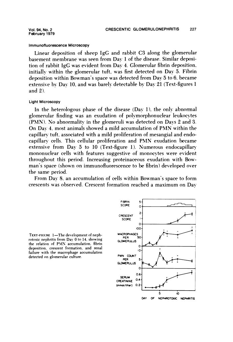
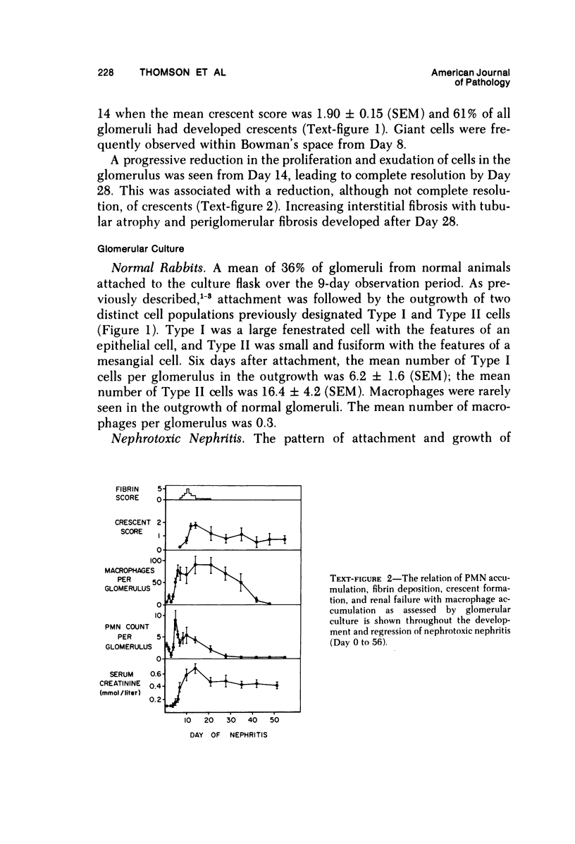
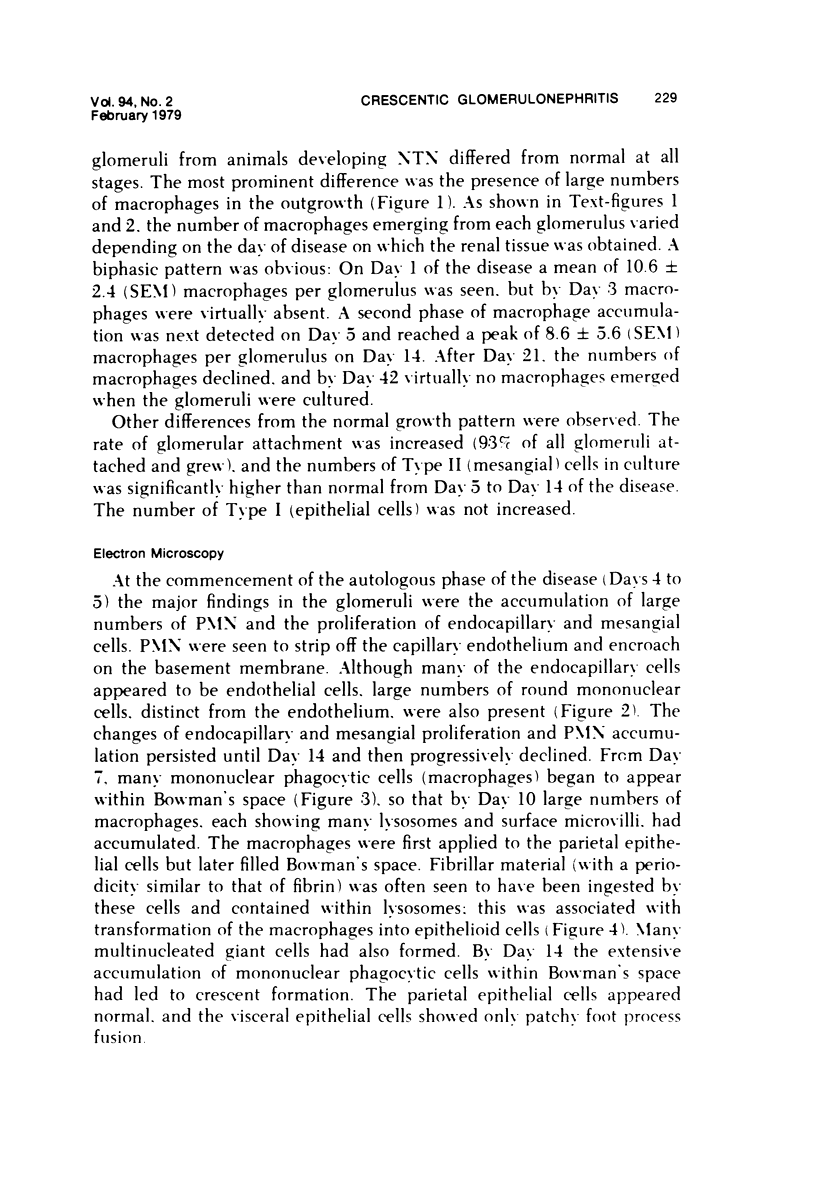
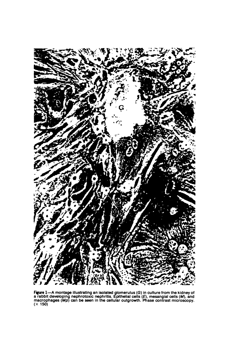
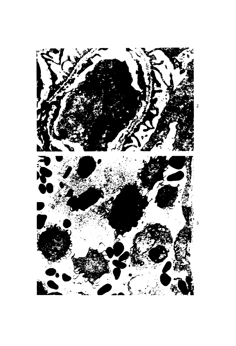
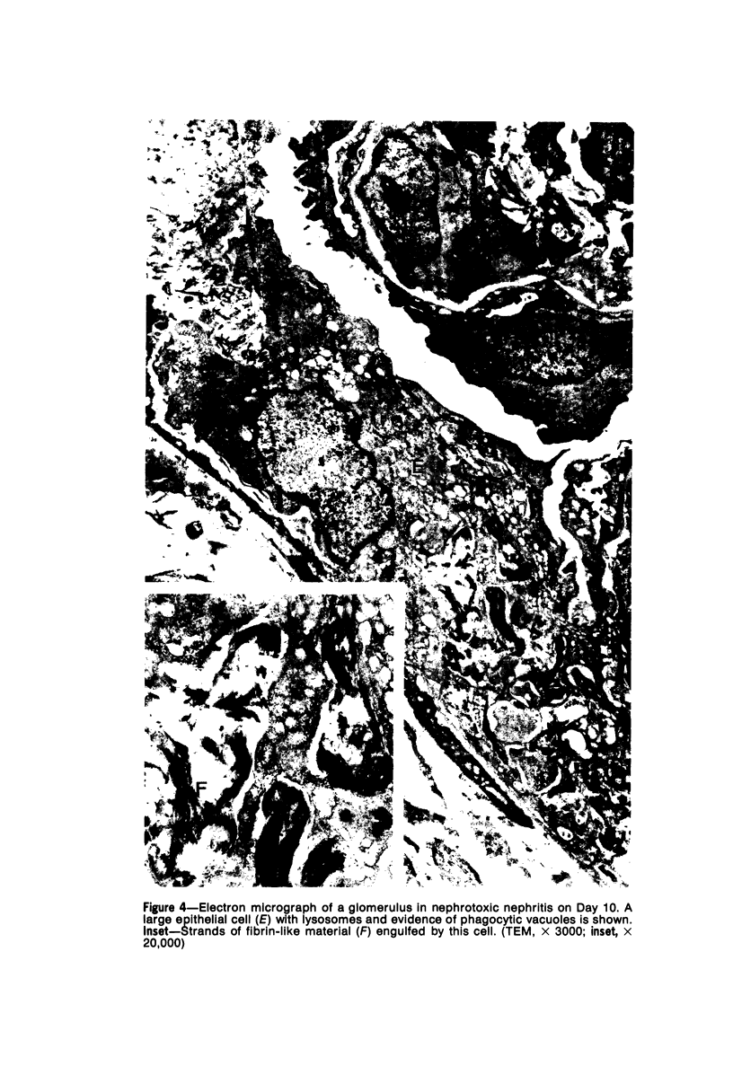
The role played by the macrophage in the development of injury in rabbit nephrotoxic nephritis (NTN) has been assessed by electron microscopy and glomerular culture of renal tissue obtained by several biopsies during the course of the disease. These observations have been correlated with the other immune, cellular, and biochemical events occurring in the glomerulus, ie, deposition of immunoglobulin and complement, proteinuria, polymorphonuclear leukocyte (PMN) exudation, fibrin deposition, crescent formation, and renal failure. A biphasic macrophage accumulation was detected, corresponding to the heterologous and autologous phases of the disease. In the autologous or crescentic phase, macrophages accumulated within the glomerular tuft from Day 5; their appearance coincided with the accumulation of PMN and development of proteinuria. Fibrin deposition in Bowman's space, which commenced on Days 6 and 7, was rapidly followed by the migration of macrophages from the glomeruli into Bowman's space. Within Bowman's space, macrophages were observed to phagocytose fibrin, transform into epithelioid and giant cells, and accumulate to form a substantial proportion of the cells forming the crescent. The inflammatory process of PMN exudation, macrophage accumulation, fibrin deposition, and crescent formation and the degree of renal failure reached a maximum by Days 12 to 14. Thereafter, resolution of the inflammatory process occurred so that by Day 40 macrophages had disappeared from the glomeruli. However, varying degrees of glomerular damage and renal failure persisted, occurring largely as a result of glomerulosclerosis and sclerosis of crescents. The tissue culture studies also demonstrated mesangial cell proliferation during the inflammatory process but did not show any abnormality of epithelial cell activity. This study demonstrates that the macrophages participate in NTN by accumulating in damaged glomeruli then migrating into Bowman's space (probably in response to fibrin deposition) where they undergo granulomatous transformation and accumulate, contributing to crescent formation.
Full text
PDF

Images in this article
Selected References
These references are in PubMed. This may not be the complete list of references from this article.
- Atkins R. C., Holdsworth S. R., Glasgow E. F., Matthews F. E. The macrophagen in human rapidly progressive glomerulonephritis. Lancet. 1976 Apr 17;1(7964):830–832. doi: 10.1016/s0140-6736(76)90480-3. [DOI] [PubMed] [Google Scholar]
- Bray M. A., Gordon D., Morley J. Proceedings: Role of prostaglandins in reactions of cellular immunity. Br J Pharmacol. 1974 Nov;52(3):453P–453P. [PMC free article] [PubMed] [Google Scholar]
- COCHRANE C. G., UNANUE E. R., DIXON F. J. A ROLE OF POLYMORPHONUCLEAR LEUKOCYTES AND COMPLEMENT IN NEPHROTOXIC NEPHRITIS. J Exp Med. 1965 Jul 1;122:99–116. doi: 10.1084/jem.122.1.99. [DOI] [PMC free article] [PubMed] [Google Scholar]
- Cochrane C. G., Müller-Eberhard H. J., Aikin B. S. Depletion of plasma complement in vivo by a protein of cobra venom: its effect on various immunologic reactions. J Immunol. 1970 Jul;105(1):55–69. [PubMed] [Google Scholar]
- Dinarello C. A., Goldin N. P., Wolff S. M. Demonstration and characterization of two distinct human leukocytic pyrogens. J Exp Med. 1974 Jun 1;139(6):1369–1381. doi: 10.1084/jem.139.6.1369. [DOI] [PMC free article] [PubMed] [Google Scholar]
- Epstein W. L., Krasnobrod H. The origin of epithelioid cells in experimental granulomas of man. Lab Invest. 1968 Feb;18(2):190–195. [PubMed] [Google Scholar]
- Holdsworth S. R., Glasgow E. F., Thomson N. M., Atkins R. C. Tissue culture of isolated human glomeruli. Pathology. 1978 Jan;10(1):59–67. doi: 10.3109/00313027809063480. [DOI] [PubMed] [Google Scholar]
- Holdsworth S. R., Thomson N. M., Glasgow E. F., Dowling J. P., Atkins R. C. Tissue culture of isolated glomeruli in experimental crescentic glomerulonephritis. J Exp Med. 1978 Jan 1;147(1):98–109. doi: 10.1084/jem.147.1.98. [DOI] [PMC free article] [PubMed] [Google Scholar]
- Naish P. F., Thomson N. M., Simpson I. J., Peters D. K. The role of polymorphonuclear leucocytes in the autologous phase of nephrotoxic nephritis. Clin Exp Immunol. 1975 Oct;22(1):102–111. [PMC free article] [PubMed] [Google Scholar]
- Papadimitriou J. M., Spector W. G. The origin, properties and fate of epithelioid cells. J Pathol. 1971 Nov;105(3):187–203. doi: 10.1002/path.1711050305. [DOI] [PubMed] [Google Scholar]
- STEBLAY R. W. Glomerulonephritis induced in sheep by injections of heterologous glomerular basement membrane and Freund's complete adjuvant. J Exp Med. 1962 Aug 1;116:253–272. doi: 10.1084/jem.116.2.253. [DOI] [PMC free article] [PubMed] [Google Scholar]
- Thomson N. M., Simpson I. J., Peters D. K. A quantitative evaluation of anticoagulants in experimental nephrotoxic nephritis. Clin Exp Immunol. 1975 Feb;19(2):301–308. [PMC free article] [PubMed] [Google Scholar]
- Unanue E. R., Dixon F. J. Experimental glomerulonephritis: immunological events and pathogenetic mechanisms. Adv Immunol. 1967;6:1–90. doi: 10.1016/s0065-2776(08)60521-0. [DOI] [PubMed] [Google Scholar]
- Unkeless J. C., Gordon S., Reich E. Secretion of plasminogen activator by stimulated macrophages. J Exp Med. 1974 Apr 1;139(4):834–850. doi: 10.1084/jem.139.4.834. [DOI] [PMC free article] [PubMed] [Google Scholar]
- Wood B. T., Thompson S. H., Goldstein G. Fluorescent antibody staining. 3. Preparation of fluorescein-isothiocyanate-labeled antibodies. J Immunol. 1965 Aug;95(2):225–229. [PubMed] [Google Scholar]