

Abstract
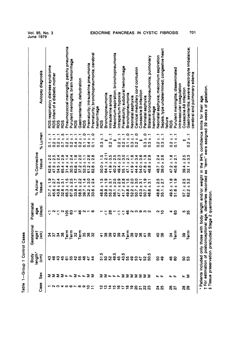
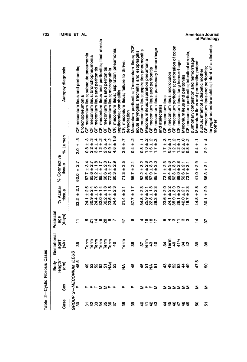
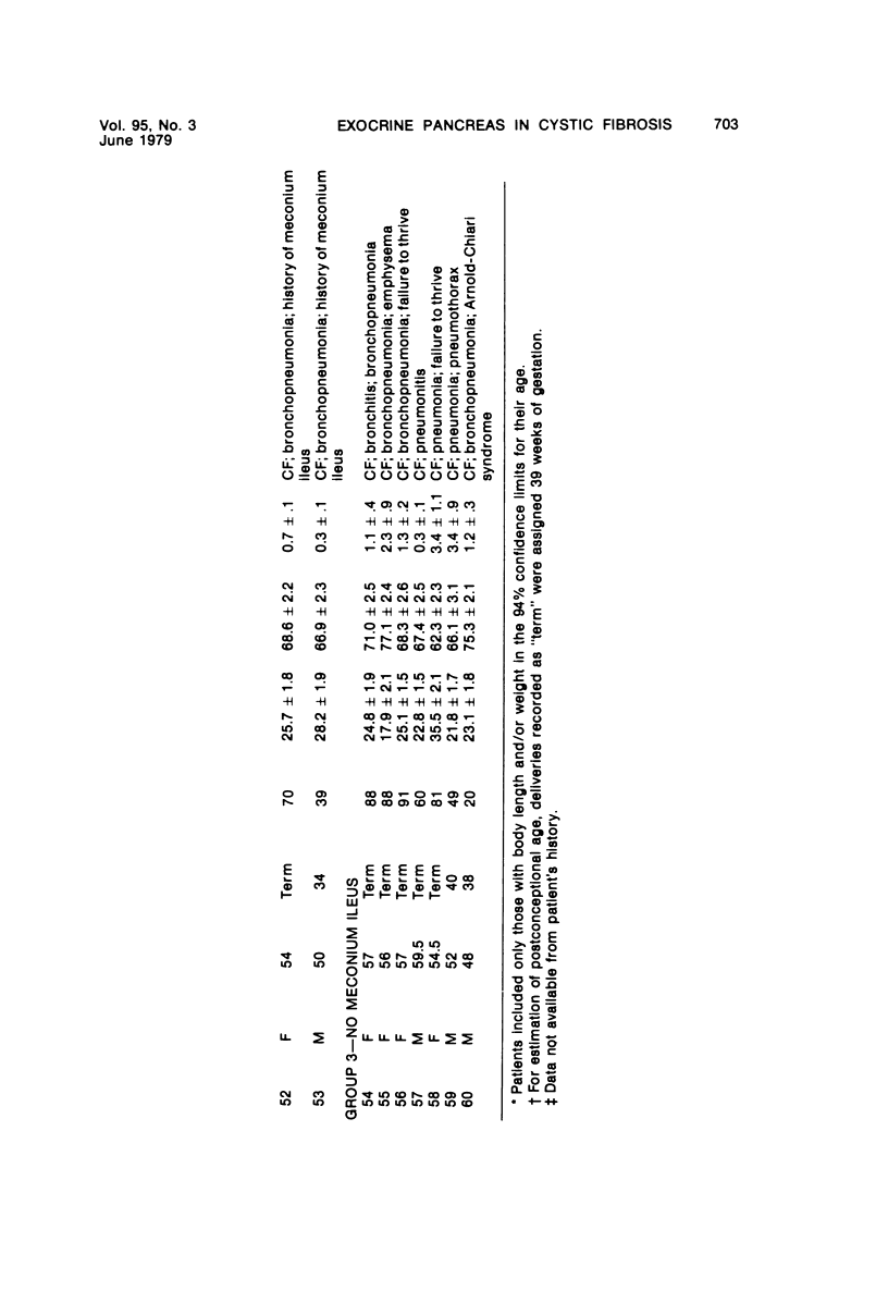
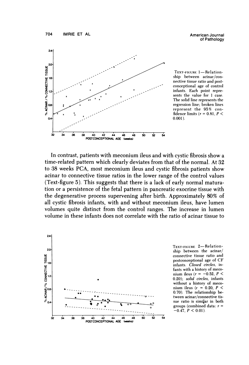
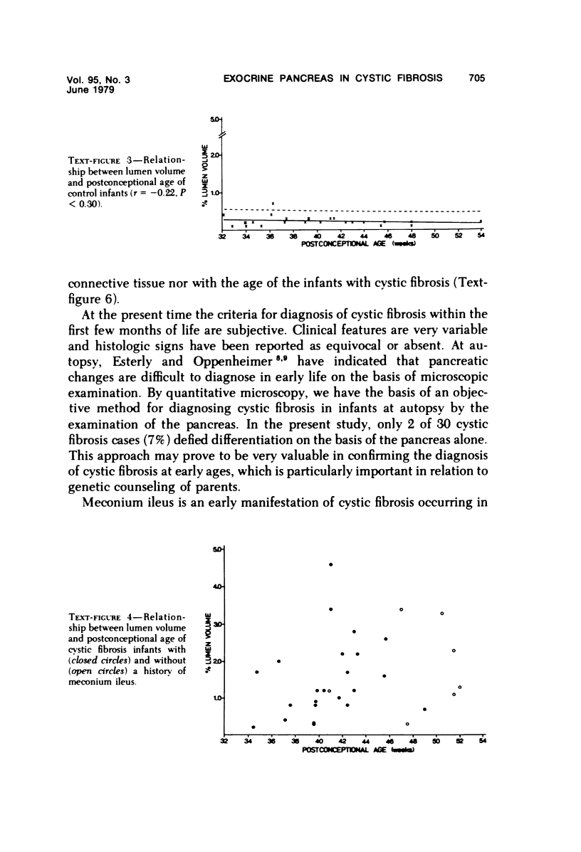
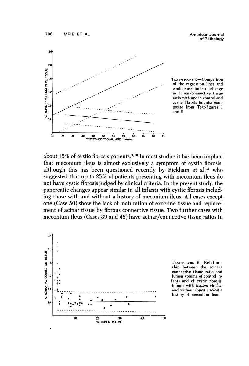
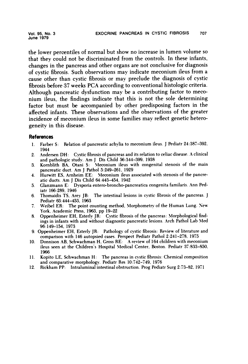
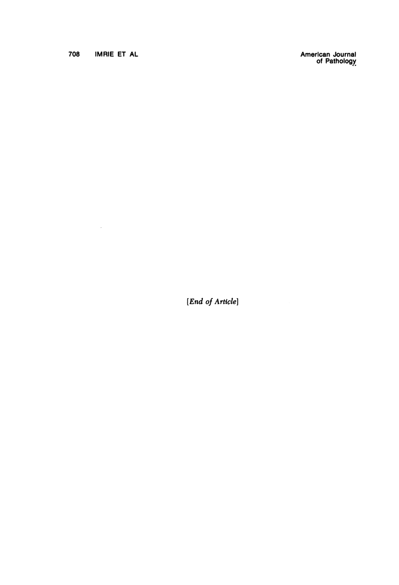
The development of the exocrine pancreas has been determined quantitatively in 31 infants with cystic fibrosis (CF) both with and without meconium ileus and in 29 control infants. In the normal pancreas, the ratio of acinar to connective tissue volume is 0.5 at 32 weeks postconceptional age (PCA) and increases linearly to 2.0 at 52 weeks PGA. In cystic fibrosis infants, with or without meconium ileus, the ration is 0.5 at 35 weeks PCA anddecreases linearly to 0.3 at 52 weeks PCA. The volume of acinar and duct lumens is greater in CF than control infants but is independent of age or acinar volume. The development of the exocrine pancreas in infants with CF with and without meconium ileus diverges from the normal pattern: There is consistent lack of exocrine tissue before or a full-term birth, which persists throghout the age range of this study. CF infants above 42 weeks PCA can be discriminated from controls on the basis of the quantitative assessment of acinar volume.
Full text
PDF
Selected References
These references are in PubMed. This may not be the complete list of references from this article.
- Donnison A. B., Shwachman H., Gross R. E. A review of 164 children with meconium ileus seen at the Children's Hospital Medical Center, Boston. Pediatrics. 1966 May;37(5):833–850. [PubMed] [Google Scholar]
- Kopito L. E., Shwachman H. The pancreas in cystic fibrosis: chemical composition and comparative morphology. Pediatr Res. 1976 Aug;10(8):742–749. doi: 10.1203/00006450-197608000-00010. [DOI] [PubMed] [Google Scholar]
- Oppenheimer E. H., Esterly J. R. Cystic fibrosis of the pancreas. Morphologic findings in infants with and without diagnostic pancreatic lesions. Arch Pathol. 1973 Sep;96(3):149–154. [PubMed] [Google Scholar]
- Oppenheimer E. H., Esterly J. R. Pathology of cystic fibrosis review of the literature and comparison with 146 autopsied cases. Perspect Pediatr Pathol. 1975;2:241–278. [PubMed] [Google Scholar]
- Rickham P. P. Intralumenal intestinal obstruction. Prog Pediatr Surg. 1971;2:73–82. [PubMed] [Google Scholar]
- THOMAIDIS T. S., AREY J. B. THE INTESTINAL LESIONS IN CYSTIC FIBROSIS OF THE PANCREAS. J Pediatr. 1963 Sep;63:444–453. doi: 10.1016/s0022-3476(63)80434-5. [DOI] [PubMed] [Google Scholar]