Abstract
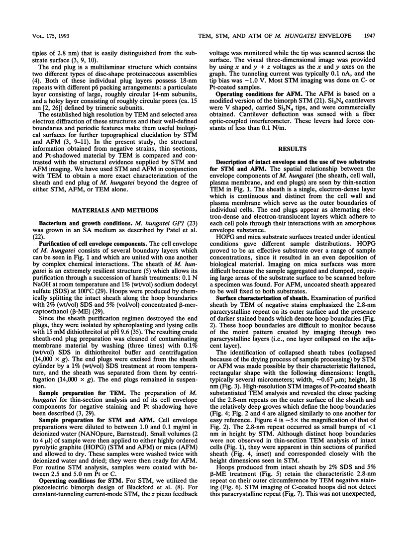
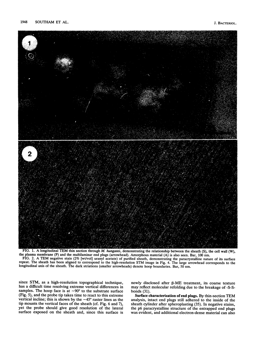
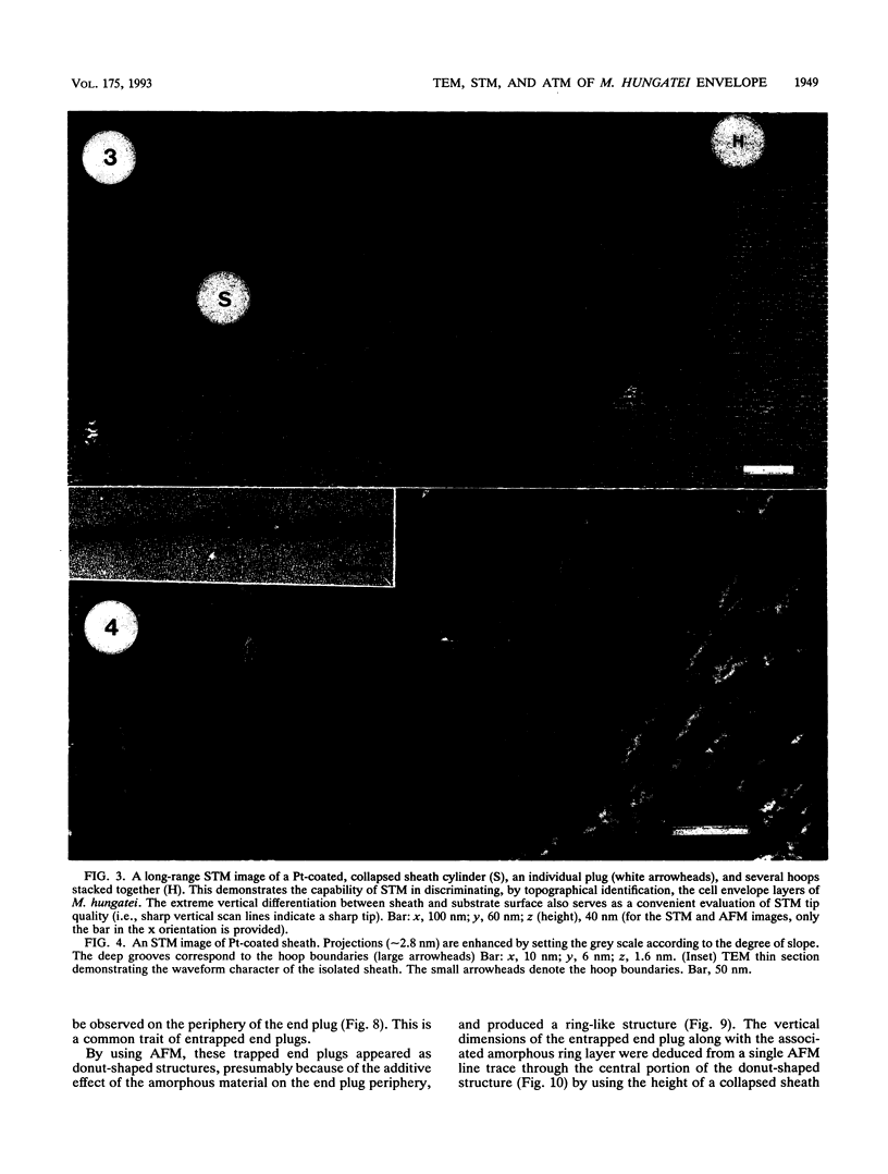
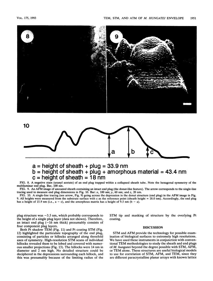
Methanospirillum hungatei GP1 possesses paracrystalline cell envelope components including end plugs and a sheath formed from stacked hoops. Both negative-stain transmission electron microscopy (TEM) and scanning tunneling microscopy (STM) distinguished the 2.8-nm repeat on the outer surface of the sheath, while negative-stain TEM alone demonstrated this repeat around the outer circumference of individual hoops. Thin sections revealed a wave-like outer sheath surface, while STM showed the presence of deep grooves that precisely defined the hoop-to-hoop boundaries at the waveform nodes. Atomic force microscopy of sheath tubes containing entrapped end plugs emphasized the end plug structure, suggesting that the sheath was malleable enough to collapse over the end plugs and deform to mimic the shape of the underlying structure. High-resolution atomic force microscopy has revised the former idea of end plug structure so that we believe each plug consists of at least four discs, each of which is approximately 3.5 nm thick. PT shadow TEM and STM both demonstrated the 14-nm hexagonal, particulate surface of an end plug, and STM showed the constituent particles to be lobed structures with numerous smaller projections, presumably corresponding to the molecular folding of the particle.
Full text
PDF



Images in this article
Selected References
These references are in PubMed. This may not be the complete list of references from this article.
- Beveridge T. J., Southam G., Jericho M. H., Blackford B. L. High-resolution topography of the S-layer sheath of the archaebacterium Methanospirillum hungatei provided by scanning tunneling microscopy. J Bacteriol. 1990 Nov;172(11):6589–6595. doi: 10.1128/jb.172.11.6589-6595.1990. [DOI] [PMC free article] [PubMed] [Google Scholar]
- Beveridge T. J., Sprott G. D., Whippey P. Ultrastructure, inferred porosity, and gram-staining character of Methanospirillum hungatei filament termini describe a unique cell permeability for this archaeobacterium. J Bacteriol. 1991 Jan;173(1):130–140. doi: 10.1128/jb.173.1.130-140.1991. [DOI] [PMC free article] [PubMed] [Google Scholar]
- Beveridge T. J., Stewart M., Doyle R. J., Sprott G. D. Unusual stability of the Methanospirillum hungatei sheath. J Bacteriol. 1985 May;162(2):728–737. doi: 10.1128/jb.162.2.728-737.1985. [DOI] [PMC free article] [PubMed] [Google Scholar]
- Binnig G, Quate CF, Gerber C. Atomic force microscope. Phys Rev Lett. 1986 Mar 3;56(9):930–933. doi: 10.1103/PhysRevLett.56.930. [DOI] [PubMed] [Google Scholar]
- Drake B., Prater C. B., Weisenhorn A. L., Gould S. A., Albrecht T. R., Quate C. F., Cannell D. S., Hansma H. G., Hansma P. K. Imaging crystals, polymers, and processes in water with the atomic force microscope. Science. 1989 Mar 24;243(4898):1586–1589. doi: 10.1126/science.2928794. [DOI] [PubMed] [Google Scholar]
- Dunlap D. D., Bustamante C. Images of single-stranded nucleic acids by scanning tunnelling microscopy. Nature. 1989 Nov 9;342(6246):204–206. doi: 10.1038/342204a0. [DOI] [PubMed] [Google Scholar]
- Edstrom R. D., Yang X. R., Lee G., Evans D. F. Viewing molecules with scanning tunneling microscopy and atomic force microscopy. FASEB J. 1990 Oct;4(13):3144–3151. doi: 10.1096/fasebj.4.13.2120098. [DOI] [PubMed] [Google Scholar]
- Fisher K. A. Monolayer freeze-fracture and scanning tunneling microscopy. J Electron Microsc Tech. 1989 Dec;13(4):355–371. doi: 10.1002/jemt.1060130408. [DOI] [PubMed] [Google Scholar]
- Fisher K. A., Yanagimoto K. C., Whitfield S. L., Thomson R. E., Gustafsson M. G., Clarke J. Scanning tunneling microscopy of planar biomembranes. Ultramicroscopy. 1990 Aug;33(2):117–126. doi: 10.1016/0304-3991(90)90014-d. [DOI] [PubMed] [Google Scholar]
- Henderson R., Unwin P. N. Three-dimensional model of purple membrane obtained by electron microscopy. Nature. 1975 Sep 4;257(5521):28–32. doi: 10.1038/257028a0. [DOI] [PubMed] [Google Scholar]
- Patel G. B., Roth L. A., van den Berg L., Clark D. S. Characterization of a strain of Methanospirillum hungatti. Can J Microbiol. 1976 Sep;22(9):1404–1410. doi: 10.1139/m76-208. [DOI] [PubMed] [Google Scholar]
- Ruben G. C. Ultrathin (1 nm) vertically shadowed platinum-carbon replicas for imaging individual molecules in freeze-etched biological DNA and material science metal and plastic specimens. J Electron Microsc Tech. 1989 Dec;13(4):335–354. doi: 10.1002/jemt.1060130407. [DOI] [PubMed] [Google Scholar]
- Shaw P. J., Hills G. J., Henwood J. A., Harris J. E., Archer D. B. Three-dimensional architecture of the cell sheath and septa of Methanospirillum hungatei. J Bacteriol. 1985 Feb;161(2):750–757. doi: 10.1128/jb.161.2.750-757.1985. [DOI] [PMC free article] [PubMed] [Google Scholar]
- Smith D. P., Bryant A., Quate C. F., Rabe J. P., Gerber C., Swalen J. D. Images of a lipid bilayer at molecular resolution by scanning tunneling microscopy. Proc Natl Acad Sci U S A. 1987 Feb;84(4):969–972. doi: 10.1073/pnas.84.4.969. [DOI] [PMC free article] [PubMed] [Google Scholar]
- Smith D. P., Hörber H., Gerber C., Binnig G. Smectic liquid crystal monolayers on graphite observed by scanning tunneling microscopy. Science. 1989 Jul 7;245(4913):43–45. doi: 10.1126/science.245.4913.43. [DOI] [PubMed] [Google Scholar]
- Southam G., Beveridge T. J. Characterization of novel, phenol-soluble polypeptides which confer rigidity to the sheath of Methanospirillum hungatei GP1. J Bacteriol. 1992 Feb;174(3):935–946. doi: 10.1128/jb.174.3.935-946.1992. [DOI] [PMC free article] [PubMed] [Google Scholar]
- Southam G., Beveridge T. J. Detection of growth sites in and protomer pools for the sheath of Methanospirillum hungatei GP1 by use of constituent organosulfur and immunogold labeling. J Bacteriol. 1992 Oct;174(20):6460–6470. doi: 10.1128/jb.174.20.6460-6470.1992. [DOI] [PMC free article] [PubMed] [Google Scholar]
- Southam G., Beveridge T. J. Dissolution and immunochemical analysis of the sheath of the archaeobacterium Methanospirillum hungatei GP1. J Bacteriol. 1991 Oct;173(19):6213–6222. doi: 10.1128/jb.173.19.6213-6222.1991. [DOI] [PMC free article] [PubMed] [Google Scholar]
- Sprott G. D., Colvin J. R., McKellar R. C. Spheroplasts of Methanospirillum hungatii formed upon treatment with dithiothreitol. Can J Microbiol. 1979 Jun;25(6):730–738. doi: 10.1139/m79-106. [DOI] [PubMed] [Google Scholar]
- Stemmer A., Engel A. Imaging biological macromolecules by STM: quantitative interpretation of topographs. Ultramicroscopy. 1990 Dec;34(3):129–140. doi: 10.1016/0304-3991(90)90067-v. [DOI] [PubMed] [Google Scholar]
- Stemmer A., Hefti A., Aebi U., Engel A. Scanning tunneling and transmission electron microscopy on identical areas of biological specimens. Ultramicroscopy. 1989 Jul-Aug;30(3):263–280. doi: 10.1016/0304-3991(89)90056-9. [DOI] [PubMed] [Google Scholar]
- Stewart M., Beveridge T. J., Sprott G. D. Crystalline order to high resolution in the sheath of Methanospirillum hungatei: a cross-beta structure. J Mol Biol. 1985 Jun 5;183(3):509–515. doi: 10.1016/0022-2836(85)90019-1. [DOI] [PubMed] [Google Scholar]
- Zeikus J. G., Bowen V. G. Fine structure of Methanospirillum hungatii. J Bacteriol. 1975 Jan;121(1):373–380. doi: 10.1128/jb.121.1.373-380.1975. [DOI] [PMC free article] [PubMed] [Google Scholar]


