Abstract
The polyketide ambruticin is an attractive candidate for drug development as an antifungal agent, but its mechanism of action has not yet been elucidated. Here we present evidence that ambruticin exerts its effect by targeting HOG, the osmotic stress control pathway, through Hik1, a group III histidine kinase.
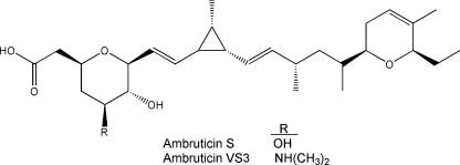
Ambruticins are polyketides of unusual structure produced by strains of Sorangium (Polyangium) cellulosum (4, 9, 17). The S and VS congeners (Fig. 1) have potent antifungal activity and might provide new drugs to treat certain fungal infections. Ambruticin S and two derivatives of ambruticin VS3 have been shown to cure mice of acute pulmonary coccidodiomycosis or histoplasmosis with twice-a-day oral dosing (6, 18). Similarly, oral administration of an analog of ambruticin VS3 significantly reduced pulmonary fungal burdens and promoted improved survival in a murine model of invasive pulmonary aspergillosis (3).
FIG. 1.
Chemical structures of ambruticins S and VS3.
A number of fungicides, including phenylpyrroles (e.g., pyrrolnitrin and fludioxonil), dicarboximides (e.g., vinclozolin, iprodione, and procymidone), and aromatic hydrocarbons (e.g., chloroneb and quintozene), used commercially to protect crops from fungal disease, exert their antifungal effects by inducing the high-osmolarity glycerol (HOG) signaling pathway, resulting in the accumulation of glycerol, with a concomitant accumulation of free fatty acids. In the absence of high external osmolarity, intracellular accumulation of glycerol causes leakage of cellular contents and ultimately results in cell death (11). Previous work has shown that ambruticin may also mediate its effect through the HOG signaling pathway. Fungal strains resistant to jerangolid, a compound similar in structure to ambruticin, are cross-resistant to pyrrolnitrin (8), and accumulation of glycerol occurs when Hansenula anomala is treated with ambruticin or pyrrolnitrin (11). The HOG pathway for osmoregulation has been characterized in considerable detail in Saccharomyces cerevisiae (10) and also in the filamentous fungus Neurospora crassa (12). Osmotic stress is thought to be sensed by one or more histidine kinases in the fungal membrane or cytosol, and the expression of many genes is activated through a signal transduction pathway homologous to the mitogen-activated kinase (MAPK) pathway of animals (15). In S. cerevisiae, the sensor is believed to be Sln1, a group VI histidine kinase (2). Sln1 transmits the stress response signal through a two-component system (Sln1-Ypd1-Ssk1) and MAPK cascade (Ssk2/Ssk22-Pbs2-Hog1). Under high-osmolarity conditions, Ssk1 is dephosphorylated; dephospho-Ssk1 activates the downstream MAPK cascade. Phospho-Hog1 is necessary to turn on the genes for high-osmolarity adaptation, resulting in a number of events, including glycerol accumulation. Filamentous fungi have similar signal transduction pathways, and group III histidine kinases (Os-1 orthologues) and group VI histidine kinases are thought to sense osmotic stress. Mutations that knock out the HOG pathway in N. crassa cause both hypersensitivity to osmotic stress and resistance to phenylpyrroles (20). The fungicide dicarboximide also seems to act via the HOG pathway, and several reports have suggested that the target of both dicarboximides and phenylpyrroles is Os-1 or an orthologue (1, 5, 7, 16, 19). In the rice BLAST fungus Magnaporthe grisea, the Os-1 orthologue is named Hik1 (13).
Many yeasts, including S. cerevisiae, are naturally resistant to phenylpyrroles, while many filamentous fungi are susceptible. The basis of this difference is not fully understood but is believed to be related, in part, to the presence of the Os-1 sensor. In a recent report, expression of the HIK1 gene from M. grisea in S. cerevisiae made the strain susceptible to phenylpyrroles and other antifungal agents (14). These observations suggest that Hik1 is either a direct target of the fungicide or a mediator of its action, which is transmitted to the HOG pathway to produce intracellular glycerol accumulation in the absence of high external osmolarity.
In order to analyze whether the expression of the HIK1 gene confers ambruticin susceptibility to S. cerevisiae, the S. cerevisiae strains employed by Motoyama et al. (14) were tested for susceptibility to ambruticin and other antifungal agents. Table 1 shows that the pYES2-HIK1-containing strain is highly susceptible to ambruticin only when Hik1 is produced. Production of active Hik1 also confers susceptibility to the phenylpyrrole fludioxonil and the dicarboximide vinclozolin, as described previously (14). To investigate whether the regions required for histidine kinase function are required for the Hik1-dependent susceptibility to ambruticin, two plasmids for the expression of mutated versions of Hik1 were used. The vector pYES-HIK1-H736V encodes a Hik1p defective in autophosphorylation, making the histidine kinase domain nonfunctional, while the plasmid pYES-HIK1-D1153E encodes Hik1p with a mutation in the phosphoacceptor residue, thereby inactivating the response regulator domain (13). S. cerevisiae strains carrying either plasmid and induced to express the mutated HIK1 gene showed no susceptibility to any of the fungicides, suggesting that both the histidine kinase and the response regulator domains are required to confer susceptibility to the tested compounds. These findings indicate that functional Hik1 (and, most likely, phosphotransfer from Hik1) is required to confer ambruticin susceptibility to S. cerevisiae.
TABLE 1.
Susceptibility of S. cerevisiae ATCC 201388 to antifungal compoundsa
| Plasmid | Drug | MIC-0 (μg/ml)
|
|
|---|---|---|---|
| SD (no induction) | SG (induction) | ||
| pYES2 | Ambruticin VS3 | >32 | >32 |
| Fludioxonil | >32 | >32 | |
| Vinclozolin | >32 | >32 | |
| pYES2-HIK1 | Ambruticin VS3 | >32 | 0.5 |
| Fludioxonil | >32 | 2 | |
| Vinclozolin | >32 | 16 | |
| pYES2-hik1-H736V | Ambruticin VS3 | >32 | >32 |
| Fludioxonil | >32 | >32 | |
| Vinclozolin | >32 | >32 | |
| pYES2-hik1-D1153E | Ambruticin VS3 | >32 | >32 |
| Fludioxonil | >32 | >32 | |
| Vinclozolin | >32 | >32 | |
The strains and plasmids are described in reference 14. MICs were determined using CLSI (formerly NCCLS) protocol M27, except that inocula were prepared using SD/−Ura or SG/−Ura medium (absence of uracil from medium selects for maintenance of plasmid) and RPMI was replaced with either SD/−Ura or SG/−Ura as the test medium. MICs were determined after 48 h of incubation. MIC-0 is defined as the concentration of antifungal that caused a 90% reduction in growth relative to the untreated control. SD medium contains yeast nitrogen base without amino acids (6.7 g/liter), glucose (20 g/liter), and 1× dropout solution (Clontech). SG medium is identical to SD, except that glucose was replaced with galactose (20 g/liter).
The level of phosphorylation of Hog1 in the presence of ambruticin was also examined. It was recently described that fungicides from the groups of phenylpyrroles, dicarboximides, and aromatic hydrocarbons mediate their effects on the HOG pathway by promoting double phosphorylation of the T174 and Y176 residues of Hog1 (13). To determine whether exposure of the Hik1-expressing S. cerevisiae strain to ambruticin resulted in phosphorylation of Hog1, an antibody (anti-p38) that detects Hog1 in the doubly phosphorylated state was employed. The total level of Hog1 was determined using an anti-Hog1 antibody. As can be seen in Fig. 2A, an elevated level of phosphorylated Hog1 in the pYES2-HIK1-containing strain was detected when HIK1 expression was induced and the cells were either treated with ambruticin or exposed to 0.5 M NaCl. In cells carrying the empty pYES2 plasmid, phosphorylation was detected only upon exposure to 0.5 M NaCl but not after treatment with ambruticin. No signal was detected in any of the Western blots for the isogenic Δhog1 mutant strain used as a control.
FIG. 2.
Western blot analysis of phospho-Hog1 and total Hog1 from S. cereviseae strains using (A) anti-p38 antibody, raised to a synthetic doubly phosphorylated segment of the mammalian protein P38 corresponding to T180/Y182 (Cell Signaling Technology), or (B) anti C-terminal Hog1 antibody (Santa Cruz Biotechnology). S. cerevisiae ATCC 201388/pYES2 (SC/pYES2) and S. cerevisiae ATCC 201388/pYES2-HIK1 (SC/pYES2-HIK1) are described in Table 1. SC Δ hog1 is an isogenic derivative of ATCC 201388 but carries a deletion of the HOG1 gene (14). S. cerevisiae strain ATCC 201388 was transformed with either pYES or pYES-HIK1, as described in Table 1, or disrupted for HOG1 (Δhog1), as described in reference 14. Cells from an overnight culture in SD/−URA were resuspended in fresh SD/−URA and grown for 4 h. The cells were treated for 10 min with NaCl (0.5 M) or ambruticin (25 ppm) or were not treated (NT) and cell extracts were prepared. Soluble protein was used for Western blot analysis. Antibody binding was visualized using a horseradish peroxidase-conjugated secondary antibody employing detection with ECL Plus (Amersham).
In summary, we have shown that the expression of Hik1 confers ambruticin susceptibility to S. cerevisiae and that Hog1 is specifically phosphorylated when the Hik1-expressing cells are exposed to ambruticin. The requirement for Hik1 suggests that it is the likely molecular target of ambruticin and that binding of the drug results in an inappropriate cellular response, including intracellular accumulation of glycerol and, ultimately, cell death. Binding of ambruticin could either stimulate the normal phosphatase activity of Hik1 or induce the back transfer of phosphate from Ypd1, the small phosphotranfer protein present in fungi and yeast that carries the phosphate signal downstream from Hik1 towards Hog1. Either mechanism for ambruticin action would result in the emergence of dephosphorylated Ssk1 (active form) and overstimulation of the HOG pathway and thus result in cell death.
Acknowledgments
We thank Ralph Reid and Chris Reeves for helpful discussions and Jonathan Kennedy for help with the Western blots.
Footnotes
Published ahead of print on 13 August 2007.
REFERENCES
- 1.Avenot, H., P. Simoneau, B. Iacomi-Vasilescu, and N. Bataille-Simoneau. 2005. Characterization of mutations in the two-component histidine kinase gene AbNIK1 from Alternaria brassicicola that confer high dicarboximide and phenylpyrrole resistance. Curr. Genet. 47:234-243. [DOI] [PubMed] [Google Scholar]
- 2.Catlett, N. L., O. C. Yoder, and B. G. Turgeon. 2003. Whole-genome analysis of two-component signal transduction genes in fungal pathogens. Eukaryot. Cell 2:1151-1161. [DOI] [PMC free article] [PubMed] [Google Scholar]
- 3.Chiang, L. Y., D. E. Ejzykowicz, Z.-Q. Tian, L. Katz, and S. G. Filler. 2006. Efficacy of ambruticin analogs in a murine model of invasive pulmonary aspergillosis. Antimicrob. Agents Chemother. 50:3464-3466. [DOI] [PMC free article] [PubMed] [Google Scholar]
- 4.Connor, D. T., R. C. Greenough, and M. von Strandtmann. 1977. W-7783, a unique antifungal antibiotic. J. Org. Chem. 42:3664-3669. [DOI] [PubMed] [Google Scholar]
- 5.Cui, W., R. E. Beever, S. L. Parkes, P. L. Weeds, and M. D. Templeton. 2002. An osmosensing histidine kinase mediates dicarboximide fungicide resistance in Botryotinia fuckeliana (Botrytis cinerea). Fungal Genet. Biol. 36:187-198. [DOI] [PubMed] [Google Scholar]
- 6.Deresinski, S. C., H. B. Levine, and D. A. Stevens. 1978. Coccidioides immitis endospores: phagocytosis by human cells. Mycopathologia 64:179-181. [DOI] [PubMed] [Google Scholar]
- 7.Dry, I. B., K. H. Yuan, and D. G. Hutton. 2004. Dicarboximide resistance in field isolates of Alternaria alternata is mediated by a mutation in a two-component histidine kinase gene. Fungal Genet. Biol. 41:102-108. [DOI] [PubMed] [Google Scholar]
- 8.Gerth, K., P. Washausen, G. Hofle, H. Irschik, and H. Reichenbach. 1996. The jerangolids: a family of new antifungal compounds from Sorangium cellulosum (Myxobacteria). Production, physico-chemical and biological properties of jerangolid A. J. Antibiot. (Tokyo) 49:71-75. [DOI] [PubMed] [Google Scholar]
- 9.Hoefle, G., H. Steinmetz, K. Gerth, and H. Reichenbach. 1991. Ambruticins VS: new members of the antifungal ambruticin family from Sorangium cellulosum. Leibigs Ann. Chem. 1991:941-945. [Google Scholar]
- 10.Hohmann, S. 2002. Osmotic stress signaling and osmoadaptation in yeasts. Microbiol. Mol. Biol. Rev. 66:300-372. [DOI] [PMC free article] [PubMed] [Google Scholar]
- 11.Knauth, P., and H. Reichenbach. 2000. On the mechanism of action of the myxobacterial fungicide ambruticin. J. Antibiot. (Tokyo) 53:1182-1190. [DOI] [PubMed] [Google Scholar]
- 12.Krantz, M., E. Becit, and S. Hohmann. 2006. Comparative analysis of HOG pathway proteins to generate hypotheses for functional analysis. Curr. Genet. 49:152-165. [DOI] [PubMed] [Google Scholar]
- 13.Motoyama, T., K. Kadokura, T. Ohira, A. Ichiishi, M. Fujimura, I. Yamaguchi, and T. Kudo. 2005. A two-component histidine kinase of the rice blast fungus is involved in osmotic stress response and fungicide action. Fungal Genet. Biol. 42:200-212. [DOI] [PubMed] [Google Scholar]
- 14.Motoyama, T., T. Ohira, K. Kadokura, A. Ichiishi, M. Fujimura, I. Yamaguchi, and T. Kudo. 2005. An Os-1 family histidine kinase from a filamentous fungus confers fungicide-sensitivity to yeast. Curr. Genet. 47:298-306. [DOI] [PubMed] [Google Scholar]
- 15.Noguchi, R., S. Banno, R. Ichikawa, F. Fukumori, A. Ichiishi, M. Kimura, I. Yamaguchi, and M. Fujimura. 2007. Identification of OS-2 MAP kinase-dependent genes induced in response to osmotic stress, antifungal agent fludioxonil, and heat shock in Neurospora crassa. Fungal Genet. Biol. 44:208-218. [DOI] [PubMed] [Google Scholar]
- 16.Ochiai, N., M. Fujimura, T. Motoyama, A. Ichiishi, R. Usami, K. Horikoshi, and I. Yamaguchi. 2001. Characterization of mutations in the two-component histidine kinase gene that confer fludioxonil resistance and osmotic sensitivity in the os-1 mutants of Neurospora crassa. Pest Manag. Sci. 57:437-442. [DOI] [PubMed] [Google Scholar]
- 17.Ringel, S. M., R. C. Greenough, S. Roemer, D. Connor, A. L. Gutt, B. Blair, G. Kanter, and M. von Strandtmann. 1977. Ambruticin (W7783), a new antifungal antibiotic. J. Antibiot. (Tokyo) 30:371-375. [DOI] [PubMed] [Google Scholar]
- 18.Shubitz, L. F., J. N. Galgiani, Z.-Q. Tian, Z. Zhong, P. Timmermans, and L. Katz. 2006. Efficacy of ambruticin analogs in a murine model of coccidioidomycosis. Antimicrob. Agents Chemother. 50:3467-3469. [DOI] [PMC free article] [PubMed] [Google Scholar]
- 19.Yoshimi, A., M. Tsuda, and C. Tanaka. 2004. Cloning and characterization of the histidine kinase gene Dic1 from Cochliobolus heterostrophus that confers dicarboximide resistance and osmotic adaptation. Mol. Genet. Genomics 271:228-236. [DOI] [PubMed] [Google Scholar]
- 20.Zhang, Y., R. Lamm, C. Pillonel, S. Lam, and J.-R. Xu. 2002. Osmoregulation and fungicide resistance: the Neurospora crassa os-2 gene encodes a HOG1 mitogen-activated protein kinase homologue. Appl. Environ. Microbiol. 68:532-538. [DOI] [PMC free article] [PubMed] [Google Scholar]


