Full text
PDF
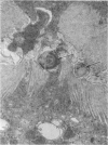
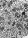
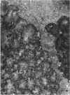
Images in this article
Selected References
These references are in PubMed. This may not be the complete list of references from this article.
- Bullivant C. M. Accidental poisoning by paraquat: Report of two cases in man. Br Med J. 1966 May 21;1(5498):1272–1273. doi: 10.1136/bmj.1.5498.1272. [DOI] [PMC free article] [PubMed] [Google Scholar]
- Campbell S. Death from paraquat in a child. Lancet. 1968 Jan 20;1(7534):144–144. doi: 10.1016/s0140-6736(68)92754-2. [DOI] [PubMed] [Google Scholar]
- Clark D. G., McElligott T. F., Hurst E. W. The toxicity of paraquat. Br J Ind Med. 1966 Apr;23(2):126–132. doi: 10.1136/oem.23.2.126. [DOI] [PMC free article] [PubMed] [Google Scholar]
- Cuppage F. E., Tate A. Repair of the nephron following injury with mercuric chloride. Am J Pathol. 1967 Sep;51(3):405–429. [PMC free article] [PubMed] [Google Scholar]
- DAEMS W. T., van RIJSSEL T. The fine structure of the peribiliary dense bodies in mouse liver tissue. J Ultrastruct Res. 1961 Jun;5:263–290. doi: 10.1016/s0022-5320(61)90020-x. [DOI] [PubMed] [Google Scholar]
- Fennelly J. J., Gallagher J. T., Carroll R. T. Paraquat poisoning in a pregnant woman. Br Med J. 1968 Sep 21;3(5620):722–723. doi: 10.1136/bmj.3.5620.722. [DOI] [PMC free article] [PubMed] [Google Scholar]
- Goldstone A., Szabo E., Koenig H. Isolation and characterization of acidic lipoprotein in renal and hepatic lysosomes. Life Sci II. 1970 Jun 8;9(11):607–616. doi: 10.1016/0024-3205(70)90211-0. [DOI] [PubMed] [Google Scholar]
- Gritzka T. L., Trump B. F. Renal tubular lesions caused by mercuric chloride. Electron microscopic observations: degeneration of the pars recta. Am J Pathol. 1968 Jun;52(6):1225–1277. [PMC free article] [PubMed] [Google Scholar]
- HERDSON P. B., GARVIN P. J., JENNINGS R. B. FINE STRUCTURAL CHANGES IN RAT LIVER INDUCED BY PHENOBARBITAL. Lab Invest. 1964 Sep;13:1032–1037. [PubMed] [Google Scholar]
- Holgersen O., Gloor F., Rohr H. P., Torhorst J. Frühveränderungen an der proximalen Tubuluszelle der Rattenniere nach Sublimatvergiftung: Degenerative und adaptive Phänomene in den proximalen Tubuluszellen. Virchows Arch B Cell Pathol. 1969 Sep 16;3(4):324–338. [PubMed] [Google Scholar]
- Hutterer F., Schaffner F., Klion F. M., Popper H. Hypertrophic, hypoactive smooth endoplasmic reticulum: a sensitive indicator of hepatotoxicity exemplified by dieldrin. Science. 1968 Sep 6;161(3845):1017–1019. doi: 10.1126/science.161.3845.1017. [DOI] [PubMed] [Google Scholar]
- KOENIG H. Histological distribution of brain gangliosides: lysosomes as glycolipoprotein granules. Nature. 1962 Aug 25;195:782–784. doi: 10.1038/195782a0. [DOI] [PubMed] [Google Scholar]
- LUFT J. H. Improvements in epoxy resin embedding methods. J Biophys Biochem Cytol. 1961 Feb;9:409–414. doi: 10.1083/jcb.9.2.409. [DOI] [PMC free article] [PubMed] [Google Scholar]
- Leak L. V., Rosen V. J., Jr Early ultrastructural alterations in proximal tubular cells after unilateral nephrectomy and x-irradiation. J Ultrastruct Res. 1966 Jun;15(3):326–348. doi: 10.1016/s0022-5320(66)80112-0. [DOI] [PubMed] [Google Scholar]
- MILLER F., PALADE G. E. LYTIC ACTIVITIES IN RENAL PROTEIN ABSORPTION DROPLETS. AN ELECTRON MICROSCOPICAL CYTOCHEMICAL STUDY. J Cell Biol. 1964 Dec;23:519–552. doi: 10.1083/jcb.23.3.519. [DOI] [PMC free article] [PubMed] [Google Scholar]
- Oreopoulos D. G., Soyannwo M. A., Sinniah R., Fenton S. S., Bruce J. H., McGeown M. G. Acute renal failure in case of Paraquat poisoning. Br Med J. 1968 Mar 23;1(5594):749–750. doi: 10.1136/bmj.1.5594.749. [DOI] [PMC free article] [PubMed] [Google Scholar]
- REYNOLDS E. S. The use of lead citrate at high pH as an electron-opaque stain in electron microscopy. J Cell Biol. 1963 Apr;17:208–212. doi: 10.1083/jcb.17.1.208. [DOI] [PMC free article] [PubMed] [Google Scholar]
- RICHARDSON K. C., JARETT L., FINKE E. H. Embedding in epoxy resins for ultrathin sectioning in electron microscopy. Stain Technol. 1960 Nov;35:313–323. doi: 10.3109/10520296009114754. [DOI] [PubMed] [Google Scholar]
- RODIN A. E., CROWSON C. N. Mercury nephrotoxicity in the rat. 1. Factors influencing the localization of the tubular lesions. Am J Pathol. 1962 Sep;41:297–313. [PMC free article] [PubMed] [Google Scholar]
- STOECKENIUS W. An electron microscope study of myelin figures. J Biophys Biochem Cytol. 1959 May 25;5(3):491–500. doi: 10.1083/jcb.5.3.491. [DOI] [PMC free article] [PubMed] [Google Scholar]
- Silverblatt F., Turck M., Bulger R. Nephrotoxicity due to cephaloridine: a light- and electron-microscopic study in rabbits. J Infect Dis. 1970 Jul-Aug;122(1):33–44. doi: 10.1093/infdis/122.1-2.33. [DOI] [PubMed] [Google Scholar]
- Striker G. E., Smuckler E. A., Kohnen P. W., Nagle R. B. Structural and functional changes in rat kidney during CCl4 intoxication. Am J Pathol. 1968 Nov;53(5):769–789. [PMC free article] [PubMed] [Google Scholar]
- TAYLOR N. S. HISTOCHEMICAL STUDIES OF NEPHROTOXICITY WITH SUBLETHAL DOSES OF MERCURY IN RATS. Am J Pathol. 1965 Jan;46:1–21. [PMC free article] [PubMed] [Google Scholar]
- THOENES W. Fine structure of lipid granules in proximal tubule cells of mouse kidney. J Cell Biol. 1962 Feb;12:433–437. doi: 10.1083/jcb.12.2.433. [DOI] [PMC free article] [PubMed] [Google Scholar]
- Vilar O., Christian J. J. Light and electron microscopy of adrenocorticotrophin-induced renal glomerular disease in mice. Lab Invest. 1967 Dec;17(6):645–659. [PubMed] [Google Scholar]


