



Abstract
Cardiovascular disease continues to be the leading cause of death worldwide, and the prevelance of cardiovascular disease has reached epidemic proportions worldwide. Not surprisingly this has led to an increasing number of vascular procedures annually. Unfortunately, the success of these procedures over time continues to limit their longterm effects. Biomedical engineering approaches to improve upon current prosthetic grafts, developing new prosthetic grafts, and in creating tissue engineered blood vessels for clinical application offer hope of improving the durability of vascular interventions and improving patients’ treatment for cardiovascular disease.
INTRODUCTION
When Alexis Carrel, the father of vascular surgery, first described the potential and limitations of autogenous and synthetic grafts, he noted that small diameter grafts were fraught with thrombotic complications;[1, 2]this limitation to technical application of prosthetic grafts has continued to plague vascular surgery, and in some ways there has been little improvement in long-term patency of small diameter prosthetic bypass grafting over the past 100 years. [3]
Pathophysiologically, denuded intima or exposed luminal area of a graft may lead to thrombosis via platelet deposition and activation of the coagulation cascade, and over time it promotes pathologic smooth muscle cell (SMC) migration, proliferation, and extracellular matrix (ECM) deposition, leading to intimal hyperplasia (IH). IH in turn narrows the vessel lumen (re-stenosis) decreasing blood flow to the point that it may promote local thrombosis or lead to symptomatic ischemia in the relevant distal end organs like the brain (cardiovascular accidents), heart (myocardial infarctions), and extremities (critical limb ischemia).
Since thrombogenicity and intimal hyperplasia represent the most common causes of graft failure and are both mediated at least in part at the luminal interface of the vessel or graft, the successful creation of a biologically compatible graft inner lining is critical to long-term patency. To this end there has been a shift in the field away from developing an inert prosthetic graft to creating a graft that supports tissue ingrowth in order to better mimic the functional properties of the blood vessel. This has been achieved to some degree using certain biomaterials that limit thrombogenicity (e.g. biomaterials that adsorb phospholipids from the blood improving blood compatibility) or stimulate tissue ingrowth and endothelialization while supporting host collagen deposition within the vessel wall.[4, 5]
Clinical support of this idea has been demonstrated by Zilla et al., who have seeded endothelial cells (EC) onto commercially available prosthetic grafts and demonstrated long-term patency rates that rival that of autogenous vein grafts.[6, 7] However it is recognized that simply applying ECs to a lumen does not necessarily obviate myointimal hyperplasia, [8] and the successful orchestration of a stable intima will likely require greater complexity than endothelial seeding alone.
Augmenting ingrowth through biologicals and bioresorbable graft scaffolds
The physical dimensions of open spaces through which the inner and outer surfaces of the graft directly communicate determines the porosity of a scaffold or synthetic graft through which cellular ingrowth can occur while the permeability of a graft is defined by its ability to permit passage of a substance (i.e. growth factor) through itself. Expanded polytetrafluoroethylene (ePTFE) is a common bypass graft that is composed of a number of solid nodes inter-connected by a matrix of thin fibrils with no uninterrupted transmural spaces. The distance between these nodes is defined as the internodal distance (IND), and this spacing allows for cellular ingrowth. However transinterstitial ingrowth is not strictly a function of porosity and architecture. We have shown that the extent of ingrowth varies greatly among different biomaterials (e.g. PGA and Dacron) despite these biomaterials having similar porosity. [9] Still, the rate of tissue ingrowth can be improved by optimizing graft porosity or permeability. Clowes et al. have demonstrated enhanced tissue ingrowth and complete reendothelialization of 60 μm or 90 μm internodal distance ePTFE grafts in a baboon model. [10] Unfortunately transinterstitial capillary ingrowth was not seen in this baboon model with the more commonly used 30 μinternodal distance ePTFE, and human trials using ePTFE with these expanded 60 μm internodal distances failed to show any advantage in platelet deposition as compared to standard 30 μm internodal distance ePTFE grafts. [11]
Since ECs have only limited capacity for regeneration, re-endothelialization of the relatively large surface areas encountered clinically exceeds the normal ingrowth capacity of ECs from the adjacent vascular surfaces (anastomotic ingrowth). Thus endothelialization requires the recruitment of ECs from sites beyond the anastomotic border via the circulation (circulating endothelial progenitor cells or ECs) or through transinterstitial migration (via angiogenesis) from the surrounding tissue and/or the vasa vasorum. (Figure 1) This is possible under the direction of localized angiogenic stimuli. The delivery of potent angiogenic proteins or genes that promote EC-specific mitogenesis or chemotaxis upon native vascular surfaces or prosthetic surfaces (grafts or stents) may be used to stimulate the generation of an endothelium upon vascular grafts or treated blood vessels after vascular interventions.[12]
Figure 1. Mechanisms of endothelial cell ingrowth.

1. Circulating endothelial cells and endothelial progenitor cells can be recruited from the bloodstream to areas of endothelial injury or exposed graft; however in general this does not lead to complete endothelialization. 2. Endothelial cell migration and/or proliferation from the anastomotic edges is generally limited to 1–2cm. 3. However transinterstitial capillary ingrowth from the adventitia or peri-graft tissue can promote endothelialization at multiple sites circumferentially along the length of the graft.
Fibroblast growth factors, notably FGF-1 (acidic FGF) and FGF-2 (basic FGF) have potent mitogenic, chemotactic, and angiogenic activity on vascular cells, but in order to deliver them locally to direct intimal regeneration, they require a delivery system that provides predictable local release with bioactivities preserved over a specific interval of time. Their ability to promote endothelial ingrowth has been tested experimentally after application to grafts. Our lab has evaluated the affixation of FGF-1 to synthetic and native vascular surfaces. In early attempts, FGF-1 was applied to various synthetic grafts via a fibrin glue delivery system that due to its structural orientation and state of polymerization had been found not to be thrombogenic. [13] After delivering FGF-1 from fibrin glue to 60 μm IND ePTFE in both canine aorto-iliac and thoracoabdominal aortic models, there was a significant increase in luminal EC proliferation as assayed by en face autoradiography, and a more rapid development of a confluent Factor VIII positive endothelial blood contacting surface. [14, 15] There was also extensive transinterstitial capillary ingrowth observed throughout the graft wall (Figure 2). Cross-sectional autoradiography did find a significant increase in subendothelial myofibroblast proliferation in these treatment grafts at one month, but this returned to baseline at later time points. However treated grafts developed a significantly thicker pseudointima than fibrin glue or untreated grafts at 140 days (139μm±178μm versus 93 μm±89μm and 67μm±151μm).
Figure 2. Transmural Induction of Endothelialization by FGF-1 on Prosthetic Graft.

Fibrin glue delivery of exogenous FGF-1 promotes ePTFE endothelialization. The untreated control is pictured on the left side at 117x, and the FGF-1 treated graft is seen on the right at 486x. The treated graft demonstrates robust capillary in-growth and cellular coverage not seen in the control graft.
“Reprinted from the Journal of Surgical Research, Vol. 57; JL Gray, SS Kang, GC Zenni, DU Kim, PI Kim, WH Burgess, W Drohan, JA Winkles, CC Haudenschild, HP Greisler; FGF-1 affixation stimulates ePTFE endothelialization without intimal hyperplasia, pp. 600, 602; 1994, with permission from Elsevier.”
In order to limit this IH response, we have developed site-directed mutants based on the FGF-1 angiogen. Unlike the wild-type FGF-1, one of these mutants, S130K, has heparin independent activity.[16] This characteristic enables the use of the protein without heparin, thereby diminishing the mitogenic activity of the protein toward vascular SMCs. We have ligated S130K to a syndecan receptor binding protein (heparin-binding growth associated molecule (HBGAM) because of its relative abundance on ECs compared to VSMCs. This mutant and other chimeric proteins that we have generated promote favorable characteristics including prolonged bioactivity, EC specificity, or increased potency, while removing unfavorable characteristics such as heparin-dependent activity and susceptibility to thrombin-induced proteolysis.[16, 17] The chimeric growth factors, HBGAM/S130K and HBGAM/FGF-1, have demonstrated beneficial attributes superior to that of FGF-1 in that they increase endothelialization in a canine carotid artery model without stimulating IH. In vivo both HBGAM/S130K and HBGAM/FGF-1 increased endothelialization of denuded canine carotid arteries at a time-point prior to spontaneous re-endothelialization when compared to a clinically relevant control (normal saline) [(85%±10% and 84%±10% versus 58%±27%;P<.01)]. This increase in re-endothelialization rate did not lead to a significant decrease in myointimal hyperplasia, likely because the stimuli for initiation of the myointimal hyperplastic response occurs early, prior to the benefit gained by establishment of a confluent quiescent endothelialized luminal surface. Nonetheless the impregnation of the arterial wall with the chimeric mitogens and fibrin did not lead to any increase in myointimal hyperplasia..
Bioresorbable grafts may also faciliatate local angiogenic responses by stimulating macrophage release of angiogenic proteins, but when grafts are constructed totally from a single bioresorbable formulation they have been associated with prohibitory aneurysmal dilatation. This can be significantly reduced by engineering the prosthesis to include either a non-bioresorbable component or two different bioresorbable formulation which resorb with different kinetics.
However the use of bioresorbable material for intravascular stents does not have to withstand the perfusion pressure that a graft requires. Rather the stent is supported mechanically by the outlying vessel wall. Still the development of biodegradable stents has progressed slowly, mostly due to difficulties in replicating the structural properties of stainless steel stents.[18] However, the promise of cell-demanded sustained drug delivery in biodegradable stents has led to a renewed interest in this approach. Currently preliminary evidence supports the short-term stability of these stents and the ability of these stents to deliver bioactive agents to the vessel lumen, providing proof of concept for this approach. The current limitation to widespread application is structural integrity, early degradation, and myointimal hyperplasia.[19, 20] Much progress in this approach is expected in coming years.
Tissue Engineered Blood Vessels
It is tempting to assume that many of the problems encountered with prosthetic and bioresorbable grafts could be overcome with tissue engineered blood vessels (TEBV), but the same challenges of integrating the biologic and technologic phases of creation that are facing the field of tissue engineering in general,[21] are facing the creation of a TEBV in particular. Specific limitations currently include the limited regenerative capacity of autogenous blood vessel cells in aged and/or diseased persons and the ability to create TEBV with the requisite bursting strength needed for application in the arterial system. Aging is directly related to decreased doubling capacity. In part this may be due to the loss of telomere length that occurs with aging. In support of this hypothesis, Niklason et al. have demonstrated an increase in the population doublings of adult VSMCs through retroviral infection with the telomerase reverse transcriptase subunit (hTERT) without evidence of inducing cellular transformation. [22] However these arteries have unacceptably low bursting strength, and this is probably because hTERT infection did not remediate the stunted production of ECM proteins (e.g. collagen) that are essential to vessel integrity; i.e. old cells’ matrix production is insufficient to the requirements of arterial pressures.
Using a clinically relevant aged fibroblast-derived media, L’Heureux has constructed a TEBV that has suitable bursting strengths at insertion, has a functional endothelium, and demonstrates mechanical stability in a variety of animal models out to 8 months. [23] The use of these grafts for dialysis access is currently being studied. This approach requires an ex vivo incubation period prior to implantation, and the ability of these TEBV to withstand in vivo remodeling is unknown.
Decellularized tissue scaffolds are an appealing alternative approach to increasing initial burst strength at implantation. These scaffolds’ compositions begin with a reasonable structural integrity and matrix composition. that may provide instructive cues for cellular ingrowth and in vivo remodeling over time. By incubating bone marrow derived cells in decellularized canine carotid arteries, Byung-Soo Kim et al. demonstrated cellular incorporation into the scaffold and subsequent differentiation of these cells into endothelial and vascular smooth muscle cells and subsequently into 3 distinct vessel layers. [24] Derivations of this approach may circumvent the need for ex vivo incubation with autogenous blood vessel cells and provide for an off-the-shelf clinical application. Using biologic gels as vessel scaffolds, one can promote tissue ingrowth and direct remodeling in a bioreactor, thereby promoting mechanical strength or vessel reactivity over time; [25] such approaches may be augmented through the addition of growth factors via sustained delivery systems in order to enhance and sustain cellular ingrowth. [26] Combinatorial strategies utilizing several of these approaches may be required prior to widespread clinical application of TEBVs.
Perfusing the Cellular Component of TEBV
TEBVs have many cellular layers that will require perfusion for sustained cellular viability and optimal phenotypic characterization. Furthermore, when in vivo tissue engineering approaches are used, autogenous cells and capillaries cannot populate a scaffold by migration or proliferation alone and thus must be recruited from the vessel lumen via circulating ECs or EPCs or from the abluminal portion of the vessel from the surrounding tissue through the angiogenic mechanism of transinterstitial capillary ingrowth. Transinterstitial capillary ingrowth from the tissue surrounding and reacting to the implanted TEBV may be able to provide perfusion for cells that are beyond the range (100 μm) of simple diffusion from the bloodstream itself. We are currently designing 3-dimensional capillary constructs (Figures 3 and 4) that could provide these grafts with the cellular and metabolic infrastructure requisite for the creation of a living TEBV. The capillary constructs could form a functional vaso vasorum that would communicate throughout the TEBV and incorporate flow into pre-existing capillary networks (inosculation). Proof of concept of this approach has been demonstrated in cardiac sheet grafts [27]. Capillary induction can then be directed by the delivery of angiogenic proteins or genes spatially within these constructs.
Figure 3. Three Dimensional in vitro Induction of a Capillary Network.

Co-cultured aggregates of vascular smooth muscle cells and endothelial cells embedded within a 3-dimensional fibrin hydrogel demonstrate directed invasion towards one another. This spatial control of vascular network development can also be modified to include many aggregates increasing its complexity and vascular network. 4x magnification.
Figure 4. Directed differentiation of Endothelial and Vascular Smooth Muscle Cells in 3-D Co-culture.
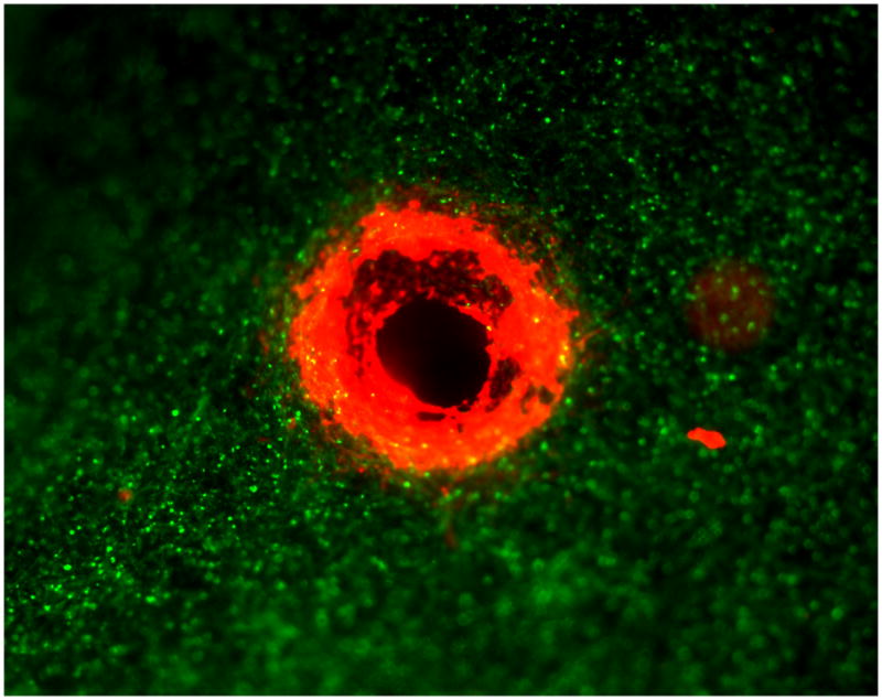
Directed spatial differentiation of an endothelial cell lined (red) tubule from an aggregate of endothelial cells with surrounding vascular smooth muscle cells (green) in 3-D co-culture after VEGF-165 treatment. 4x magnification.
Conclusion
Much of the work to date developing biocompatible bypass grafts has looked at sustaining or overdriving inherent reparative mechanisms of vascular cells in order to improve cellular incorporation into grafts rather than understanding and recreating the developmental mechanisms of blood vessel creation. Novel approaches replicating developmental patterns may be a requisite to TEBV development. Further no single scaffold, cell type, or growth factor will be sufficient to create a TEBV that acts like a blood vessel. Rather we must pursue complex approaches that work within the genetic framework seen in development to create vessels that not only look like blood vessels but also act like them.
Footnotes
Publisher's Disclaimer: This is a PDF file of an unedited manuscript that has been accepted for publication. As a service to our customers we are providing this early version of the manuscript. The manuscript will undergo copyediting, typesetting, and review of the resulting proof before it is published in its final citable form. Please note that during the production process errors may be discovered which could affect the content, and all legal disclaimers that apply to the journal pertain.
References
- 1.Carrel A. Results of permanent intubation of the thoracic aorta. Surg Gynecol Obstet. 1912;15:245–248. [Google Scholar]
- 2.Carrel A, Guthrie C. Uniterminal and biterminal venous transplantations. Surg Gynecol Obstet. 1906;2:266–286. [Google Scholar]
- 3.Veith FJ, Gupta SK, Ascer E, et al. Six-year prospective multicenter randomized comparison of autologous saphenous vein and expanded polytetrafluoroethylene grafts in infrainguinal arterial reconstructions. J Vasc Surg. 1986;3:104–114. doi: 10.1067/mva.1986.avs0030104. [DOI] [PubMed] [Google Scholar]
- 4.Werkmeister JA, Edwards GA, White JF, et al. In vivo evaluation of modified mandrel-grown vascular prostheses. J Biomed Mater Res. 1999;47:316–323. doi: 10.1002/(sici)1097-4636(19991205)47:3<316::aid-jbm5>3.0.co;2-w. [DOI] [PubMed] [Google Scholar]
- 5.Nakabayashi N, Williams DF. Preparation of non-thrombogenic materials using 2-methacryloyloxyethyl phosphorylcholine. Biomaterials. 2003;24:2431–2435. doi: 10.1016/s0142-9612(03)00113-3. [DOI] [PubMed] [Google Scholar]
- 6.Deutsch M, Meinhart J, Fischlein T, Preiss P, Zilla P. Clinical autologous in vitro endothelialization of infrainguinal ePTFE grafts in 100 patients: a 9-year experience. Surgery. 1999;126:847–855. [PubMed] [Google Scholar]
- 7.Meinhart JG, Deutsch M, Fischlein T, Howanietz N, Froschl A, Zilla P. Clinical autologous in vitro endothelialization of 153 infrainguinal ePTFE grafts. Ann Thorac Surg. 2001;71:S327–331. doi: 10.1016/s0003-4975(01)02555-3. [DOI] [PubMed] [Google Scholar]
- 8.Aoki J, Serruys PW, van Beusekom H, et al. Endothelial progenitor cell capture by stents coated with antibody against CD34: the HEALING-FIM (Healthy Endothelial Accelerated Lining Inhibits Neointimal Growth-First In Man) Registry. J Am Coll Cardiol. 2005;45:1574–1579. doi: 10.1016/j.jacc.2005.01.048. [DOI] [PubMed] [Google Scholar]
- 9.Greisler HP, Kim DU, Price JB, Voorhees AB., Jr Arterial regenerative activity after prosthetic implantation. Arch Surg. 1985;120:315–323. doi: 10.1001/archsurg.1985.01390270055010. [DOI] [PubMed] [Google Scholar]
- 10.Clowes AW, Kirkman TR, Reidy MA. Mechanisms of arterial graft healing. Rapid transmural capillary ingrowth provides a source of intimal endothelium and smooth muscle in porous PTFE prostheses. Am J Pathol. 1986;123:220–230. [PMC free article] [PubMed] [Google Scholar]
- 11.Clowes AW, Kohler T. Graft endothelialization: the role of angiogenic mechanisms. J Vasc Surg. 1991;13:734–736. doi: 10.1016/0741-5214(91)90367-4. [DOI] [PubMed] [Google Scholar]
- 12.Walter DH, Cejna M, Diaz-Sandoval L, et al. Local gene transfer of phVEGF-2 plasmid by gene-eluting stents: an alternative strategy for inhibition of restenosis. Circulation. 2004;110:36–45. doi: 10.1161/01.CIR.0000133324.38115.0A. [DOI] [PubMed] [Google Scholar]
- 13.Zarge JI, Gosselin C, Huang P, Vorp DA, Severyn DA, Greisler HP. Platelet deposition on ePTFE grafts coated with fibrin glue with or without FGF-1 and heparin. J Surg Res. 1997;67:4–8. doi: 10.1006/jsre.1996.4903. [DOI] [PubMed] [Google Scholar]
- 14.Gray JL, Kang SS, Zenni GC, et al. FGF-1 affixation stimulates ePTFE endothelialization without intimal hyperplasia. J Surg Res. 1994;57:596–612. doi: 10.1006/jsre.1994.1189. [DOI] [PubMed] [Google Scholar]
- 15.Greisler HP, Cziperle DJ, Kim DU, et al. Enhanced endothelialization of expanded polytetrafluoroethylene grafts by fibroblast growth factor type 1 pretreatment. Surgery. 1992;112:244–254. discussion 254–245. [PubMed] [Google Scholar]
- 16.Shireman PK, Xue L, Maddox E, Burgess WH, Greisler HP. The S130K fibroblast growth factor-1 mutant induces heparin-independent proliferation and is resistant to thrombin degradation in fibrin glue. J Vasc Surg. 2000;31:382–390. doi: 10.1016/s0741-5214(00)90168-x. [DOI] [PubMed] [Google Scholar]
- 17.Brewster LP, Brey EM, Tassiopoulos AK, et al. Heparin-independent mitogenicity in an endothelial and smooth muscle cell chimeric growth factor (S130K-HBGAM) Am J Surg. 2004;188:575–579. doi: 10.1016/j.amjsurg.2004.07.012. [DOI] [PubMed] [Google Scholar]
- 18.Serruys PW, Kutryk MJ, Ong AT. Coronary-artery stents. N Engl J Med. 2006;354:483–495. doi: 10.1056/NEJMra051091. [DOI] [PubMed] [Google Scholar]
- 19.Ramcharitar S, Vaina S, Serruys PW. The Next Generation of Drug-Eluting Stents : What’s on the Horizon? Am J Cardiovasc Drugs. 2007;7:81–93. doi: 10.2165/00129784-200707020-00001. [DOI] [PubMed] [Google Scholar]
- 20.Tanimoto S, Serruys PW, Thuesen L, et al. Comparison of in vivo acute stent recoil between the bioabsorbable everolimus-eluting coronary stent and the everolimus-eluting cobalt chromium coronary stent: Insights from the ABSORB and SPIRIT trials. Catheter Cardiovasc Interv. 2007 doi: 10.1002/ccd.21136. [DOI] [PubMed] [Google Scholar]
- 21.Williams DF. To engineer is to create: the link between engineering and regeneration. Trends Biotechnol. 2006;24:4–8. doi: 10.1016/j.tibtech.2005.10.006. [DOI] [PubMed] [Google Scholar]
- 22.Poh M, Boyer M, Solan A, et al. Blood vessels engineered from human cells. Lancet. 2005;365:2122–2124. doi: 10.1016/S0140-6736(05)66735-9. [DOI] [PubMed] [Google Scholar]
- 23.L’Heureux N, Dusserre N, Konig G, et al. Human tissue-engineered blood vessels for adult arterial revascularization. Nat Med. 2006;12:361–365. doi: 10.1038/nm1364. [DOI] [PMC free article] [PubMed] [Google Scholar]
- 24.Cho SW, Lim SH, Kim IK, et al. Small-diameter blood vessels engineered with bone marrow-derived cells. Ann Surg. 2005;241:506–515. doi: 10.1097/01.sla.0000154268.12239.ed. [DOI] [PMC free article] [PubMed] [Google Scholar]
- 25.Swartz DD, Russell JA, Andreadis ST. Engineering of fibrin-based functional and implantable small-diameter blood vessels. Am J Physiol Heart Circ Physiol. 2005;288:H1451–1460. doi: 10.1152/ajpheart.00479.2004. [DOI] [PubMed] [Google Scholar]
- 26.Ehrbar M, V, Djonov G, Schnell C, et al. Cell-demanded liberation of VEGF121 from fibrin implants induces local and controlled blood vessel growth. Circ Res. 2004;94:1124–1132. doi: 10.1161/01.RES.0000126411.29641.08. [DOI] [PubMed] [Google Scholar]
- 27.Sekiya S, Shimizu T, Yamato M, Kikuchi A, Okano T. Bioengineered cardiac cell sheet grafts have intrinsic angiogenic potential. Biochem Biophys Res Commun. 2006;341:573–582. doi: 10.1016/j.bbrc.2005.12.217. [DOI] [PubMed] [Google Scholar]