Abstract
Purpose:
This study continued our previous investigations of the ligaments stabilizing the scaphoid and lunate in which we examined the scapholunate interosseous ligament, the radioscaphocapitate, and the scaphotrapezial ligament. In this current study, we examined the effects of sectioning the dorsal radiocarpal ligament, dorsal intercarpal ligament, scapholunate interosseous ligament, radioscaphocapitate, and scaphotrapezial ligaments. In the current study, the scapholunate interosseous ligament, radioscaphocapitate, and scaphotrapezial ligaments were sectioned in a different order than performed previously.
Methods:
Three sets of 8 cadaver wrists were tested in a wrist joint motion simulator. In each set of wrists, only 3 of the 5 ligaments were cut in specific sequences. Each wrist was moved in continuous cycles of flexion-extension and radial-ulnar deviation. Kinematic data for the scaphoid and lunate were recorded for each wrist in the intact state, after the 3 ligaments were sectioned in various sequences and after the wrist was moved through 1,000 cycles of motion.
Results:
Dividing the dorsal intercarpal or scaphotrapezial ligaments did not alter the motion of the scaphoid or lunate. Dividing the dorsal radiocarpal ligament alone caused a slight statistical increase in lunate radial deviation. Dividing the scapholunate interosseous ligament after first dividing the dorsal intercarpal, dorsal radiocarpal, or scaphotrapezial ligaments caused large increases in scaphoid flexion and lunate extension.
Conclusions:
Based on these findings, we concluded that the scapholunate interosseous ligament is the primary stabilizer and that the other ligaments are secondary stabilizers of the scapholunate articulation. Dividing the dorsal radiocarpal, dorsal intercarpal, or scaphotrapezial ligaments after cutting the scapholunate interosseous ligament produces further changes in scapholunate instability or results in changes in the kinematics for a larger portion of the wrist motion cycle.
Keywords: Scapholunate instability, wrist, biomechanics
Treatment of soft tissue injuries of the wrist is clinically frustrating. The diagnosis of which specific ligaments are injured is difficult. The most common instability problem occurs at the scapholunate (SL) joint. Many surgical procedures have been advocated, but the long-term outcomes of these procedures are undetermined.
One of the primary difficulties in understanding this problem is that the specific function of the carpal ligaments is not known. It is important to define the function and relative importance of these structures to develop better surgical treatments for the injured SL joint. This biomechanical study is one of a series of experiments that evaluates the importance of ligaments that attach to the scaphoid or lunate.1,2 According to previous anatomical studies3-9 there are 7 ligaments that crossover or attach to the scaphoid or lunate. These are the scapholunate interosseous ligament (SLIL), the radioscaphocapitate ligament (RSC), the long radiolunate ligament, the short radiolunate ligament, the scaphotrapezial ligament (ST), the dorsal radiocarpal ligament (DRC), and the dorsal intercarpal ligament (DIC). These ligaments have been or will be evaluated systematically to determine their relative importance in stabilizing the SL joint.
Our previous investigations of the ligaments stabilizing the scaphoid and lunate examined the SLIL, RSC, and ST ligaments. The purpose of this study was to examine the effects of cutting the DRC and the DIC ligaments, in addition to the SLIL, RSC, and ST ligaments on SL articulation. The SLIL, RSC, and the ST ligaments were sectioned in a different order than performed previously. The stabilizing role of these ligaments was determined by sequentially cutting these ligaments and evaluating their effect on the motions of the scaphoid and lunate during the wrist motions of flexion-extension and radial-ulnar deviation. If dividing a ligament produced no change in scaphoid and lunate motion it was presumed that this ligament had no effect on SL stability. The hypothesis of this study was that the SLIL is the primary stabilizer of the SL articulation and that the other structures do not significantly stabilize this joint.
Materials and Methods
Twenty-four cadaver upper extremities (average age, 72.4 y; range, 47−98 y; 11 women, 13 men) were used to evaluate the effect of cutting the SLIL, DRC, DIC, RSC, and ST in described sequences to determine their effect on altering the motion and stability of the SL joint. The methodology of this study was similar to that published previously.1,2 The right upper extremities of each cadaver specimen were arthroscoped to confirm that there was no intra-articular pathology. Only normal specimens or specimens with a degenerative tear of the triangular fibrocartilage complex were included. The skin and muscle were excised. The tendons of the wrist flexors, extensors, and the thumb abductor were left intact. The remaining tendons were excised. All ligamentous structures about the wrist were left intact. Electromagnetic sensors (Polhemus Fastrak; Polhemus, Colchester, VT) were attached indirectly to the scaphoid, lunate, third metacarpal, and radius through carbon fiber rods and acrylic platforms. The specimen then was placed in the wrist joint simulator.10 This device produces repetitive cyclic motion of flexion-extension, radial-ulnar deviation, or a combination of these motions such as wrist circumduction by pulling on the appropriate tendons with physiologic forces. After the specimen was placed in the simulator, the elbow was fixed at 90° of flexion by pinning the ulnohumeral joint. The electromagnetic sources for the electromagnetic sensors (Fastrak) were attached to an acrylic plate that was attached to the proximal ulna. One source was affiliated with the sensor and was used to monitor third metacarpal motion, which is defined as global wrist motion. The other source was associated with sensors for the radial, scaphoid, and lunate motions.
After the specimen was placed in the wrist joint simulator, static kinematic data were collected with the wrist in neutral position and load was applied to the tendons. The wrist then was moved in sinusoidal cyclic motions of 30° extension to 50° flexion for 6 cycles. During this time, force data were collected for the tendons and motion and positional data were collected for the scaphoid, lunate, third metacarpal, and distal radius. Data also were collected during cyclic wrist motion of 10° radial deviation to 20° ulnar deviation. Specimens then were assigned randomly to 1 of 3 ligament-sectioning groups. Each group contained 8 specimens.
In the first group the kinematic data for the third metacarpal, scaphoid, lunate, and radius were collected in the intact specimen in the neutral wrist position and for cyclic wrist flexion-extension and cyclic radial-ulnar deviation motions. The ST ligament as described by Drewniany et al7 then was cut. Data then were collected as during the neutral and cyclic motions as for the intact specimen. The entire SLIL ligament then was cut and kinematic data again were collected in the neutral wrist position and cyclic motions. The RSC ligament then was cut between the radius and scaphoid and kinematic data were collected. This ligament was identified before the experiment during the arthroscopy of the specimen and a suture was looped around it by arthroscopic means. Sectioning the tissue within the loop of suture resulted in complete sectioning of the RSC. After these ligaments were sectioned and data were collected, the specimen was moved through 1,000 cycles of wrist flexion-extension to mimic wrist motion after injury. After the 1,000 cycles of motion, data from the electromagnetic sensors (Fastrak) again were collected as described previously.
In the second group of 8 specimens the effect of dividing the DRC, the DIC, and the SLIL on scaphoid and lunate kinematics was studied. The methodology used in group 1 was used for group 2. The data were collected in the intact specimen after dividing the DRC at its insertion on the radius; after dividing the DIC at the interval between the capitate, scaphoid, and trapezoid, as well as separating it from its attachment to the lunate; and after the entire SLIL was sectioned. The specimen was moved through 1,000 cycles of motion and kinematic data again were collected.
In the third group of 8 specimens the same methodology was used and the DIC, DRC, and SLIL again were studied, but in a different sequence. Data were collected in the intact specimen data after dividing the DIC, SLIL, and DRC, and then again after 1,000 cycles of wrist flexion/extension (Table 1).
Table 1.
Order of Ligament Sectioning in the Three Experimental Groups
| Group 1 | Group 2 | Group 3 |
|---|---|---|
| ST | DRC | DIC |
| SLIL | DIC | SLIL |
| RSC | SLIL | DRC |
After kinematic data collection was completed, the specimen was placed in a rigid foam box and encased in expandable polyurethane foam to rigidly immobilize the specimen. The specimen then was scanned by computed tomography (CT) at 1-mm intervals for the first 8 arms and at 0.7-mm intervals for the remaining 16 arms. Electromagnetic sensor (Fastrak) kinematic data were recorded at the time of the CT scan. This combination of data determined the position of each sensor relative to the bone being monitored and aided in the construction of 3-dimensional models.
These 3-dimensional models were constructed in the same manner as described previously.2,11 The CT slices were exported into segmentation software (Sliceomatic; Tomovision, Montreal, Canada). These models were exported into another software package (3D Studio Max; Discreet, Montreal, Canada) for rendering. This software also allowed animation of the objects based on the kinematic data. The resulting animation allowed a 3-dimensional visualization of the carpal bones moving and a direct comparison of the kinematics before and after ligament sectioning.
In addition, the minimum distance between the scaphoid and the lunate was calculated.11 The models were exported to another software program (Geomagic; Raindrop Geomagic, Research Park Triangle, NC) that converted each surface-rendered model into solid objects that could be viewed in computer-aided design software (Solid Works; Solid Works, Concord, MA). By using Solid Works and in-house written software, the minimum distance between the scaphoid and lunate in a static neutral position and during all flexion-extension and radial-ulnar deviation positions was calculated for the intact case and after dividing the various ligaments. Because of the movement of the sensor posts during encapsulation of the arm with the polyurethane foam before CT scanning or CT scanning problems, the minimum distances were calculated in only 4 arms from group 1, in 5 arms from group 2, and in 5 arms from group 3.
At the conclusion of the experiment the specimen was dissected to verify that the sensors were placed accurately and that there was complete sectioning of the ligaments.
Changes in carpal motion caused by ligament sectioning were analyzed using a 1-way repeated-measures analysis of variance (Duncan’s method) at each degree of wrist motion at a p value of less than .05. Changes in the minimum distance between the bones caused by ligament sectioning also were analyzed using a repeated-measures analysis of variance method at each degree of wrist motion.
Results
During wrist flexion-extension and radial-ulnar deviation there were many statistically significant changes in carpal kinematics after ligament sectioning. Some of these significant changes were less than 1°. Because 1° of change seemed to be small from a clinical perspective, we decided to present only changes that were significant and greater than 2°.
Group 1 Sequence of Ligament Sectioning
In the first group of arms the ST ligament was sectioned first, followed by the SLIL, then the RSC, and then 1,000 cycles of motion. The following results pertain to the wrist motion of flexion/extension and were statistically significant (Table 2; Figures 1, 2; Appendix Figures A1, A2). Dividing the ST ligament had no effect on carpal kinematics. Dividing the SLIL after the ST ligament resulted in increased scaphoid flexion and increased lunate extension during wrist flexion. Dividing the RSC ligament in this sequence produced no additional changes in scaphoid flexion and resulted in no change in scaphoid radial-ulnar deviation. Radioscaphocapitate sectioning produced increased lunate extension during more of each cycle of wrist flexion-extension. The addition of 1,000 cycles of motion after all 3 ligaments were sectioned resulted in increased scaphoid flexion compared with the intact group or after the ST was cut. There was also an increase in scaphoid ulnar deviation during wrist flexion-extension compared with the intact group or after dividing any ligaments. Ligament sectioning during wrist flexion/extension altered the lunate radial-ulnar deviation motion, but the changes were less than 2°.
Table 2.
Group 1 Statistically Significant Carpal Bone Motion Changes With Sequential Ligament Sectioning During Wrist Flexion/Extension
| Carpal Motion | Statistical Comparison of Ligament Sectioning | Portion of Motion Affected | Maximum Angular Difference, ° |
|---|---|---|---|
| Scaphoid flexion-extension | |||
| I ≠ C2 | From flexion to neutral | 3.8 | |
| I ≠ C3 | From flexion to neutral | 3.9 | |
| I ≠ C4 | During flexion | 5.7 | |
| C1 ≠ C4 | During flexion | 5.6 | |
| Scaphoid radial-ulnar deviation | |||
| All motions except maximum flexion and maximum extension | |||
| I ≠ C4 | 4.2 | ||
| All motions except maximum flexion and maximum extension | |||
| C1 ≠ C4 | 4.2 | ||
| C2 ≠ C4 | Near neutral flexion-extension | 3.1 | |
| C3 ≠ C4 | Near neutral flexion-extension | 3.1 | |
| Lunate flexion-extension | |||
| I ≠ C2 | From slight extension to maximum flexion | 5.1 | |
| I ≠ C3 | All of flexion | 6.4 | |
| I ≠ C4 | Entire motion | 10.6 | |
| C1 ≠ C2 | Neutral into maximum flexion | 5.1 | |
| C1 ≠ C3 | All of wrist flexion | 6.5 | |
| C1 ≠ C4 | Entire motion | 10.6 | |
| C2 ≠ C4 | Entire motion | 5.9 | |
| C3 ≠ C4 | In extension | 4.5 | |
| Lunate radial-ulnar deviation | |||
| I ≠ C2 | But changes were < 2° | <2 | |
| I ≠ C3 | |||
| I ≠ C4 | |||
| C1 ≠ C3 | |||
| C1 ≠ C4 |
I, intact specimen; C1, ST ligament cut; C2, ST and SLIL ligaments cut; C3, ST, SLIL, and RSC ligaments cut; C4, ST, SLIL, and RSC ligaments cut plus 1,000 cycles of motion; ≠, statistically different from each other.
Figure 1.
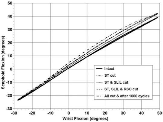
Average scaphoid flexion and extension as a function of wrist flexion and extension in the intact wrist and after sequential ligamentous sectioning in the first group of arms. The graph represents the motion of the scaphoid during a complete cycle of wrist motion from flexion to extension to flexion. Flexion is positive.
Figure 2.
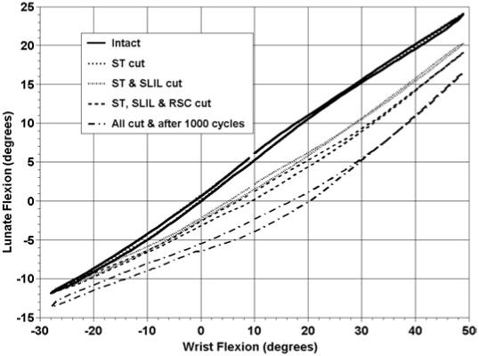
Average lunate flexion and extension as a function of wrist flexion and extension in the intact wrist and after sequential ligamentous sectioning in the first group of arms. The graph represents the motion of the lunate during a complete cycle of wrist motion from flexion to extension to flexion. Flexion is positive.
Figure A1.
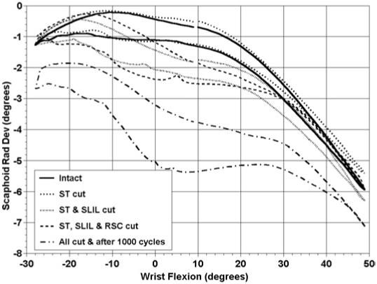
Average scaphoid radial and ulnar deviation as a function of wrist flexion and extension in the intact wrist and after sequential ligamentous sectioning in the first group of arms. The graph represents the motion of the scaphoid during a complete cycle of wrist motion from flexion to extension to flexion. Flexion and radial deviation are positive.
Figure A2.
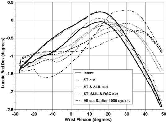
Average lunate radial and ulnar deviation as a function of wrist flexion and extension deviation in the intact wrist and after sequential ligamentous sectioning in the first group of arms. The graph represents the motion of the lunate during a complete cycle of wrist motion from flexion to extension to flexion. Flexion and radial deviation are positive.
The following results were statistically significant and pertain to the effect of group 1 ligament sectioning during the wrist motion of radial-ulnar deviation (Table 3; Appendix Figures A3-A6). Dividing the ST ligament had no effect on scaphoid or lunate kinematics during wrist radial-ulnar deviation. Dividing the ST and SLIL resulted in increased lunate extension when the wrist was moving from ulnar to radial deviation. Dividing the RSC resulted in increased lunate extension when compared with only the ST cut, but no additional changes when compared with the ST and SLIL cut. One thousand cycles of motion after ligament sectioning resulted in additional increases in lunate extension when compared with all other groups. Ligament sectioning plus 1,000 cycles of motion also resulted in increased lunate radial deviation compared with the intact specimen or after the SLIL ligament was cut, but these changes were less than 2°.
Table 3.
Group 1 Statistically Significant Carpal Bone Motion Changes With Sequential Ligament Sectioning During Wrist Radial/Ulnar Deviation
| Carpal Motion | Statistical Comparison of Ligament Sectioning | Portion of Motion Affected | Maximum Angular Difference, ° |
|---|---|---|---|
| Scaphoid flexion-extension | |||
| I ≠ C4 | Radial to ulnar deviation | 4.6 | |
| C1 ≠ C4 | Radial to ulnar deviation | 4.1 | |
| Scaphoid radial-ulnar deviation | |||
| I ≠ C4 | Except in maximum radial deviation and in maximum ulnar deviation | 3.5 | |
| C1 ≠ C4 | Except in maximum radial deviation and in maximum ulnar deviation | 3.3 | |
| Lunate flexion-extension | |||
| I ≠ C2 | From ulnar deviation to radial deviation | 3.2 | |
| I ≠ C3 | All motion except maximum ulnar deviation | 4.0 | |
| I ≠ C4 | Entire motion | 6.8 | |
| C1 ≠ C2 | Going into radial deviation | 3.0 | |
| C1 ≠ C3 | All of radial deviation | 3.8 | |
| C1 ≠ C4 | Entire motion | 6.5 | |
| C2 ≠ C4 | Entire motion | 3.9 | |
| C3 ≠ C4 | From radial deviation to ulnar deviation | 3.1 | |
| Lunate radial-ulnar deviation | |||
| I ≠ C4 | But changes < 2° | <2 | |
| C1 ≠ C4 | But changes < 2° | <2 |
I, intact specimen; C1, ST ligament cut; C2, ST and SLIL ligaments cut; C3, ST, SLIL, and RSC ligaments cut; C4, ST, SLIL, and RSC ligaments cut plus 1,000 cycles of motion; ≠, statistically different from each other.
Figure A3.
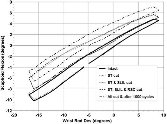
Average scaphoid flexion and extension as a function of wrist radial and ulnar deviation in the intact wrist and after sequential ligamentous sectioning in the first group of arms. The graph represents the motion of the scaphoid during a complete cycle of wrist motion from radial deviation to ulnar deviation to radial deviation. Flexion and radial deviation are positive. The irregular ends of the curves are the result of including only the data points common to all 8 arms during a range of motion. Some arms continued beyond the shown end points.
Figure A6.
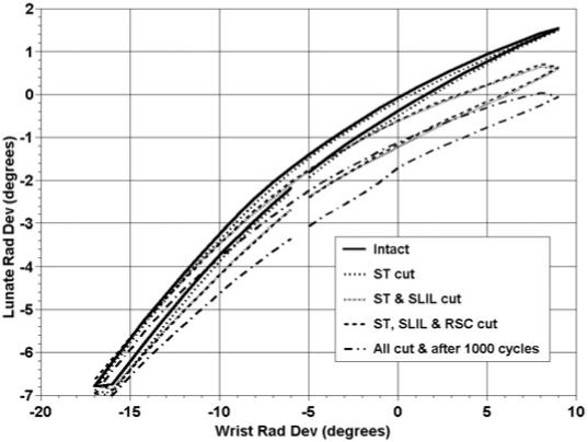
Average lunate radial and ulnar deviation as a function of wrist radial and ulnar deviation in the intact wrist and after sequential ligamentous sectioning in the first group of arms. The graph represents the motion of the lunate during a complete cycle of wrist motion from radial deviation to ulnar deviation to radial deviation. Flexion and radial deviation are positive. The irregular ends of the curves are the result of including only the data points common to all 8 arms during a range of motion. Some arms continued beyond the shown end points.
By using the CAD models generated from the kinematic data and CT scans, the minimum distance between the scaphoid and lunate at each degree of flexion-extension and radial-ulnar deviation was calculated (Table 4, Figures 3, 4). The SL gap statistically increased during wrist flexion-extension after ligament sectioning plus 1,000 cycles of motion compared with the intact specimen or after the ST or ST and SLIL ligaments were cut. During radial-ulnar deviation, the gap statistically increased after all ligaments were sectioning plus 1,000 cycles of motion compared with the intact specimen or after the ST or ST and SLIL ligaments were cut. The gap also was increased after all of these ligaments were cut compared with the intact specimen or after the ST ligament was cut.
Table 4.
Group 1 Comparison of Which Ligament Sectioning Produced Increased Scapholunate Gap
| Wrist Motion | Statistical Comparison of Ligament Sectioning | Portion of Motion Affected |
|---|---|---|
| Wrist flexion-extension | All motion except extreme extension | |
| I ≠ C4 | ||
| C1 ≠ C4 | Wrist flexion | |
| C2 ≠ C4 | Wrist flexion | |
| Wrist radial-ulnar deviation | I ≠ C4 | Nearly all of wrist motion |
| C1 ≠ C4 | Nearly all of wrist motion | |
| C2 ≠ C4 | Nearly all of wrist motion | |
| I ≠ C3 | Wrist ulnar deviation | |
| C1 ≠ C3 | Wrist ulnar deviation |
I, intact specimen; C1, ST ligament cut; C2, ST and SLIL ligaments cut; C3, ST, SLIL, and RSC ligaments cut; C4, ST, SLIL, and RSC ligaments cut plus 1,000 cycles of motion; ≠, statistically different from each other.
Figure 3.
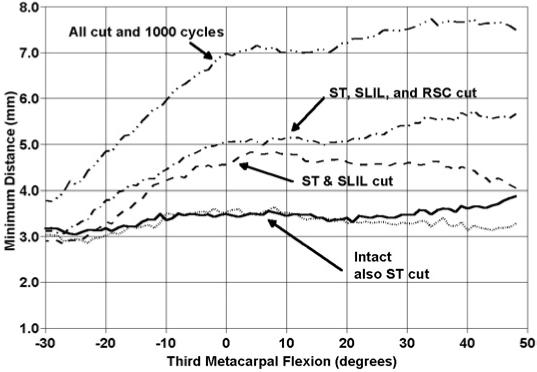
Average minimum distance between the scaphoid and lunate in the intact wrist and after sequential sectioning in the first group of arms. The graph represents the portion of the wrist motion cycle as the wrist is moving from wrist flexion to extension.
Figure 4.
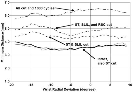
Average minimum distance between the scaphoid and lunate in the intact wrist and after sequential ligament sectioning in the first group of arms. The graph represents the portion of the wrist motion cycle as the wrist is moving from radial to ulnar deviation.
Group 2 Sequence of Ligament Sectioning
In the second group of arms, different carpal ligaments were sectioned to evaluate their affect on SL instability. The DRC was cut first, followed by the DIC, and then the SLIL. The following results pertain to the kinematic changes that were statistically significant during wrist flexion-extension (Table 5; Figures 5, 6; Appendix Figures A7, A8). Dividing the DRC alone caused a slight increase in lunate radial deviation in wrist flexion. Dividing the DIC after cutting the DRC had either no effect or no additional effect on scaphoid or lunate motion; however, dividing the SLIL after the DRC and DIC increased scaphoid flexion, scaphoid ulnar deviation, and lunate extension compared with the intact condition or after the DRC or the DRC and DIC had been sectioned. The addition of 1,000 cycles of wrist motion produced no additional changes in these 3 motions. Dividing all 3 ligaments plus 1,000 cycles of motion produced a further increase in lunate radial deviation compared with the DRC and DIC sectioned condition.
Table 5.
Group 2 Statistically Significant Carpal Bone Motion Changes With Sequential Ligament Sectioning During Wrist Flexion/Extension
| Carpal Motion | Statistical Comparison of Ligament Sectioning | Portion of Motion Affected | Maximum Angular Difference, ° |
|---|---|---|---|
| Scaphoid flexion-extension | |||
| C3 ≠ I | During wrist flexion | 11.0 | |
| C3 ≠ C1 | During wrist flexion | 11.3 | |
| C3 ≠ C2 | During wrist flexion | 11.5 | |
| C4 ≠ I | During wrist flexion | 11.9 | |
| C4 ≠ C1 | During wrist flexion | 12.2 | |
| C4 ≠ C2 | During wrist flexion | 12.4 | |
| Scaphoid radial-ulnar deviation | |||
| C3 ≠ I | During wrist flexion | 7.7 | |
| C3 ≠ C1 | During wrist flexion | 9.2 | |
| C3 ≠ C2 | During wrist flexion | 9.3 | |
| C4 ≠ I | During wrist flexion | 7.6 | |
| C4 ≠ C1 | During wrist flexion | 9.2 | |
| C4 ≠ C2 | During wrist flexion | 9.3 | |
| Lunate flexion-extension | |||
| C3 ≠ I | Entire wrist motion | 15.5 | |
| C3 ≠ C1 | Entire wrist motion | 15.2 | |
| C3 ≠ C2 | Entire wrist motion | 14.9 | |
| C4 ≠ I | Entire wrist motion | 17.1 | |
| C4 ≠ C1 | Entire wrist motion | 17.2 | |
| C4 ≠ C2 | Entire wrist motion | 16.9 | |
| Lunate radial-ulnar deviation | |||
| I ≠ C1 | During maximum flexion | 2.8 | |
| I ≠ C2 | During maximum flexion | 2.9 | |
| I ≠ C3 | During maximum flexion | 4.7 | |
| I ≠ C4 | During maximum flexion | 4.9 | |
| C3 ≠ C2 | During maximum flexion | 2.2 | |
| C4 ≠ C2 | During maximum flexion | 2.1 |
I, intact specimen; C1, DRC ligament cut; C2, DRC and DIC ligaments cut; C3, DRC, DIC, and SLIL ligaments cut; C4, DRC, DIC, and SLIL ligaments cut plus 1,000 cycles of motion; ≠, statistically different from each other.
Figure 5.
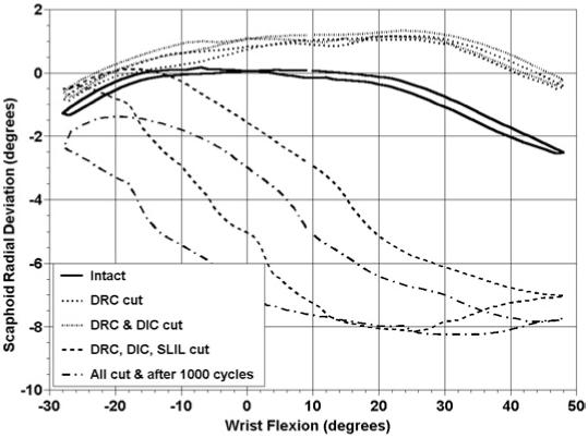
Average scaphoid radial and ulnar deviation as a function of wrist flexion and extension in the intact wrist and after sequential ligamentous sectioning in the second group of arms. The graph represents the motion of the scaphoid during a complete cycle of wrist motion from flexion to extension to flexion. Flexion and radial deviation are positive.
Figure 6.
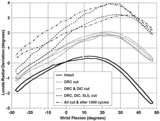
Average lunate radial and ulnar deviation as a function of wrist flexion and extension in the intact wrist and after sequential ligamentous sectioning in the second group of arms. The graph represents the motion of the lunate during a complete cycle of wrist motion from flexion to extension to flexion. Flexion and radial deviation are positive.
Figure A7.
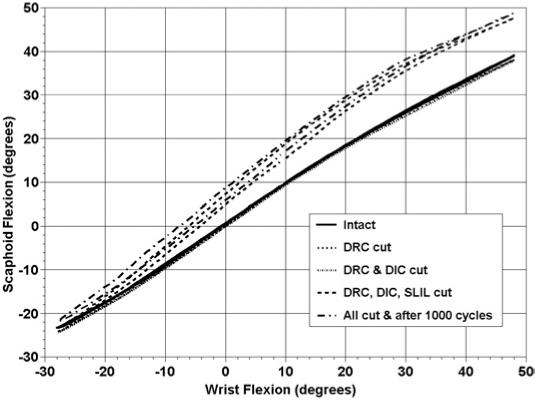
Average scaphoid flexion and extension as a function of wrist flexion and extension in the intact wrist and after sequential ligamentous sectioning in the second group of arms. The graph represents the motion of the scaphoid during a complete cycle of wrist motion from flexion to extension to flexion. Flexion is positive.
Figure A8.
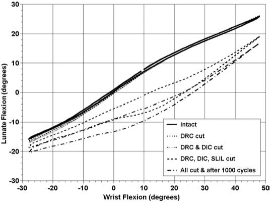
Average lunate flexion and extension as a function of wrist flexion and extension in the intact wrist and after sequential ligamentous sectioning in the second group of arms. The graph represents the motion of the lunate during a complete cycle of wrist motion from flexion to extension to flexion. Flexion is positive.
The following results occurred during wrist radial-ulnar deviation and were statistically significant (Table 6; Figure 7; Appendix Figures A9-A11). Dividing the DRC or the DRC and DIC had no effect on scaphoid or lunate motion. Dividing the SLIL after the DRC and DIC resulted in increased scaphoid flexion and ulnar deviation during wrist ulnar deviation. The addition of 1,000 cycles of motion also resulted in increased scaphoid flexion and ulnar deviation during almost all of wrist radial-ulnar deviation. Dividing the DRC, DIC, and SLIL resulted in increased lunate extension during all of wrist radial-ulnar deviation. The addition of 1,000 cycles of motion resulted in further statistical changes. Ligament sectioning and 1,000 cycles of motion increased lunate radial deviation compared with the intact specimen or after the DRC is cut.
Table 6.
Group 2 Statistically Significant Carpal Bone Motion Changes With Sequential Ligament Sectioning During Wrist Radial/Ulnar Deviation
| Carpal Motion | Statistical Comparison of Ligament Sectioning | Portion of Motion Affected | Maximum Angular Difference, ° |
|---|---|---|---|
| Scaphoid flexion-extension | |||
| C3 ≠ I | During wrist ulnar deviation | 11.8 | |
| C3 ≠ C1 | During wrist ulnar deviation | 12.0 | |
| C3 ≠ C2 | During wrist ulnar deviation | 12.8 | |
| During nearly all of wrist radial-ulnar deviation | |||
| C4 ≠ I | 17.7 | ||
| During nearly all of wrist radial-ulnar deviation | |||
| C4 ≠ C1 | 17.9 | ||
| During nearly all of wrist radial-ulnar deviation | |||
| C4 ≠ C2 | 18.7 | ||
| Scaphoid radial-ulnar deviation | |||
| C3 ≠ I | During wrist ulnar deviation | 4.9 | |
| C3 ≠ C1 | During wrist ulnar deviation | 5.1 | |
| C3 ≠ C2 | During wrist ulnar deviation | 5.7 | |
| During nearly all of wrist radial-ulnar deviation | |||
| C4 ≠ I | 10.2 | ||
| During nearly all of wrist radial-ulnar deviation | |||
| C4 ≠ C1 | 10.3 | ||
| During nearly all of wrist radial-ulnar deviation | |||
| C4 ≠ C2 | 11 | ||
| C3 ≠ C4 | During wrist ulnar deviation | 5.3 | |
| Lunate flexion-extension | |||
| C3 ≠ I | Entire wrist motion | 7.9 | |
| C3 ≠ C1 | Entire wrist motion | 8.1 | |
| C3 ≠ C2 | Entire wrist motion | 7.3 | |
| C4 ≠ I | Entire wrist motion | 11.6 | |
| C4 ≠ C1 | Entire wrist motion | 11.8 | |
| C4 ≠ C2 | Entire wrist motion | 11.1 | |
| C3 ≠ C4 | Entire wrist motion | 4.0 | |
| Lunate radial-ulnar deviation | |||
| C4 ≠ I | Entire wrist motion | 2.7 | |
| C4 ≠ C1 | Entire wrist motion | 2.2 |
I, intact specimen; C1, DRC ligament cut; C2, DRC and DIC ligaments cut; C3, DRC, DIC, and SLIL ligaments cut; C4, DRC, DIC, and SLIL ligaments cut plus 1,000 cycles of motion; ≠, statistically different from each other.
Figure 7.
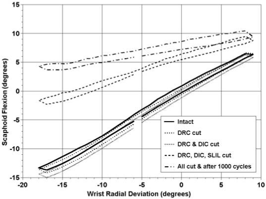
Average scaphoid flexion and extension as a function of wrist radial and ulnar deviation in the intact wrist and after sequential ligamentous sectioning in the second group of arms. The graph represents the motion of the scaphoid during a complete cycle of wrist motion from radial deviation to ulnar deviation to radial deviation. Flexion and radial deviation are positive. The irregular ends of the curves are the result of including only the data points common to all 8 arms during a range of motion. Some arms continued beyond the shown end points.
Figure A9.
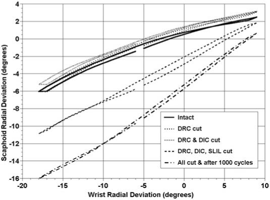
Average scaphoid radial and ulnar deviation as a function of wrist radial and ulnar deviation in the intact wrist and after sequential ligamentous sectioning in the second group of arms. The graph represents the motion of the scaphoid during a complete cycle of wrist motion from radial deviation to ulnar deviation to radial deviation. Flexion and radial deviation are positive.
Figure A11.
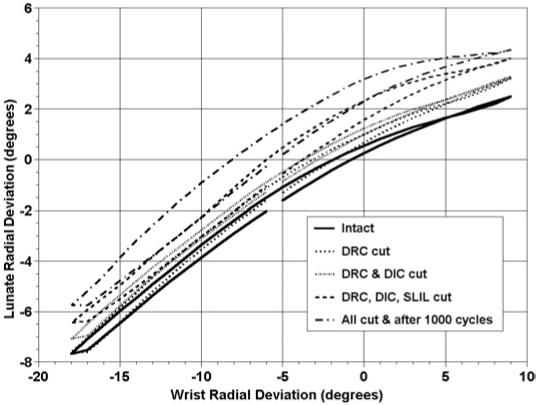
Average lunate radial and ulnar deviation as a function of wrist radial and ulnar deviation in the intact wrist and after sequential ligamentous sectioning in the second group of arms. The graph represents the motion of the lunate during a complete cycle of wrist motion from radial deviation to ulnar deviation to radial deviation. Flexion and radial deviation are positive.
The minimum distance also was calculated between the scaphoid and lunate (Table 7; Figure 8; Appendix Figure A12). During wrist flexion-extension dividing all 3 ligaments produced a significantly increased SL gap during wrist flexion. During wrist radial-ulnar deviation the sectioning of all 3 ligaments plus 1,000 cycles of motion statistically increased the SL gap.
Table 7.
Group 2 Comparison of Which Ligament Sectioning Produced Increased Scapholunate Gap
| Wrist Motion | Statistical Comparison of Ligament Sectioning | Portion of Motion Affected |
|---|---|---|
| Wrist flexion-extension | I ≠ C3 | During wrist flexion |
| I ≠ C4 | During wrist flexion | |
| C1 ≠ C3 | During wrist flexion | |
| C1 ≠ C4 | During wrist flexion | |
| C2 ≠ C3 | During wrist flexion | |
| C2 ≠ C4 | During wrist flexion | |
| Wrist radial-ulnar deviation | C4 ≠ I | Entire wrist motion |
| C4 ≠ C1 | Entire wrist motion | |
| C4 ≠ C2 | Entire wrist motion | |
| C4 ≠ C3 | Entire wrist motion |
I, intact specimen; C1, DRC ligament cut; C2, DRC and DIC ligaments cut; C3, DRC, DIC, and SLIL ligaments cut; C4, DRC, DIC, and SLIL ligaments cut plus 1,000 cycles of motion; ≠, statistically different from each other.
Figure 8.
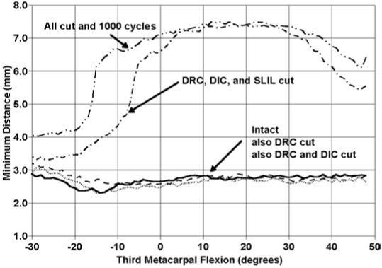
Average minimum distance between the scaphoid and lunate in the intact wrist and after sequential ligamentous sectioning in the second group of arms. The graph represents the portion of the wrist motion cycle as the wrist is moving from wrist flexion to extension.
Figure A12.
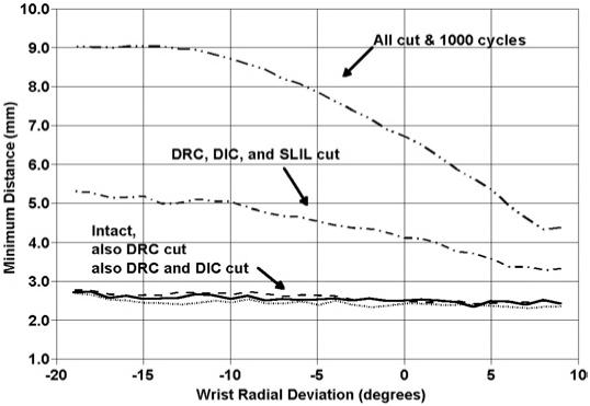
Average minimum distance between the scaphoid and lunate in the intact wrist and after sequential ligamentous sectioning in the second group of arms. The graph represents the portion of the wrist motion cycle as the wrist is moving from radial to ulnar deviation.
Group 3 Sequence of Ligament Sectioning
In the third group of 8 arms the DIC was sectioned first, followed by the SLIL, and then the DRC. The following results pertain to wrist flexion-extension and were statistically significant (Table 8; Figure 9; Appendix Figures A13-A15). Dividing the DIC had no effect on scaphoid or lunate motion during wrist flexion-extension. Dividing the SLIL after the DIC increased scaphoid flexion during wrist flexion. Further sectioning of the DRC or 1,000 cycles of motion produced no further changes in scaphoid flexion during wrist flexion-extension. The scaphoid ulnarly deviated more after the DIC and SLIL were cut during wrist flexion. There was some additional scaphoid ulnar deviation after all ligaments were cut and 1,000 cycles of motion compared with the case in which only the DIC was cut. The lunate extended after the DIC and SLIL were cut during wrist flexion. Dividing all ligaments plus 1,000 cycles of motion resulted in increased lunate extension during the entire wrist motion compared with the intact specimen or after the DIC was cut. Dividing all ligaments plus 1,000 cycles of motion resulted in increased lunate extension during wrist extension compared with the case in which the DIC and SLIL were cut. Dividing the SLIL after the DIC resulted in increased lunate radial deviation during maximum wrist flexion compared with the intact specimen or after the DIC was cut. Dividing the DIC, SLIL, and DRC produced increased lunate radial deviation during wrist flexion compared with the intact specimen, after the DIC was cut, or after the DIC and SLIL were cut. Cutting all ligaments plus 1,000 cycles of motion produced increased lunate radial deviation compared with the intact specimen or after the DIC was cut during all of the wrist flexion-extension motion. When this condition was compared with the DIC and SLIL sectioned specimen there was increased lunate radial deviation except when the wrist was in a neutral position.
Table 8.
Group 3 Statistically Significant Carpal Bone Motion Changes With Sequential Ligament Sectioning During Wrist Flexion/Extension
| Carpal Motion | Statistical Comparison of Ligament Sectioning | Portion of Motion Affected | Maximum Angular Difference, ° |
|---|---|---|---|
| Scaphoid flexion-extension | |||
| I ≠ C2 | During wrist flexion | 11.9 | |
| I ≠ C3 | During wrist flexion | 12.8 | |
| I ≠ C4 | During wrist flexion | 12.3 | |
| C1 ≠ C2 | During wrist flexion | 12.0 | |
| C1 ≠ C3 | During wrist flexion | 12.8 | |
| C1 ≠ C4 | During wrist flexion | 12.3 | |
| Scaphoid radial-ulnar deviation | |||
| I ≠ C2 | During wrist flexion | 8.9 | |
| I ≠ C3 | During wrist flexion | 10.1 | |
| I ≠ C4 | During wrist flexion and some extension | 11.0 | |
| C1 ≠ C2 | During wrist flexion | 8.9 | |
| C1 ≠ C3 | During wrist flexion | 10.1 | |
| C1 ≠ C4 | During wrist flexion and some extension | 11.0 | |
| Lunate flexion-extension | |||
| I ≠ C2 | During wrist flexion | 11.3 | |
| I ≠ C3 | During wrist flexion | 12.7 | |
| I ≠ C4 | During entire wrist motion | 15.9 | |
| C1 ≠ C2 | During wrist flexion | 11.1 | |
| C1 ≠ C3 | During wrist flexion | 12.5 | |
| C1 ≠ C4 | During entire wrist motion | 15.6 | |
| C2 ≠ C4 | During some of wrist extension | 5.6 | |
| Lunate radial-ulnar deviation | |||
| I ≠ C2 | During maximum flexion | <2 | |
| During all of wrist motion except maximum extension | |||
| I ≠ C3 | 4.9 | ||
| I ≠ C4 | During entire wrist motion | 5.6 | |
| C1 ≠ C2 | During maximum flexion | 2.2 | |
| During all of wrist motion except maximum extension | |||
| C1 ≠ C3 | 5.1 | ||
| C1 ≠ C4 | During entire wrist motion | 5.8 | |
| C3 ≠ C2 | During wrist flexion | 3.2 | |
| C4 ≠ C2 | During all wrist motion except neutral | 3.9 | |
| C4 ≠ C3 | During wrist extension | <2 |
I, intact specimen; C1, DIC ligament cut; C2, DIC and SLIL ligaments cut; C3, DIC, SLIL, and DRC cut; C4, DIC, SLIL, and DRC ligaments cut plus 1,000 cycles of motion; ≠, statistically different from each other.
Figure 9.
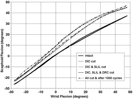
Average scaphoid flexion and extension as a function of wrist flexion and extension in the intact wrist and after sequential ligamentous sectioning in the third group of arms. The graph represents the motion of the scaphoid during a complete cycle of wrist motion from flexion to extension to flexion. Flexion is positive.
Figure A13.
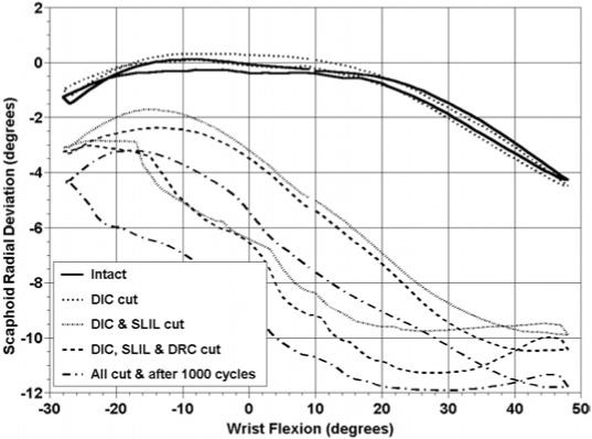
Average scaphoid radial and ulnar deviation as a function of wrist flexion and extension in the intact wrist and after sequential ligamentous sectioning in the third group of arms. The graph represents the motion of the scaphoid during a complete cycle of wrist motion from flexion to extension to flexion. Flexion and radial deviation are positive.
Figure A15.
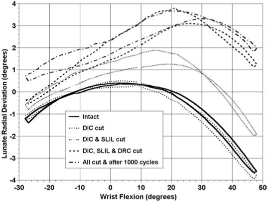
Average lunate radial and ulnar deviation as a function of wrist flexion and extension deviation in the intact wrist and after sequential ligamentous sectioning in the third group of arms. The graph represents the motion of the lunate during a complete cycle of wrist motion from flexion to extension to flexion. Flexion and radial deviation are positive. The irregular ends of the curves are the result of including only the data points common to all 8 arms during a range of motion. Some arms continued beyond the shown end points.
The following results pertain to the wrist motion of radial-ulnar deviation and are statistically significant (Table 9; Figure 10; Appendix Figures A16-A18). There was increased scaphoid flexion during all of wrist radial-ulnar deviation after cutting the DIC and SLIL; the DIC, SLIL, and DRC; or cutting all 3 ligaments plus 1,000 cycles of motion compared with the intact specimen or after the DIC alone was cut. There was increased scaphoid ulnar deviation after cutting the DIC and SLIL; or the DIC, SLIL, and DRC; or after cutting all 3 ligaments plus 1,000 cycles of motion during wrist ulnar deviation compared with the intact specimen. Dividing the DIC and SLIL, or the DIC, SLIL, and DRC, increased scaphoid ulnar deviation when compared with cutting only the DIC during wrist ulnar deviation. Cutting all ligaments plus 1,000 cycles of motion increased scaphoid ulnar deviation compared with cutting only the DIC during nearly all of wrist radial-ulnar deviation.
Table 9.
Group 3 Statistically Significant Carpal Bone Motion Changes With Sequential Ligament Sectioning During Wrist Radial/Ulnar Deviation
| Carpal Motion | Statistical Comparison of Ligament Sectioning | Portion of Motion Affected | Maximum Angular Difference, ° |
|---|---|---|---|
| Scaphoid flexion-extension | |||
| I ≠ C2 | During entire wrist motion | 10.1 | |
| I ≠ C3 | During entire wrist motion | 9.5 | |
| I ≠ C4 | During entire wrist motion | 13.7 | |
| C1 ≠ C2 | During entire wrist motion | 9.5 | |
| C1 ≠ C3 | During entire wrist motion | 9.0 | |
| C1 ≠ C4 | During entire wrist motion | 13.2 | |
| Scaphoid radial-ulnar deviation | |||
| I ≠ C2 | During wrist ulnar deviation | 5.5 | |
| I ≠ C3 | During wrist ulnar deviation | 5.7 | |
| I ≠ C4 | During wrist ulnar deviation | 8.2 | |
| C1 ≠ C2 | During wrist ulnar deviation | 6.0 | |
| C1 ≠ C3 | During wrist ulnar deviation | 6.3 | |
| C1 ≠ C4 | During entire wrist motion | 8.8 | |
| Lunate flexion-extension | |||
| I ≠ C3 | During wrist radial deviation | 9.4 | |
| I ≠ C4 | During entire wrist motion | 12.7 | |
| C1 ≠ C3 | During wrist radial deviation | 9.4 | |
| C1 ≠ C4 | During entire wrist motion | 12.6 | |
| C2 ≠ C4 | During wrist ulnar deviation | 7.2 | |
| Lunate radial-ulnar deviation | |||
| I ≠ C2 | During wrist ulnar deviation | <2 | |
| During wrist ulnar deviation and some radial deviation | |||
| I ≠ C3 | <2 | ||
| I ≠ C4 | During entire wrist motion | 2.3 | |
| C1 ≠ C2 | During wrist ulnar deviation | <2 | |
| During wrist ulnar deviation and some radial deviation | |||
| C1 ≠ C3 | <2 | ||
| C1 ≠ C4 | During entire wrist motion | 2.2 | |
| C2 ≠ C4 | During wrist radial deviation | <2 |
I, intact specimen; C1, DIC ligament cut; C2, DIC and SLIL ligaments cut; C3, DIC, SLIL, and DRC cut; C4, DIC, SLIL, and DRC ligaments cut plus 1,000 cycles of motion; ≠, statistically different from each other.
Figure 10.
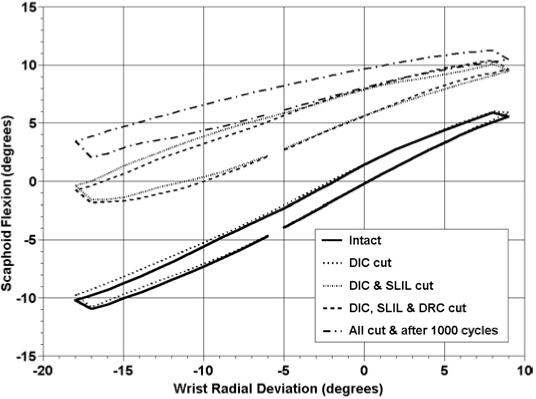
Average scaphoid flexion and extension as a function of wrist radial and ulnar deviation in the intact wrist and after sequential ligamentous sectioning in the third group of arms. The graph represents the motion of the scaphoid during a complete cycle of wrist motion from radial deviation to ulnar deviation to radial deviation. Flexion and radial deviation are positive. The irregular ends of the curves are the result of including only the data points common to all 8 arms during a range of motion. Some arms continued beyond the shown end points.
Figure A16.
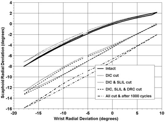
Average scaphoid radial and ulnar deviation as a function of wrist radial and ulnar deviation in the intact wrist and after sequential ligamentous sectioning in the third group of arms. The graph represents the motion of the scaphoid during a complete cycle of wrist motion from radial deviation to ulnar deviation to radial deviation. Flexion and radial deviation are positive.
Figure A18.
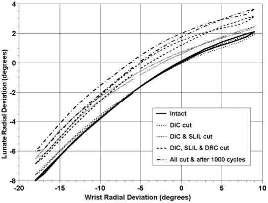
Average lunate radial and ulnar deviation as a function of wrist radial and ulnar deviation in the intact wrist and after sequential ligamentous sectioning in the third group of arms. The graph represents the motion of the lunate during a complete cycle of wrist motion from radial deviation to ulnar deviation to radial deviation. Flexion and radial deviation are positive.
Cutting the DIC, SLIL, and DRC resulted in increased lunate extension during wrist radial deviation compared with the intact or DIC-sectioned specimen. Dividing the DIC, SLIL, and DRC and 1,000 cycles of motion resulted in increased lunate extension during the entire wrist radial-ulnar deviation motion compared with the intact or DIC-sectioned specimen. Cutting all ligaments plus 1,000 cycles of motion resulted in increased lunate extension during wrist ulnar deviation compared with the case in which the DIC and SLIL were cut.
Dividing the DIC and SLIL increased the lunate radial deviation compared with the intact or DIC-sectioned specimen in wrist ulnar deviation. Dividing the DIC, SLIL, and DRC resulted in increased lunate radial deviation compared with the intact or DIC-sectioned specimen during all of wrist ulnar deviation. Cutting all ligaments plus 1,000 cycles of motion increased the lunate radial deviation during all of wrist radial-ulnar deviation compared with the intact or DIC-sectioned specimen.
The minimum distance between the scaphoid and lunate statistically increased after dividing the DIC and SLIL; the DIC, SLIL, and DRC; or all 3 ligaments plus 1,000 cycles of motion compared with the intact specimen or after cutting only the DIC except during wrist extension (Table 10; Appendix Figures A19, A20). Cutting all 3 ligaments plus 1,000 cycles of motion increased the SL gap during all of wrist radial-ulnar deviation compared with the intact or DIC-sectioned groups. Cutting all 3 ligaments plus 1,000 cycles of motion increased the SL gap during wrist ulnar deviation compared with the DIC and SLIL sectioned group.
Table 10.
Group 3 Comparison of Which Ligament Sectioning Produced Increased Scapholunate Gap
| Wrist Motion | Statistical Comparison of Ligament Sectioning | Portion of Motion Affected |
|---|---|---|
| Wrist flexion-extension | I ≠ C2 | During wrist flexion |
| I ≠ C3 | During wrist flexion | |
| I ≠ C4 | During wrist flexion | |
| C1 ≠ C2 | During wrist flexion | |
| C1 ≠ C3 | During wrist flexion | |
| C1 ≠ C4 | During wrist flexion | |
| Wrist radial-ulnar deviation | I ≠ C4 | During entire wrist motion |
| C1 ≠ C4 | During entire wrist motion | |
| C2 ≠ C4 | During wrist ulnar deviation |
I, intact specimen; C1, DIC ligament cut; C2, DIC and SLIL ligaments cut; C3, DIC, SLIL, and DRC ligaments cut; C4, DIC, SLIL, and DRC ligaments cut plus 1,000 cycles of motion; ≠, statistically different from each other.
Figure A19.
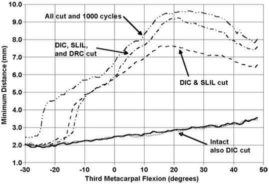
Average minimum distance between the scaphoid and lunate in the intact wrist and after sequential ligamentous sectioning in the third group of arms. The graph represents the portion of the wrist motion cycle as the wrist is moving from wrist flexion to extension.
Figure A20.
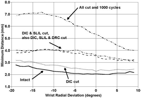
Average minimum distance between the scaphoid and lunate in the intact wrist and after sequential ligamentous sectioning in the third group of arms. The graph represents the portion of the wrist motion cycle as the wrist is moving from radial to ulnar deviation.
Discussion
This study continued our evaluation of sequential ligament sectioning and cyclic wrist motion on SL kinematics. The reason we sectioned the RSC and ST ligaments was to continue to evaluate various sequences of volar ligament sectioning on SL instability. The DIC and the DRC were chosen because of the current interest in these ligaments.9 Some investigators12 have advocated sparing these structures during a dorsal approach to the wrist. Others13,14 have suggested using the DIC to stabilize chronic SLIL instability. Sequential sectioning of these structures helps our understanding of their relative importance in SL kinematics.
Evaluation of the data supports the hypothesis that the SLIL is the primary stabilizer of the SL joint. Dividing the DIC alone or the ST alone had no effect on scaphoid and lunate kinematics during either wrist flexion/extension or wrist radial/ulnar deviation. Dividing the DRC alone did cause increased lunate radial deviation when the wrist was in maximum wrist flexion. Dividing the SLIL after any of the ligaments tested produced increased scaphoid flexion and ulnar deviation while the lunate extended and under some circumstances radially deviated. No other ligament or combination of ligament sectioning produced this magnitude of change. Additional ligament sectioning after the SLIL was cut resulted in altered SL kinematics over a larger range of wrist motion. The addition of 1,000 cycles of motion typically produced additional incremental changes in carpal bone kinematics compared with after the SLIL was cut. Our interpretation of these findings is that the SLIL is a prime stabilizer of the SL joint. There is relatively little change in the motion pattern until this structure is cut. The other ligaments tested play a secondary role as a supplement to the SLIL and cannot take over the function of this primary stabilizer. Cyclic motion after ligament sectioning appears to cause further deterioration in carpal kinematics. It is our hypothesis that the cyclic motion causes plastic deformation in the remaining structures, which helps stabilize the SL joint.
Berger et al12 advocated a surgical approach to the dorsal wrist joint that spares the DRC and DIC by making a V-shaped incision in the wrist. Our biomechanical study supports the conclusion that as long as the SLIL is intact, the DRC and DIC can be cut without altering SL kinematics. A dorsal capsular incision that cuts these 2 structures should not alter scaphoid and lunate motion and may facilitate the ease of performing a surgical procedure on the dorsal wrist.
Szabo et al14 advocated the use of the DIC to reconstruct chronic SL dissociation. Based on our biomechanical study the use of the DIC in this manner should not further destabilize the carpus, although our study was not intended to evaluate this reconstruction in stabilizing the SL articulation. Based on our results, however, it would seem that redirecting this structure without reattaching the SL ligament or reconstructing this ligament would not fully restore normal carpal kinematics.
Mitsuyasu et al15 performed a biomechanical study to evaluate the DIC and SLIL. They found that with static loading of the wrist joint, held in neutral wrist flexion-extension and neutral radial-ulnar deviation, dividing the SLIL and the DIC from off of both the scaphoid and the lunate resulted in an increased SL gap. A relative increase in lunate extension was observed only after also dividing the attachment of the DIC to the lunate. The results of our study support the hypothesis that dividing the SLIL results in an increase in the SL gap and an increase in lunate extension. The difference in results may be attributable to the technique, such as how the wrist was loaded, or the order of ligament sectioning.
Elsaidi et al16 also evaluated the SLIL and many other ligaments about the wrist and their effect on SLIL stability. Radiographic measurements were used to detect changes after ligamentous sectioning and with axial load being applied to the wrist. They found that cutting the palmar extrinsic ligaments plus the SLIL did not alter the position of the scaphoid. After the additional sectioning of the DIC from the lunate and the DRC, a static SL instability pattern was observed. These results are in contrast to our results presented here or previously,1,2 in which we concluded that the RSC, ST, DIC, and DRC provide only a secondary influence on SL stability. We observed that cutting the SLIL either before or after these structures were cut produced significant changes in scaphoid and lunate kinematics. The differences in our results as compared with those of Elsaidi et al,16 may in part be a result of differences in technique. For example, many of our observations were quite apparent in wrist flexion but not in extension, or occur in neutral flexion-extension, only after the wrist is dynamically coming into neutral after being flexed. In addition, our study used electromagnetic tracking devices that allowed 3-dimensional viewing of the carpal bone changes with an accuracy of 0.2 mm and 0.2°.17
The limitations of our methodology were similar to any biomechanical study using cadaver material. To circumvent these deficiencies all specimens were arthroscoped to evaluate structures without injuring ligaments. The experiment was completed in 1 day and each specimen was used as its own control. Changes caused by ligament sectioning were compared with the same specimen in the intact state. Another limitation to this study was that the motions examined were limited to 30° of wrist extension, 50° of flexion, 20° of ulnar deviation, and 10° of radial deviation. In some arms, these were the limits of wrist motion in these planes. The ligaments noted here to have minimal stabilizing roles may have more significant roles in the extremes of motion not studied in this experiment. In fact, the DIC, DRC, RSC, and ST should be expected to have important roles in stabilizing other portions of the carpus or for other activities.
Figure A4.
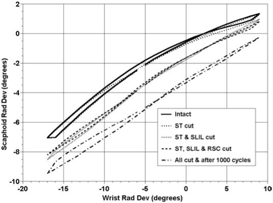
Average scaphoid radial and ulnar deviation as a function of wrist radial and ulnar deviation in the intact wrist and after sequential ligamentous sectioning in the first group of arms. The graph represents the motion of the scaphoid during a complete cycle of wrist motion from radial deviation to ulnar deviation to radial deviation. Flexion and radial deviation are positive. The irregular ends of the curves are the result of including only the data points common to all 8 arms during a range of motion. Some arms continued beyond the shown end points.
Figure A5.
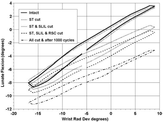
Average lunate flexion and extension as a function of wrist radial and ulnar deviation in the intact wrist and after sequential ligamentous sectioning in the first group of arms. The graph represents the motion of the lunate during a complete cycle of wrist motion from radial deviation to ulnar deviation to radial deviation. Flexion and radial deviation are positive. The irregular ends of the curves are the result of including only the data points common to all 8 arms during a range of motion. Some arms continued beyond the shown end points.
Figure A10.
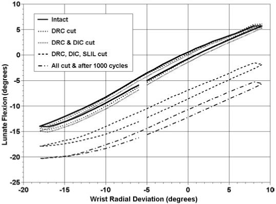
Average lunate flexion and extension as a function of wrist radial and ulnar deviation in the intact wrist and after sequential ligamentous sectioning in the second group of arms. The graph represents the motion of the lunate during a complete cycle of wrist motion from radial deviation to ulnar deviation to radial deviation. Flexion and radial deviation are positive.
Figure A14.
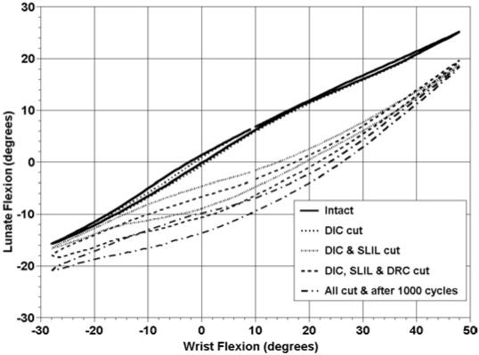
Average lunate flexion and extension as a function of wrist flexion and extension in the intact wrist and after sequential ligamentous sectioning in the third group of arms. The graph represents the motion of the lunate during a complete cycle of wrist motion from flexion to extension to flexion. Flexion is positive.
Figure A17.
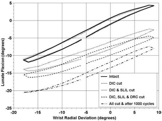
Average lunate flexion and extension as a function of wrist radial and ulnar deviation in the intact wrist and after sequential ligamentous sectioning in the third group of arms. The graph represents the motion of the lunate during a complete cycle of wrist motion from radial deviation to ulnar deviation to radial deviation. Flexion and radial deviation are positive. The irregular ends of the curves are the result of including only the data points common to all 8 arms during a range of motion. Some arms continued beyond the shown end points.
Acknowledgments
Supported by the National Institutes of Health and a National Institute of Arthritis and Musculoskeletal and Skin Diseases grant (1R01 AR050099).
Footnotes
No benefits in any form have been received or will be received from a commercial party related directly or indirectly to the subject of this article.
References
- 1.Short WH, Werner FW, Green JK, Masaoka S. Biomechanical evaluation of ligamentous stabilizers of the scaphoid and lunate. J Hand Surg. 2002;27A:991–1002. doi: 10.1053/jhsu.2002.35878. [DOI] [PMC free article] [PubMed] [Google Scholar]
- 2.Short WH, Werner FW, Green JK, Masaoka S. Biomechanical evaluation of the ligamentous stabilizers of the scaphoid and lunate: part II. J Hand Surg. 2005;30A:24–34. doi: 10.1016/j.jhsa.2004.09.015. [DOI] [PubMed] [Google Scholar]
- 3.Berger RA. The gross and histologic anatomy of the scapholunate interosseous ligament. J Hand Surg. 1996;21A:170–178. doi: 10.1016/S0363-5023(96)80096-7. [DOI] [PubMed] [Google Scholar]
- 4.Berger RA. The ligaments of the wrist. A current overview of anatomy with considerations of their potential functions. Hand Clin. 1997;13:63–82. [PubMed] [Google Scholar]
- 5.Berger RA, Landsmeer JM. The palmar radiocarpal ligaments: a study of adult and fetal human wrist joints. J Hand Surg. 1990;15A:847–854. doi: 10.1016/0363-5023(90)90002-9. [DOI] [PubMed] [Google Scholar]
- 6.Moritomo H, Viegas SF, Nakamura K, Dasilva MF, Patterson RM. The scaphotrapezio-trapezoidal joint. Part 1: an anatomic and radiographic study. J Hand Surg. 2000;25A:899–910. doi: 10.1053/jhsu.2000.4582. [DOI] [PubMed] [Google Scholar]
- 7.Drewniany JJ, Palmer AK, Flatt AE. The scaphotrapezial ligament complex: an anatomic and biomechanical study. J Hand Surg. 1985;10A:492–498. doi: 10.1016/s0363-5023(85)80070-8. [DOI] [PubMed] [Google Scholar]
- 8.Viegas SF. The dorsal ligaments of the wrist. Hand Clin. 2001;17:65–75. [PubMed] [Google Scholar]
- 9.Viegas SF, Yamaguchi S, Boyd NL, Patterson RM. The dorsal ligaments of the wrist: anatomy, mechanical properties, and function. J Hand Surg. 1999;24A:456–468. doi: 10.1053/jhsu.1999.0456. [DOI] [PubMed] [Google Scholar]
- 10.Werner FW, Palmer AK, Somerset JH, Tong JJ, Gillison DB, Fortino MD, Short WH. Wrist joint motion simulator. J Orthop Res. 1996;14:639–646. doi: 10.1002/jor.1100140420. [DOI] [PubMed] [Google Scholar]
- 11.Green JK, Werner FW, Wang H, Weiner MM, Sacks JS, Short WH. Three-dimensional modeling and animation of two carpal bones: a technique. J Biomech. 2004;37:757–762. doi: 10.1016/j.jbiomech.2003.10.001. [DOI] [PubMed] [Google Scholar]
- 12.Berger RA, Bishop AT, Bettinger PC. New dorsal capsulotomy for the surgical exposure of the wrist. Ann Plast Surg. 1995;35:54–59. doi: 10.1097/00000637-199507000-00011. [DOI] [PubMed] [Google Scholar]
- 13.Moran SL, Cooney WP, Berger RA, Strickland J. Capsulodesis for the treatment of chronic scapholunate instability. J Hand Surg. 2005;30A:16–23. doi: 10.1016/j.jhsa.2004.07.021. [DOI] [PubMed] [Google Scholar]
- 14.Szabo RM, Slater RR, Jr, Palumbo CF, Gerlach T. Dorsal intercarpal ligament capsulodesis for chronic, static scapholunate dissociation: clinical results. J Hand Surg. 2002;27A:978–984. doi: 10.1053/jhsu.2002.36523. [DOI] [PubMed] [Google Scholar]
- 15.Mitsuyasu H, Patterson RM, Shah MA, Buford WL, Iwamato Y, Viegas SF. The role of the dorsal intercarpal ligament in dynamic and static scapholunate instability. J Hand Surg. 2004;29A:279–288. doi: 10.1016/j.jhsa.2003.11.004. [DOI] [PubMed] [Google Scholar]
- 16.Elsaidi GA, Ruch DS, Kuzma GR, Smith BP. Dorsal wrist ligament insertions stabilize the scapholunate interval. Clin Orthop. 2004;425:152–157. doi: 10.1097/01.blo.0000136836.78049.45. [DOI] [PubMed] [Google Scholar]
- 17.Short WH, Werner FW, Green JK, Weiner MM, Masaoka S. The effect of sectioning the dorsal radiocarpal ligament and insertion of a pressure sensor into the radiocarpal joint on scaphoid and lunate kinematics. J Hand Surg. 2002;27A:68–76. doi: 10.1053/jhsu.2002.30074. [DOI] [PubMed] [Google Scholar]


