Abstract
Cross talk between NF-κB and c-Jun N-terminal kinases (JNKs) has been implicated in the cell life and death decision under various stresses. Functional suppression of JNK activation by NF-κB has recently been proposed as a key cellular survival mechanism and contributes to cancer cells escaping from apoptosis. We provide a novel scenario of the proapoptotic role of IκB kinase β (IKKβ)–NF-κB, which can act as the activator of the JNK pathway through the induction of GADD45α for triggering MKK4/JNK activation, in response to the stimulation of arsenite, a cancer therapeutic reagent. This effect of IKKβ–NF-κB is dependent on p50 but not the p65/relA NF-κB subunit, which can increase the stability of GADD45α protein through suppressing its ubiquitination and proteasome-dependent degradation. IKKβ–NF-κB can therefore either activate or suppress the JNK cascade and consequently mediate pro- or antiapoptotic effects, depending on the manner of its induction. Furthermore, the NF-κB p50 subunit can exert a novel regulatory function on protein modification independent of the classical NF-κB transcriptional activity.
Introduction
The transcription factor NF-κB is homo- or heterodimers formed from a multigene family that encodes five structure-related proteins: p50 (NF-κB1), p52 (NF-κB2), p65 (RelA), c-Rel (Rel), and RelB. p50/p65 heterodimer is the predominantly, although not exclusively, detectable form of NF-κB in various cells. Normally, NF-κB is sequestered in the cytoplasm in an inactive form by binding to the IκB inhibitors. Activation of NF-κB requires IκB kinase (IKK) to mediate IκB phosphorylation, an event leading to IκB degradation and consequently freeing NF-κB to translocate into the nucleus for regulating the transcription of its target genes. The IKK complex contains two catalytic subunits, IKKα and -β, and a regulatory subunit, IKKγ. The classical manner for NF-κB activation is mainly dependent on the IKKβ subunit induction (Ghosh and Karin, 2002; Hayden and Ghosh, 2004).
Induction of the IKK–NF-κB pathway has been observed under various cellular stresses. One important role of NF-κB activation in these biological processes is to modulate the cellular apoptotic response (Barkett and Gilmore, 1999; Lee et al., 2000; Baldwin, 2001; De Smaele et al., 2001; Tang et al., 2001; Papa et al., 2004a). Many antiapoptotic genes, such as Bcl-XL, XIAP (X chromosome–linked inhibitor of apoptosis), IAP1 and -2, c-FLIP, and Bfl-1/A1, have κB elements in their promoter or enhancer regions and therefore are inducible by NF-κB to protect cells from apoptosis under diverse stimulations (Barkett and Gilmore, 1999; Baldwin, 2001). In addition, functional suppression of the JNK cascade, a key intrinsic cell death machinery programming cell apoptotic response to environmental changes (Davis, 2000; Weston and Davis, 2002; Lin, 2003), has recently been proposed as a key mechanism for the antiapoptotic action of NF-κB under multiple cellular stresses, including the transformation conditions (Bubici et al., 2004; Nakano, 2004; Papa et al., 2004b). NF-κB suppresses the JNK cell death pathway either through the transcriptional up-regulation of a set of its targeted genes, such as the caspase inhibitor XIAP, the zinc-finger protein A20, or GADD (growth arrest and DNA damage inducible) 45β, which can act as the blockers of the JNK cascade (Lee et al., 2000; Tang et al., 2001; Papa et al., 2004a), or through the transcriptional suppression of GADD45α/γ, a potent activator for the JNK upstream kinase MKK4/JNKK1 (Zerbini et al., 2004; Zerbini and Libermann, 2005).
Although antiapoptosis represents a fundamental role of NF-κB in cellular stress responses, NF-κB is also capable of mediating a proapoptotic response in certain circumstances (Ghosh and Karin, 2002; Campbell et al., 2004; Hayden and Ghosh, 2004; Thyss et al., 2005). It has been shown that UVC and some anticancer drugs (daunorubicin/doxorubicin) induce NF-κB, especially the p65/RelA subunit, to recruit histone deacetylases to the promoter regions of some NF-κB–dependent antiapoptotic genes, actively suppress the expression of these genes, and promote cell death under these stress conditions (Campbell et al., 2004). In the case of UVB radiation, NF-κB is induced to selectively up-regulate the expression of the transcription factor and tumor suppressor Egr-1, which in turn transcriptionally activates GADD45α to trigger cell apoptosis (Thyss et al., 2005). Fas and FasL induction is also implicated in the NF-κB–mediated cell apoptotic process (Kasibhatla et al., 1998, 1999). Therefore, molecular mechanisms underlying the proapoptotic action of NF-κB may be diverse, depending on the nature of the stimuli. Notably, to date, both anti- and proapoptotic effects of NF-κB are shown to rely on the p65/RelA subunit, which contains a transcriptional activation domain toward its C terminus (Campbell et al., 2004; Papa et al., 2004a; Zerbini et al., 2004; Thyss et al., 2005). Little is known about the role of another ubiquitously expressed subunit, p50, which lacks the transcriptional activation domain and, thus, does not have the intrinsic ability to drive transcription, like its p65 counterpart, in the course of the NF-κB–relevant biological processes.
Arsenic is a kind of environmental carcinogen involved in the incidence of multiple human cancers (Huang et al., 1999b; Simeonova and Luster, 2000; Bode and Dong, 2002; Dong, 2002). Meanwhile, arsenic-containing compounds have long been used as a therapeutic regimen for the treatment of human leukemia (Chen et al., 1997). How arsenic engages in the promotion of oncogenesis or performs an anticancer effect still remains an enigma. With regard to its effects in tumorigenesis, cell transformation can only be observed with exposure to lower concentrations of arsenite. In contrast, high doses of arsenite stimulation appear to induce the cytotoxic effect (Huang et al., 1999a, 1999b; Bode and Dong, 2002; Dong, 2002). Induction of JNK activation has been proven to be involved in both arsenite-induced killing and cell transformation (Huang et al., 1999a, 1999b). However, the roles of the IKKβ–NF-κB signaling pathway in the cellular arsenite response remains controversial, depending on the doses and cell types used (Chen et al., 2001; Bode and Dong, 2002).
Here, we show that IKKβ–NF-κB can be induced by a higher concentration of arsenite to transduce a cell apoptotic signal through up-regulation of GADD45α and, subsequently, activation of the MKK4–JNK cell death pathway. The NF-κB activity in arsenite response is specifically linked to the p50 but not the p65/RelA subunit, which mediates the function of increasing GADD45α protein stability through prevention of its ubiquitination and proteasome-dependent degradation. Our results, together with other reports (Papa et al., 2004a, 2004b; Zerbini et al., 2004; Zerbini and Libermann, 2005), indicate a dual role of NF-κB on the regulation of the intrinsic JNK cell death cascade. Most important, for the first time, to the best of our knowledge, we suggest a new model of NF-κB for regulating cellular apoptotic response through an action independent of its transcriptional activity, which is mediated by the p50 but not the p65 NF-κB subunit.
Results
IKKβ–NF-κB mediates the cellular apoptotic response to arsenite exposure
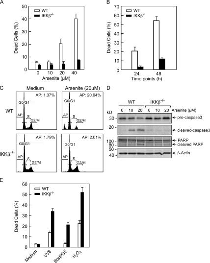
IKKβ is the dominant kinase responsible for NF-κB activation under various conditions (Hayden and Ghosh, 2004). To characterize the roles of the IKKβ–NF-κB pathway in the arsenite response, mouse embryonic fibroblasts (MEFs) derived from wild-type (WT) or IKKβ gene knockout (IKKβ−/−) mice were exploited, and their responses to two doses (10 and 20 μM) of arsenite stimulations were compared. As shown in Fig. 1 A, exposure to 10 μM of arsenite did not cause detectable cytotoxic effects to both types of MEFs within 24 h. However, a considerable increase in cell death was observed for WT MEFs at 24 h under the treatment of 20 μM arsenite, but no increase in cell death for IKKβ−/− MEFs was observed under the same conditions, assayed by both the trypan blue staining and flow cytometric analysis (Fig. 1, A and C). The difference of cell death in response to the 20 μM arsenite stimulation between WT and IKKβ−/− cells was more obvious at 48 h after the treatment, when >54% of cell death was detected for WT MEFs, versus only ∼10% for IKKβ−/− MEFs (Fig. 1 B). Accordingly, the induction of the cleavage of caspase3 and poly (ADP-ribose) polymerase (PARP), two indicators of apoptosis (Mullen, 2004), was readily detectable in WT MEFs but substantially reduced in IKKβ−/− MEFs (Fig. 1 D). The same cells were also exposed to other cytotoxic stimuli, including UVB, benzo-[a]pyrene-7,8-diol-9,10-epoxide (B[a]PDE), and hydrogen peroxide. In contrast to this observation, IKKβ−/− MEFs showed more sensitivity to apoptosis under these stress conditions (Fig. 1 E). These results suggest that the apoptotic-resistant phenotype of IKKβ−/− MEFs is specifically exhibited under the arsenite stress.
Figure 1.
Arsenite-induced apoptosis is suppressed in IKKβ−/− MEFs. (A and B) Cell death of WT and IKKβ−/− MEFs exposed to different doses of arsenite for 24 h (A) or to a single dose of 20 μM arsenite for the times indicated (B) were detected by the trypan blue exclusion assay. At least 500 cells were counted for each group, and results are presented as mean ± SD from three independent experiments. (C) Flow cytometric analysis for the apoptosis of WT and IKKβ−/− MEFs with or without 24 h of arsenite treatment. A representative result is shown. AP represents the percentage of the apoptotic cells. (D) WT and IKKβ−/− MEFs were treated with arsenite for 24 h, and the inducible cleavage of both caspase3 and PARP was detected by immunoblot assay. (E) WT and IKKβ−/− MEFs were exposed to 2 kJ/m2 UVB, 4 μM B(a)PDE, or 50 μM H2O2 for 24 h, and cell death was monitored by the trypan blue exclusion assay.
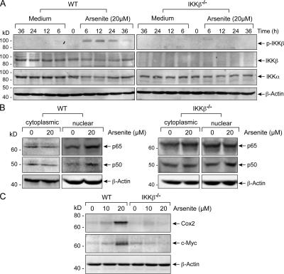
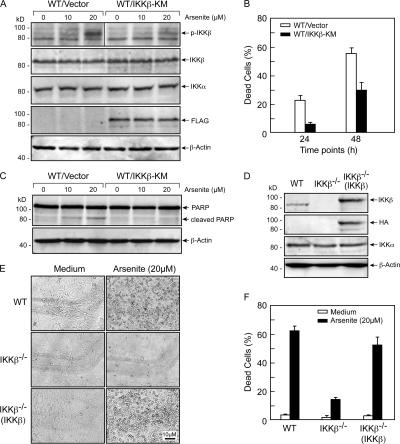
To confirm that the IKKβ-dependent pathway is explored to transduce a cell death signal in arsenite response, we first showed that efficient induction of IKKβ phosphorylation (Fig. 2 A), NF-κB component (p50 and p65) nuclear translocation (Fig. 2 B), and expression of two NF-κB–targeted genes, c-Myc and Cox-2 (Fig. 2 C), were readily detectable in WT cells but not in IKKβ−/− cells under 20 μM arsenite exposure, indicating that arsenite stimulation has the effect of activating the NF-κB pathway, and such an induction is dependent on the existence and activity of IKKβ. We next stably expressed a dominant-negative IKKβ mutant (IKKβ-KM; Geleziunas et al., 1998; Chu et al., 1999; Chen et al., 2001) in WT MEFs to block the IKKβ–NF-κB pathway (Fig. 3 A). Meanwhile, an HA-tagged IKKβ construct was introduced into IKKβ−/− MEFs to reconstitute the IKKβ–NF-κB pathway. As expected, stable expression of IKKβ-KM considerably suppressed the arsenite-induced cell death (Fig. 3 B) and PARP cleavage (Fig. 3 C) in WT MEFs; however, stable overexpression of HA-IKKβ in IKKβ-null MEFs (Fig. 3 D) remarkably sensitized these cells to the arsenite-induced cell death (Fig. 3, E and F). Collectively, we propose that induction of the IKKβ–NF-κB pathway programs a cellular apoptotic response to arsenite stimulation.
Figure 2.
Arsenite-induced IKKβ activation and NF-κB components (p50 and p65/RelA) nuclear translocation were blocked in IKKβ−/− MEFs. (A) WT and IKKβ−/− MEFs were treated with 20 μM arsenite for the indicated times, and the activation of IKKβ was detected by immunoblot assay. (B) WT and IKKβ−/− MEFs were treated with 20 μM arsenite for 12 h, and the distribution of p50 and p65 in the cytoplasmic and nuclear fractions was detected. (C) WT and IKKβ−/− MEFs were treated as described in B, and the expression levels of two NF-κB targeted genes, c-Myc and Cox2, in the whole cell extract were detected by immunoblot assay.
Figure 3.
Evidence for the involvement of the IKKβ–NF-κB pathway in mediating arsenite-induced cell apoptosis. (A) WT cells stably transfected with the construct containing dominant-negative kinase mutant of IKKβ (FLAG-IKKβ-KM) or the control vector were treated with arsenite for 12 h, and the activation of IKKβ was detected. (B) Cells described in A were treated with 20 μM arsenite for the times indicated, and cell death was measured by the trypan blue viability assay. (C) Cells described in A were treated with 20 μM arsenite for 24 h, and the cleavage of PARP was detected. (D) Stable expression of a recombinant IKKβ (HA-IKKβ) in IKKβ−/− MEFs. (E and F) WT, IKKβ−/−, and the reconstituted IKKβ−/−(IKKβ) MEFs were exposed to 20 μM arsenite for 48 h, and cell death was detected by both the microscopic observation (E) and the trypan blue viability assay (F). Error bars indicate mean ± SD.
IKKβ–NF-κB mediates arsenite-induced apoptosis via JNK activation
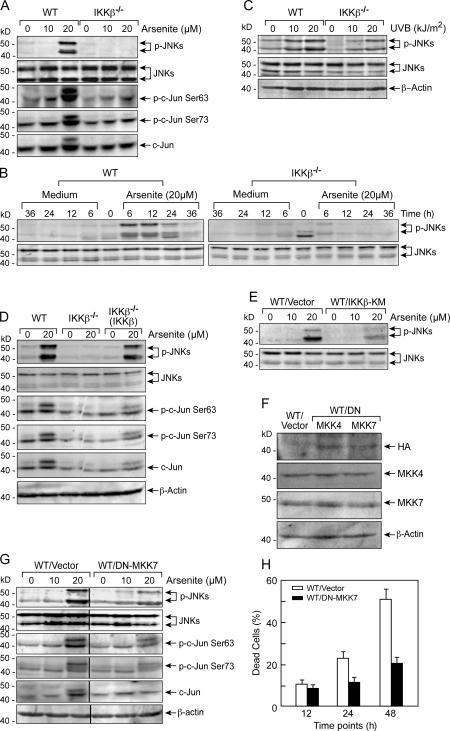
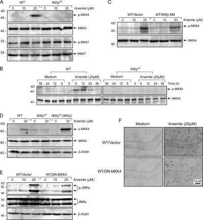
Our previous studies have demonstrated that induction of JNKs contributes greatly to arsenite-induced cell apoptosis (Huang et al., 1999b). We thus tested whether induction of IKKβ–NF-κB under arsenite stress has any relevance to JNK activation. As shown in Fig. 4 A, induction of marked phosphorylation of JNK and its substrate c-Jun in WT MEFs was only observed under 20 μM arsenite stimulation, a dose with an obvious cytotoxic effect on MEFs (Fig. 1, A–C). The time course–dependent experiment indicated that the arsenite-induced JNK activation in WT MEFs occurred as early as 6 h and was sustained up to 24 h after the treatment (Fig. 4 B). In contrast to the observation for WT cells, no obvious induction of JNK phosphorylation was found in IKKβ−/− MEFs in both time- and dose-dependent experiments (Fig. 4, A and B). However, effective induction of JNK phosphorylation was still observed in IKKβ−/− MEFs under the UVB radiation (Fig. 4 C), suggesting that genetic ablation of IKKβ selectively affected the arsenite-induced JNK response. Again, reconstitution expression of IKKβ in IKKβ−/− MEFs restored arsenite-induced activation of JNKs and c-Jun (Fig. 4 D), whereas overexpression of IKKβ-KM in WT MEFs substantially suppressed arsenite-induced JNK phosphorylation (Fig. 4 E). These results indicate that arsenite-induced JNK activation is dependent on the IKKβ–NF-κB signaling pathway.
Figure 4.
IKKβ–NF-κB mediates arsenite-induced apoptosis via activation of the JNK pathway. (A and B) The same cell lysates as in Fig. 2 C (A) or Fig. 2 A (B) were examined for JNKs and/or c-Jun activation. (C) WT and IKKβ−/− MEFs were exposed to different doses of UVB for 2 h, and the activation of JNKs was detected. (D) WT, IKKβ−/−, and the reconstituted IKKβ−/−(IKKβ) MEFs were exposed to 20 μM arsenite for 12 h, and the activation of JNKs and c-Jun were detected. (E) The same cell lysates as in Fig. 3 A were examined for JNK phosphorylation. (F) Stable expression of a recombinant dominant-negative MKK7 (HA-DN-MKK7) or dominant-negative MKK4 (HA-DN-MKK4) in WT MEFs. (G) WT/DN-MKK7 or the vector control stable transfectants were treated with 20 μM arsenite for 12 h, and the inducible phosphorylations of both JNKs and c-Jun were analyzed. (H) Cells described in G were treated with 20 μM arsenite for different periods of time, and cell death was detected by the trypan blue exclusion assay. Error bars indicate mean ± SD.
To further confirm that IKKβ–NF-κB–mediated JNK activation is attributable to the arsenite-induced proapoptotic effect, a dominant-negative mutant for a JNK-specific upstream kinase MKK7 (DN-MKK7; Tang et al., 2001) was stably transfected into WT MEFs to block JNK induction (Fig. 4 F). As shown in Fig. 4 (G and H), ectopic overexpression of DN-MKK7 in WT MEFs partially reduced arsenite-induced JNK pathway activation, associated with a remarkable attenuation of cell death in these transfected cells.
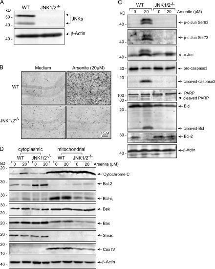
The requirement of the JNK pathway in mediating arsenite-induced apoptosis was also indicated by the observation that arsenite-induced cell death was blocked in the JNK1 and -2 double gene knockout MEFs (Fig. 5, A and B; Sabapathy et al., 2004), associated with a considerable suppression of the inducible cleavage of caspase3 and PARP in these MEFs (Fig. 5 C). Similar results were also found in JNK1−/− and JNK2−/− MEFs (unpublished data). Disturbance of mitochondrial function has been shown to be involved in JNK-mediated apoptosis under certain stresses (Davis, 2000; Tournier et al., 2000; Weston and Davis, 2002). We then examined whether this also occurred in arsenite response. As shown in Fig. 5 (C and D), a substantial down-regulation of the antiapoptotic protein Bcl-2, activation of the proapoptotic protein Bid (indicated by its inducible cleavage), and the release of cytochrome c from the mitochondria into the cytoplasm were observed in WT MEFs upon arsenite treatment, but these events were not detectable in JNK1/2−/− MEFs (Fig. 5, C and D). No obvious changes for other mitochondrial components, including Bcl-XL, Bax, Bak, and Smac, were found during the arsenite treatment (Fig. 5 D). We thus proposed that disturbance of the mitochondrial function might contribute, at least in part, to JNK-mediated apoptosis in arsenite response.
Figure 5.
JNK activation is responsible for arsenite-induced apoptosis via mitochondria-dependent pathway. (A) Identification of JNK1 and -2 expressions in the WT and JNK1/2−/− MEFs. (B) WT and JNK1/2−/− MEFs were treated with arsenite for 48 h, and cell death onset was detected by the microscopic observation. (C) WT and JNK1/2−/− MEFs were treated with 20 μM arsenite for 24 h, and the inducible c-Jun phosphorylation; cleavage of caspase3, PARP, and Bid; and Bcl-2 expression were detected. (D) Mitochondrial proteins were prepared from the cells treated as described in C. The distribution of cytochrome c, Smac, Bcl-XL, Bcl-2, Bax, and Bak in the cytoplasm and mitochondria were monitored. CoxIV was detected as a mitochondria-specific marker.
IKKβ–NF-κB mediates arsenite-induced JNK activation via targeting MKK4 phosphorylation
MKK4/JNKK1 and MKK7/JNKK2 are the two upstream kinases required for the full activation of JNKs (Davis, 2000; Weston and Davis, 2002). We next determined whether NF-κB activates JNKs via modulation of these two kinases' induction under arsenite stimulation. As shown in Fig. 6 A, phosphorylated MKK7 was present in both WT and IKKβ−/− MEFs without marked induction before and after the treatment with either 10 or 20 μM arsenite. However, after treating with 20 μM arsenite, a potent induction of MKK4 phosphorylation was found in WT MEFs, but this was not detectable in IKKβ−/− MEFs. The time course–dependent experiment indicated that the dynamics of arsenite-induced MKK4 phosphorylation was consistent with that of the JNK induction in WT MEFs (compare Fig. 6 B with Fig. 4 B), whereas no inducible activation of MKK4 was observed in IKKβ−/− MEFs at the indicated time points (Fig. 6 B). Furthermore, inhibition of the IKKβ–NF-κB pathway by IKKβ-KM remarkably suppressed arsenite-induced MKK4 and JNK phosphorylation in WT MEFs, whereas reexpression of IKKβ in IKKβ−/− MEFs restored the response of arsenite-induced MKK4 phosphorylation in these cells (Fig. 6, C and D). These data indicate that the IKKβ–NF-κB pathway signals to activate MKK4 under the arsenite response.
Figure 6.
IKKβ–NF-κB mediates arsenite-induced JNK activation via targeting modulation of MKK4 phosphorylation. (A and B) The same cell lysates described in Fig. 2 (C and A) were detected for the activation of MKK4 and/or MKK7. (C) The same cell lysates as in Fig. 3 A were examined for MKK4 phosphorylation. (D) The same cell lysates as in Fig. 4 D were examined for MKK4 activation. (E) WT/vector (the same mass as used in Fig. 4 G) and WT/DN-MKK4 stable transfectants were treated with arsenite for 12 h, and the activation of JNKs was examined. (F) Cells described in E were treated with 20 μM arsenite for 48 h, and cell death onset was detected by microscopic observation.
To determine whether the induction of MKK4 accounts for the arsenite-induced JNK activation and cell apoptosis, DN-MKK4 was stably introduced into the WT MEFs (Fig. 4 F). As indicated in Fig. 6 (E and F), expression of DN-MKK4 inhibited arsenite-induced JNK activation and considerably reduced cell death. Based on these results, we conclude that targeting induction of MKK4 links IKKβ–NF-κB on JNK functional activation in arsenite response.
IKKβ–NF-κB signals the activation of the MKK4–JNK cascade by the up-regulation of GADD45α in cellular arsenite response
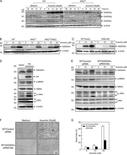
The GADD45 family proteins (GADD45α, -β, and -γ) were proven as binding partner and activator of MEKK4 MAP3K in vitro and led to the activation of the downstream target JNKs (Takekawa and Saito, 1998). We thus tested whether this family protein can act as the downstream effecter of IKKβ–NF-κB for mediating JNK activation in arsenite response. We repeatedly found that arsenite stimulation selectively up-regulated GADD45α protein levels in WT cells. However, no obvious alteration of the expression of GADD45β and -γ was observed under the same conditions (Fig. 7 A). Arsenite-induced GADD45α up-regulation was impaired in IKKβ−/− MEFs (Fig. 7 A) and was restored in IKKβ−/− cells by reconstitution of IKKβ (Fig. 7 B). In addition, ectopic overexpression of IKKβ-KM in WT MEFs blocked arsenite-induced GADD45α accumulation (Fig. 7 C). These results indicate that arsenite stimulation up-regulates GADD45α through the IKKβ–NF-κB–dependent pathway.
Figure 7.
IKKβ–NF-κB mediates arsenite-induced activation of the MKK4–JNK cascade through up-regulation of GADD45α. (A) The same cell lysates as in Fig. 2 A were examined for the expression levels of GADD45α, -β, and -γ. (B) WT, IKKβ−/−, and the reconstituted IKKβ−/−(IKKβ) MEFs were exposed to arsenite for 12 h, and the expression level of GADD45α was detected. (C) The same cell lysates as in Fig. 3 A were used to examine the expression level of GADD45α. (D) WT MEFs were transiently transfected with the expression plasmid of GADD45α (HA-GADD45α) or the control vector. Cells were harvested 36 h later, and the activation of MKK4 and JNKs under overexpression of GADD45α was detected. (E) WT MEFs were cotransfected with the mixture of two GADD45α siRNA constructs or the scrambled control siRNA constructs. The corresponding stable transfectants were treated with different doses of arsenite for 12 h, and the activation of MKK4 and JNKs was detected. (F and G) GADD45α siRNA or the control siRNA stable transfectants were treated with different doses of arsenite for 48 h, and the cell death was detected by either direct microscopic observation (F) or trypan blue exclusion assay (G). Error bars indicate mean ± SD.
To test whether GADD45α up-regulation is responsible for JNK activation in arsenite response, we first transiently transfected an HA-tagged GADD45α construct into WT MEFs and showed that overexpression of HA-GADD45α actually induced both MKK4 and JNK phosphorylation in the transfected cell population in the absence of arsenite stimulation (Fig. 7 D). We also designed a pair of siRNAs that targeted two different regions on the GADD45α mRNA. Stable transfection with a combination of these two siRNAs into WT MEFs nearly abolished arsenite-induced GADD45α up-regulation, accompanied by the blocking of arsenite-induced MKK4 and JNK phosphorylation (Fig. 7 E) and a substantial decrease of arsenite- induced cell death (Fig. 7, F and G). Therefore, we conclude that up-regulation of GADD45α is responsible for triggering JNK activation and cell apoptosis under arsenite stress.
Arsenite-induced GADD45α up-regulation, MKK4–JNK activation, and cell apoptosis are related to the p50 but not the p65/relA NF-κB component
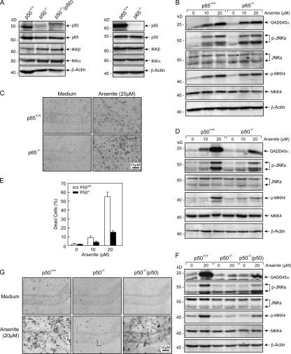
We have demonstrated that the NF-κB–GADD45α–MKK4–JNK pathway is responsible for mediating arsenite-induced cell apoptosis. p50 and p65 are two predominant NF-κB components expressed in a variety of cell types (Ghosh and Karin, 2002; Hayden and Ghosh, 2004) and have different roles in regulating the biological effects of the IKKβ–NF-κB signaling pathway under certain stimulation conditions (Abbadie et al., 1993; Beg et al., 1995; Saccani et al., 2003). To further clarify the roles of these two components in arsenite responses, MEFs from p50 and p65 gene knockout animals were exploited (Fig. 8 A). Interestingly, we found that genetic ablation of p65 did not affect arsenite-induced GADD45α up-regulation, MKK4 phosphorylation, and JNK activation (Fig. 8 B). These cells also showed sensitivity to arsenite-induced cell death (Fig. 8 C). However, all of the events of arsenite-induced GADD45α up-regulation, MKK4–JNK activation, and cell apoptosis were considerably suppressed in p50−/− MEFs (Fig. 8, D and E). Furthermore, reconstitution of p50−/− MEFs with p50 transfection (Fig. 8 A) restored arsenite-induced GADD45α up-regulation and MKK4–JNK activation (Fig. 8 F), accompanied by the sensitivity of p50−/−(p50) cells to arsenite-induced apoptosis (Fig. 8 G). These data disclose a property of the p50 NF-κB component in the induction of the GADD45α–MKK4–JNK cell death pathway under arsenite stress.
Figure 8.
The effect of NF-κB on mediating arsenite-induced activation of the GADD45α–MKK4–JNK cascade and cell apoptosis is related to the p50 but not the p65/RelA subunit. (A) Identification of p50 and p65/RelA expression in the WT, p65−/−, p50−/−, and p50−/− (p50) MEFs. (B) p65+/+ and p65−/− MEFs were treated with different doses of arsenite for 12 h, and the induction of GADD45α expression, MKK4, and JNK phosphorylation were detected. (C) p65+/+ and p65−/− MEFs were exposed to 20 μM arsenite for 24 h, and cell death onset was detected with microscopic observation. (D) p50+/+ and p50−/− MEFs were treated with arsenite as described in B, and the same immunoblot was preformed. (E) p50+/+ and p50−/− MEFs were treated with different doses of arsenite for 48 h, and cell death was analyzed by the trypan blue exclusion assay. (F) WT, p50−/−, and the reconstituted p50−/−(p50) MEFs were exposed to 20 μM arsenite for 12 h, and the expression of GADD45α and the activation of MKK4 and JNKs was detected. (G) Cells described in F were exposed to arsenite for 48 h, and the apoptotic cell death was detected with microscopic observation.
IKKβ–NF-κB p50 prevents GADD45α proteins from degradation by the ubiquitin–proteasome pathway in arsenite response
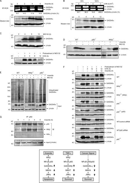
A recent study showed that UV radiation induces transcriptional up-regulation of GADD45α through an NF-κB–dependent pathway (Thyss et al., 2005). Unlike the p65 component, the p50 subunit of NF-κB does not possess the transcriptional activity for lack of the transactivation domain (Ghosh and Karin, 2002; Hayden and Ghosh, 2004). This implies that the induction of GADD45α observed in the arsenite response may be mechanically different to that in the UV radiation. Supporting this prediction, the RT-PCR assay showed that GADD45α mRNA was constitutively expressed in the resting WT MEFs and its expression levels did not exhibit detectable change over 8 h of arsenite treatment; however, up-regulation of GADD45α protein levels was detected as early as 2 h and, more significant, at 4 h after arsenite stimulation (Fig. 9 A). As a control, we observed that the up-regulation of GADD45α mRNA and its protein levels under UVR exhibited the consistent time course–dependent response (Fig. 9 B). This finding suggests that the induction of GADD45α in arsenite response might occur at the posttranscriptional level, most possibly by the modulation of the protein stabilities. To test this hypothesis, WT MEFs were treated with MG132, an established inhibitor of protein degradation by disruption of the proteasome system (Nandi et al., 2006). As indicated in Fig. 9 C, similar to that of the arsenite stimulation, MG132 treatment resulted in a remarkable accumulation of GADD45α proteins. Meanwhile, when the culture was withdrawn from MG132 and subjected to the treatment of cyclohexamide (CHX), a protein synthesis inhibitor, to block the de novo production of proteins, the preaccumulated GADD45α by MG132 could undergo a gradual degradation with the reassembly of the proteasome system (Fig. 9 C). MG132 treatment also resulted in GADD45α protein accumulation in IKKβ−/− and p50−/− MEFs, although arsenite failed to accumulate this protein in both cells (Fig. 9 D). These results together demonstrate that the expressed GADD45α is actively subjected to a proteasome-dependent degradation process in resting cells, and such a process is blocked in arsenite response via the IKKβ–p50–dependent pathway.
Figure 9.
IKKβ–NF-κB p50 mediates arsenite's effect on inhibition of ubiquitination and degradation of GADD45α proteins. (A and B) WT cells were treated with 20 μM arsenite (A) or 2 kJ/m2 UVB (B) for the times indicated, and the inducible expression of GADD45α mRNA and proteins were detected by PT-PCR and immunoblot assay, respectively. Data from one of the two independent experiments are shown, and the relative expression levels of GADD45α mRNA normalized to the β-actin control is also presented. (C) WT cells were either treated with 10 μM MG132 for the indicated times (top) or pretreated with 10 μM MG132 for 4 h, followed by exposure to 10 μM CHX for the different period of times after withdrawal of MG132 (bottom). Immunoblot assay was performed to detect the expression of GADD45α proteins. (D) WT, IKKβ−/−, and p50−/− MEFs were exposed to 20 μM arsenite or 10 μM MG132 for 12 h, and the expression of GADD45α in individual cells was detected. (E) WT, IKKβ−/−, and p50−/− MEFs were treated with 20 μM arsenite, 10 μM MG132, or the combination of these two reagents for 12 h, and the whole cell extracts were immunoprecipitated with anti-GADD45α antibody. Ubiquitinated GADD45α was detected with anti-ubiquitin antibody. Input of the proteins subjected to immunoprecipitation is also shown. (F) WT, IKKβ−/−, p50−/−, and the corresponding reconstituted MEFs, as well as p50-specific siRNA stably transfected WT cells, were pretreated with 10 μM MG132 for 4 h and exposed to 10 μM CHX in the absence or presence of 20 μM arsenite for the times indicated after removal of MG132. GADD45α expression was detected by immunoblot assay. (G) WT MEFs were treated with 20 μM arsenite for the times indicated, and the whole cell extracts were immunoprecipitated with anti-p50 antibody. The immunoprecipitated samples were detected with anti-p50, -IKKα, and -IKKβ antibodies, respectively. Input of the proteins subjected to immunoprecipitation is also shown. (H) Models of different mechanisms of NF-κB in the cell life and death control under diverse conditions mediated by GADD45 family members and the JNK pathway.
Ubiquitination is the common mechanism for dictating proteins into proteasome-dependent degradation (Nandi et al., 2006). We next determined whether arsenite induces GADD45α accumulation in WT MEFs by suppressing its ubiquitination. GADD45α was immunoprecipitated with specific anti-GADD45α antibodies, and its ubiquitination status was analyzed with anti-ubiquitin antibodies. As shown in Fig. 9 E (left), only a weak ubiquitin signal was detected in the sample of untreated WT cells. This result was predictable, as there was little GADD45α protein present in these resting cells because of its rapid degradation. However, a strong ubiquitin signal was detected in the lane for MG132-treated cells, indicating that the accumulated GADD45α protein by MG132 is highly ubiquitinated. Interestingly, the ubiquitin signal for GADD45α in the arsenite-treated cells was almost as weak as that in the untreated control sample, although its amount in these cells is the same as that in the MG132-treated cells, suggesting that the accumulated GADD45α by arsenite stimulation is less ubiquitinated. The effect of arsenite on suppressing GADD45α ubiquitination was also manifested in the MG132/arsenite double-treated cells, in which the ubiquitin signal for GADD45α was substantially decreased as compared with that in cells treated with MG132 alone. The ubiquitination status of GADD45α was also analyzed in IKKβ−/− and p50−/− MEFs (Fig. 9 E, middle and right). As expected, MG132 treatment also accumulated large amounts of highly ubiquitinated GADD45α in both cells. However, the effect of arsenite on suppressing GADD45α ubiquitination, which was observed in WT MEFs, was not exhibited in both gene knockout cells, indicated by the same high levels of GADD45α ubiquitination in MG132/arsenite and MG132-treated cells. These results demonstrate that arsenite stimulation provokes a response to suppress GADD45α ubiquitination, depending on the IKKβ–p50 signaling pathway.
To further clarify whether the action of IKKβ–p50 on preventing GADD45α degradation under arsenite stimulation is through preventing this protein from ubiquitination or by mediating a process of deubiquitinating the ubiquitinated proteins, we designed an experimental system in which the MEFs were pretreated with MG132 to accumulate a certain amount of GADD45α proteins in vivo. After the pretreatment, MG132 was removed from the medium and the protein synthesis inhibitor CHX was added to the cells alone or in combination with arsenite. In this way, we were able to analyze the effect of arsenite on the dynamic degradation of the preubiquitinated GADD45α proteins during the course of the reassembly of the proteasome system in vivo. As shown in Fig. 9 F, the preaccumulated GADD45α proteins disappeared in all tested cells within 12 h after removal of MG132 in the absence of arsenite. With arsenite stimulation, GADD45α proteins remained stable at 12 h after withdrawal of MG132 in WT and p65−/− MEFs. However, almost no GADD45α proteins were detectable at this time point in either IKKβ−/− or p50−/− MEFs under the same conditions. Again, reintroduction of IKKβ and p50 into the according gene knockout cells restored the effect of arsenite on preventing GADD45α degradation. In addition, suppression of p50 expression by its specific siRNA (unpublished data) partially disrupted the effect of arsenite in WT MEFs. Based on these results, we propose that IKKβ–p50 might exert an effect for mediating the deubiquitination of GADD45α under the arsenite response, and this might contribute, at least in part, to the phenomenon of the arsenite-induced GADD45α up-regulation.
Discussion
The IKKβ–NF-κB signaling pathway transmits signals essential for cell survival in a variety of physiological and pathological processes (Hayden and Ghosh, 2004). Deregulation of this signaling pathway has been directly implicated in the evasion of the apoptotic responses of many human cancers. As a result, targeted inhibition of the IKKβ–NF-κB pathway has been proposed as a strategy for the development of new anticancer drugs (Karin et al., 2002). Interestingly, it has recently been demonstrated that this signaling pathway can also be induced to exert proapoptotic effects in response to some apoptotic inducers (Kasibhatla et al., 1998, 1999; Campbell et al., 2004; Thyss et al., 2005). Although this finding raises caution regarding the rationale of the proposed use of IKKβ–NF-κB inhibitors in combination with other anticancer drugs in a clinical setting, it also provides an alternative for targeting IKKβ–NF-κB for cancer therapy and chemoprevention. Therefore, identification of the conditions and the according mechanisms for converting the antiapoptotic role of this signaling pathway to the proapoptotic action in cancer cells will be of medical significance.
Arsenite was regarded as both a carcinogen and tumor-therapeutic agent because of its ability to mediate cellular apoptotic or transformation effects under different conditions (Chen et al., 1997; Bode and Dong, 2002; Dong, 2002). Our previous studies have disclosed that a low dose of arsenite (1.25–5 μM) is capable of promoting the cell cycle by the induction of cyclin D1 expression through the IKKβ–NF-κB pathway (Ouyang et al., 2005). However, the precise mechanism of arsenite on the tumor therapeutic effect is not well clarified. In this study, we demonstrate that at a higher dose (20 μM) arsenite can induce an activity of the IKKβ–p50 NF-κB complex to program cell apoptosis via triggering the GADD45α–MKK4–JNK cell death cascade (Fig. 9 H). These novel findings demonstrate that IKKβ–NF-κB has the intrinsic competence to directly turn on the cell death program through the activation of JNK cascade under stress conditions and add a new content to the cross talk between NF-κB and JNK signaling pathways in cell life and death decisions (Bubici et al., 2004; Nakano, 2004; Papa et al., 2004b). As suppression of the JNK activation by NF-κB is essential for cancer cell survival (Zerbini et al., 2004; Zerbini and Libermann, 2005), our finding that arsenite is capable of subverting the inhibitory effect of NF-κB on JNK activation may be of medical importance. It merits further elucidation as to whether such a mechanism underlies the effect of this reagent in cancer therapy.
How NF-κB can be induced to functionally suppress or activate the JNK cell death pathway is an interesting question. Previous reports (Papa et al., 2004a; Zerbini et al., 2004) and the data in this study suggest that the GADD45 protein family (GADD45α, -β, and -γ) may serves as a key modulator for functionally linking IKKβ–NF-κB to the JNK pathway. In this model (Fig. 9 H), by differential regulation of GADD45 family members (up- or down-regulated), NF-κB can be induced to suppress or activate the JNK cell death pathway, thereby exerting its pro- or antiapoptotic effects according to the nature of the stresses. Our results have disclosed the predominant role of GADD45α in arsenite-induced cell death response. In addition, previous data have shown that suppression of GADD45α and -γ contributed to cancer cell survival, whereas induction of GADD45β antagonized TNFα-induced killing (Fig. 9 H; Papa et al., 2004a; Zerbini et al., 2004). These results suggest that GADD45α and -γ mainly contribute to the NF-κB–mediated cell death effect, whereas GADD45β appears to involve NF-κB–dependent cell survival.
Interestingly, we noticed that all of the previous studies regarding the regulatory effect of NF-κB on GADD45 expression are exclusively dependent on the transcriptional activity of NF-κB, such as the transcriptional suppression on GADD45α and -γ by constitutively active NF-κB in cancer cells and the transcriptional induction of GADD45β by NF-κB in response to TNFα stimulation. In addition, NF-κB activity in these responses is mediated by the cooperative action of the p65 and p50 subunits (Papa et al., 2004a; Zerbini et al., 2004). In contrast, we demonstrate that the role of NF-κB on mediating the arsenite-induced GADD45α up-regulation is by preventing this protein from the ubiquitin–proteasome–dependent degradation, instead of the transcriptional induction of the GADD45α gene. Furthermore, this effect of NF-κB only relates to the p50 but not the p65/RelA subunit. Therefore, our data, for the first time, has provided a strong evidence for a new function of the NF-κB p50 subunit, which is independent of the classical NF-κB transcriptional activity but relates to the posttranslational modification of a protein.
GADD45 is originally described as a p53-dependent and stress-inducible gene that also can be regulated by many other transcription factors (Wang et al., 1999; Zerbini et al., 2004; Thyss et al., 2005). In addition, recent evidence suggests that protein ubiquitination represents another way for GADD45 regulation (Leung et al., 2001). Here, we showed that arsenite-induced GADD45α accumulation through preventing this protein from ubiquitination-dependent degradation, further emphasizing the importance of this kind of mechanism on the functional control of GADD45 family members.
The mechanism responsible for the p50-mediated GADD45α modification is currently unknown. Clearly, it has some relevance to the presence of IKKβ protein, indicated by the similar manner of the changes on ubiquitination, degradation, and accumulation of GADD45α proteins under arsenite stimulation in IKKβ−/− and p50−/− MEFs (Fig. 9, E and F). How IKKβ functionally links to the NF-κB p50 subunit in arsenite response remains unclear. However, we have observed that a transient interaction between IKKβ and p50 can be induced by arsenite in WT MEFs in the coimmunoprecipitation assay, and the time course–dependent response of the IKKβ–p50 complex formation (between 1 and 4 h after arsenite stimulation; Fig. 9 G) is consistent with the appearance of the accumulated GADD45α proteins (2–4 h after arsenite stimulation; Fig. 9 A). In contrast, there is no obvious interaction between IKKα and p50 under the same conditions (Fig. 9 G). This result provided us with an important clue suggesting that the inducible interaction between IKKβ and p50 by arsenite might confer a novel property to this putative complex for suppressing GADD45α ubiquitination. Because arsenite stimulation increased the ubiquitination of total cellular proteins (unpublished data) while selectively decreasing the GADD45α ubiquitination level, we proposed that the most probable action of IKKβ–NF-κB p50 is to target GADD45α-specific E3 ubiquitin ligase or the cellular deubiquitination enzymes. The functional link between IKKβ and p50 and the possible mechanism responsible for the IKKβ–p50–containing complex on regulating GADD45α ubiquitination and degradation are currently under investigation.
In summary, this study demonstrates for the first time that the IKKβ–NF-κB signaling pathway can transduce the apoptotic signal through eliciting the intrinsic GADD45α–MKK4–JNK cell death route and provides a novel scenario for the implication of the cross talk between these two master pathways in the cell life and death decisions encountering diverse stresses. Furthermore, we disclosed a new function of NF-κB p50 subunit, which can act as a critical regulator of the ubiquitin–proteasome–dependent modification of GADD45α. Our study, together with other recent findings, clearly suggests the existence of different mechanisms underlying the NF-κB pathway. Elucidation of these issues should shed important insight on the understanding of how NF-κB integrates diverse stimuli to generate a unified outcome suitable for a specific situation, a question raised in the current field of NF-κB investigation.
Materials and methods
Plasmids, antibodies, and other reagents
The plasmid expressing the kinase mutant of IKKβ (Flag-IKKβ-KM) was a gift from H. Nakano (Juntendo University, Tokyo, Japan; Nakano et al., 1998). The plasmid expressing HA-tagged full-length GADD45α (HA-GADD45α) was described in a previous study (Wang et al., 1999). The plasmid containing p50 cDNA was provided by J. Ye (Louisiana State University, Baton Rouge, LA). The plasmids expressing dominant-negative MKK4 (HA-DN-MKK4) or dominant-negative MKK7 (HA-DN-MKK7) and the full-length IKKβ (HA-IKKβ) were described in a previous study (Tang et al., 2001). The antibodies against phospho-IKKα/β, IKKα, IKKβ, phospho-JNK, JNK, phospho-MKK4, MKK4, phospho-MKK7, MKK7, phospho-c-Jun, c-Jun, caspase3, PARP, Bcl-2, Bcl-XL, Bax, and Bak were purchased from Cell Signaling Technology. The antibodies against GADD45α, -β, and -γ; c-Myc; p65; p50; Bid; and the agarose-conjugated anti-p50 antibody were obtained from Santa Cruz Biotechnology, Inc. Anti–cytochrome c and Smac antibodies were purchased from BD Biosciences. Anti-HA and ubiquitin antibodies were purchased from Upstate Biotechnology; anti-FLAG and anti–β-actin antibodies were obtained from Sigma-Aldrich; and anti-Cox2 antibody was purchased from Cayman Chemical. MG132 and CHX were purchased from Calbiochem.
Cell culture and transfection
IKKβ−/− MEFs were a gift from M. Karin (University of California, San Diego, La Jolla, CA; Tang et al., 2001). The p50−/− and p65−/− MEFs and their corresponding WT MEFs were provided by J. Ye. The JNK1/2−/− MEFs were provided by K. Sabapathy (National Cancer Center, Singapore; Sabapathy et al., 2004). The WT and the gene knockout MEFs were maintained in DME (Calbiochem) supplemented with 10% FBS, 1% penicillin/streptomycin, and 2 mM l-glutamine (Life Technologies) at 37°C. Cell transfections were performed with Lipofectamine reagent (Invitrogen) according to the manufacturer's instruction. For stable transfection, cultures were subjected to either hygromycin B or G418 drug selection, and cells surviving from the drug selection were pooled as stable mass. These stable transfectants were cultured in the selective drug-free medium for at least two passages before used for according experiments. For transient transfection, cells were harvested at 36 h after transfection for immunoblot analysis.
Construction of GADD45α siRNA expression plasmids
The two sequences, 5′-gatcctgccttaagtcaa-3′ and 5′-agtcgctacatggatcagt-3′, on GADD45α mRNA were selected by the siRNA Target Finder (http://www.ambion.com/techlib/misc/siRNA_finder.html; Ambion) as siRNA target sites and were expressed by using the GeneSuppressor system (Imgenex). The constructs containing the corresponding scrambled target sequences were used as controls. The established constructs were either separately transfected or cotransfected into WT cells for stable expression.
Cell death analysis
The arsenite-induced cell death was determined by trypan blue exclusion assay and flow cytometric analysis after propidium iodide staining of the nuclei. For the live cell microscopy, the images were taken with an inverted microscope (CKX41; Olympus) equipped with the achromatic objectives (10×; NA 0.25; working distance 8.8 mm) and the digital camera (DP12-2; Olympus) at room temperature. The images were analyzed using Photoshop (Adobe).
Western blot
Whole cell extracts were prepared with the cell lysis buffer (10 mM Tris-HCl, pH 7.4, 1% SDS, and 1 mM Na3VO4). Cytoplasmic and nuclear proteins were prepared with Cellytic nuclear extraction kit (Sigma-Aldrich) following the manufacturer's protocols. The mitochondrial proteins were prepared with the Mitochondria isolation kit for mammalian cells (Pierce Chemical Co.) following the manufacturer's protocols. Protein concentrations were determined by the protein quantification assay kit (Bio-Rad Laboratories). 30 μg of proteins were resolved by SDS-PAGE, probed with the indicated primary antibodies, and incubated with the AP-conjugated second antibody. Signals were detected by the enhanced chemifluorescence Western blotting system as described in our previous reports (Huang et al., 1999a; Li et al., 2004). The images were acquired by scanning with the phosphoimager (model Storm 860; Molecular Dynamics) at room temperature.
Immunoprecipitation
For ubiquitination studies, cells were treated with 10 μM MG132, 20 μM arsenite, or the combination of these two reagents for 12 h and then lysed in the cell lysis buffer (1% Triton X-100, 150 mM NaCl, 10 mM Tris, pH 7.4, 1 mM EDTA, 1 mM EGTA, 0.2 mM Na3VO4, 0.5% NP-40, and complete protein inhibitors mixture tablet) on ice. 0.5 mg total lysate was precleared by incubation with Protein A/G plus-agarose (Santa Cruz Biotechnology, Inc.) and incubated with 2 μg anti-GADD45α monoclonal antibody for 2 h at 4°C. 40 μl Protein A/G plus-agarose were added into the mixture and incubated with agitation for an additional 4 h at 4°C. The immunoprecipitated samples were washed with the cell lysis buffer and subjected to the Western blot assay with the anti-ubiquitin antibody. To detect putative p50 binding proteins, cell lysates from arsenite-treated WT cells were incubated with agarose-conjugated anti-p50 antibody, and the immunoprecipitated samples were subjected to the Western blot assay with anti-IKKα and -IKKβ antibodies, respectively.
RT-PCR
Total RNA was extracted with Trizol reagent (Invitrogen), and cDNAs were synthesized with ThermoScript RT-PCR system (Invitrogen). Two oligonucleotides (5′-atgactttggaggaattctcg-3′ and 5′-cactgatccatgtagcgacct-3′) were used as the specific primers to amplify mouse GADD45α cDNA. The mouse β-actin cDNA fragments were amplified by the primers 5′-gacgatgatattgccgcact-3′ and 5′-gataccacgcttgctctgag-3′.
Acknowledgments
We thank Ms. Helen Duss for her editorial assistance.
This work was supported in part by grants from National Institutes of Health/National Cancer Institute (CA094964, CA112557, and CA103180) and National Institutes of Health/National Institute of Environmental Health Sciences (ES012451 and ES000260).
Abbreviations used in this paper: CHX, cyclohexamide; GADD, growth arrest and DNA damage inducible; IKK, IκB kinase; MEF, mouse embryonic fibroblast; PARP, poly (ADP-ribose) polymerase; WT, wild-type.
References
- Abbadie, C., N. Kabrun, F. Bouali, J. Smardova, D. Stehelin, B. Vandenbunder, and P.J. Enrietto. 1993. High levels of c-rel expression are associated with programmed cell death in the developing avian embryo and in bone marrow cells in vitro. Cell. 75:899–912. [DOI] [PubMed] [Google Scholar]
- Baldwin, A.S. 2001. Control of oncogenesis and cancer therapy resistance by the transcriptional factor NF-κB. J. Clin. Invest. 107:241–246. [DOI] [PMC free article] [PubMed] [Google Scholar]
- Barkett, M., and T.D. Gilmore. 1999. Control of apoptosis by Rel/NF-κB transcriptional factor. Oncogene. 18:6910–6924. [DOI] [PubMed] [Google Scholar]
- Beg, A.A., W.C. Sha, R.T. Bronson, S. Ghosh, and D. Baltimore. 1995. Embryonic lethality and liver degeneration in mice lacking the RelA component of NF-kappa B. Nature. 376:167–170. [DOI] [PubMed] [Google Scholar]
- Bode, A.M., and Z. Dong. 2002. The paradox of arsenic: molecular mechanisms of cell transformation and chemotherapeutic effects. Crit. Rev. Oncol. Hematol. 42:5–24. [DOI] [PubMed] [Google Scholar]
- Bubici, C., S. Papa, C.G. Pham, F. Zazzeroni, and G. Franzoso. 2004. NF-κB and JNK: an intricate affair. Cell Cycle. 3:1524–1529. [DOI] [PubMed] [Google Scholar]
- Campbell, K.J., S. Rocha, and N.D. Perkins. 2004. Active repression of antiapoptotic gene expression by RelA(p65) NF-κB. Mol. Cell. 13:853–865. [DOI] [PubMed] [Google Scholar]
- Chen, F., Y. Lu, Z. Zhang, V. Vallyathan, M. Ding, V. Castranova, and X. Shi. 2001. Opposite effect of NF-κB and c-Jun N-terminal kinase on p53-independent GADD45 induction by arsenite. J. Biol. Chem. 276:11414–11419. [DOI] [PubMed] [Google Scholar]
- Chen, G.Q., X.G. Shi, W. Tang, S.M. Xiong, J. Zhu, X. Cai, Z.G. Han, J.H. Ni, G.Y. Shi, P.M. Jia, et al. 1997. Use of arsenic oxide (As2O3) in the treatment of acute promyelocytic leukemia (APL) I. As2O3 exerts dose-dependent dual effects on APL cells. Blood. 89:3345–3353. [PubMed] [Google Scholar]
- Chu, W.M., D. Ostertag, Z.W. Li, L. Chang, Y. Chen, Y. Hu, B. Williams, J. Perrault, and M. Karin. 1999. JNK2 and IKKbeta are required for activating the innate response to viral infection. Immunity. 11:721–731. [DOI] [PubMed] [Google Scholar]
- Davis, R.J. 2000. Signal transduction by the JNK group of MAP kinases. Cell. 103:239–252. [DOI] [PubMed] [Google Scholar]
- De Smaele, E., F. Zazzeroni, S. Papa, D.U. Nguyen, R. Jin, J. Jones, R. Cong, and G. Franzoso. 2001. Induction of gadd45β by NF-κB downregulates pro-apoptotic JNK signaling. Nature. 414:308–317. [DOI] [PubMed] [Google Scholar]
- Dong, Z. 2002. The molecular mechanisms of arsenic-induced cell transformation and apoptosis. Environ. Health Perspect. 110:757–759. [DOI] [PMC free article] [PubMed] [Google Scholar]
- Geleziunas, R., S. Ferrell, X. Lin, Y. Mu, E.T. Cunningham, M. Jr, M.A. Grant, J. Connelly, E. Hambor, K.B. Marcu, and W.C. Greene. 1998. Human T-cell leukemia virus type 1 Tax induction of NF-kappaB involves activation of the IkappaB kinase alpha (IKKalpha) and IKKbeta cellular kinases. Mol. Cell. Biol. 18:5157–5165. [DOI] [PMC free article] [PubMed] [Google Scholar]
- Ghosh, S., and M. Karin. 2002. Missing pieces in the NF-κB puzzle. Cell. 109:S81–S96. [DOI] [PubMed] [Google Scholar]
- Hayden, M.S., and S. Ghosh. 2004. Signaling to NF-κB. Genes Dev. 18:2195–2224. [DOI] [PubMed] [Google Scholar]
- Huang, C., W.-Y. Ma, J. Li, M. Nomura, and Z. Dong. 1999. a. Arsenic induces apoptosis through a c-Jun NH2-terminal kinase-dependent, p53-independent pathway. Cancer Res. 59:3053–3058. [PubMed] [Google Scholar]
- Huang, C., W.-Y. Ma, A. Goranson, and Z. Dong. 1999. b. Requirement of Erk, but not JNK, for arsenite-induced cell transformation. J. Biol. Chem. 274:14595–14601. [DOI] [PubMed] [Google Scholar]
- Karin, M., Y. Cao, F.R. Greten, and Z.W. Li. 2002. NF-kappaB in cancer: from innocent bystander to major culprit. Nat. Rev. Cancer. 2:301–310. [DOI] [PubMed] [Google Scholar]
- Kasibhatla, S., T. Brunner, L. Genestier, F. Echeverri, A. Mahboubi, and D.R. Green. 1998. DNA damaging agents induce expression of Fas ligand and subsequent apoptosis in T lymphocytes via the activation of NF-κB and AP-1. Mol. Cell. 1:543–551. [DOI] [PubMed] [Google Scholar]
- Kasibhatla, S., L. Genestier, and D.R. Green. 1999. Regulation of Fas-ligand expression during activation-induced cell death in T lymphocyte via NF-κB. J. Biol. Chem. 274:987–992. [DOI] [PubMed] [Google Scholar]
- Lee, E.G., D.L. Boone, S. Chai, S.L. Libby, M. Chien, J.P. Lodolce, and A. Ma. 2000. Failure to regulate TNFα-induced NF-κB and cell death response in A20-deficient mice. Science. 289:2350–2354. [DOI] [PMC free article] [PubMed] [Google Scholar]
- Leung, C.H., W. Lam, W.J. Zhuang, N.S. Wong, M.S. Yang, and W.F. Fong. 2001. PKCdelta-dependent deubiquitination and stabilization of Gadd45 in A431 cells overexposed to EGF. Biochem. Biophys. Res. Commun. 285:283–288. [DOI] [PubMed] [Google Scholar]
- Li, J., G. Davidson, Y. Huang, B.H. Jiang, X. Shi, M. Costa, and C. Huang. 2004. Nickel compounds act through phosphatidylinositol-3-kinase/Akt-dependent, p70(S6k)-independent pathway to induce hypoxia inducible factor transactivation and Cap43 expression in mouse epidermal Cl41 cells. Cancer Res. 64:94–101. [DOI] [PubMed] [Google Scholar]
- Lin, A. 2003. Activation of the JNK signaling pathway: breaking the brake on apoptosis. Bioessays. 25:17–24. [DOI] [PubMed] [Google Scholar]
- Mullen, P. 2004. PARP cleavage as a means of accessing apoptosis. Methods Mol. Med. 88:171–181. [DOI] [PubMed] [Google Scholar]
- Nakano, H. 2004. Sinaling crosstalk between NF-κB and JNK. Trends Immunol. 25:402–405. [DOI] [PubMed] [Google Scholar]
- Nakano, H., M. Shindo, S. Sakon, S. Nishinaka, M. Mihara, H. Yagita, and K. Okumura. 1998. Differential regulation of IkappaB kinase alpha and beta by two upstream kinases, NF-kappaB-inducing kinase and mitogen- activated protein kinase/ERK kinase kinase-1. Proc. Natl. Acad. Sci. USA. 95:3537–3542. [DOI] [PMC free article] [PubMed] [Google Scholar]
- Nandi, D., P. Tahiliani, A. Kumar, and D. Chandu. 2006. The ubiquitin-proteasome system. J. Biosci. 31:137–155. [DOI] [PubMed] [Google Scholar]
- Ouyang, W., Q. Ma, J. Li, D. Zhang, Z.G. Liu, A.K. Rustgi, and C. Huang. 2005. Cyclin D1 induction through IkappaB kinase beta/nuclear factor-kappaB pathway is responsible for arsenite-induced increased cell cycle G1-S phase transition in human keratinocytes. Cancer Res. 65:9287–9293. [DOI] [PubMed] [Google Scholar]
- Papa, S., F. Zazzeroni, C. Bubici, S. Jayawardena, K. Alvarez, S. Matsuda, D.U. Nguyen, C.G. Pham, A.H. Nelsbach, T. Melis, et al. 2004. a. Gadd45β mediates the NF-κB suppression of JNK signaling by targeting MKK7/JNKK2. Nat. Cell Biol. 6:146–153. [DOI] [PubMed] [Google Scholar]
- Papa, S., F. Zazzeroni, C.G. Pham, C. Bubici, and G. Franzoso. 2004. b. Linking JNK signaling to NF-κB: a key to survival. J. Cell Sci. 117:5197–5208. [DOI] [PubMed] [Google Scholar]
- Sabapathy, K., K. Hochedlinger, S.Y. Nam, A. Bauer, M. Karin, and E.F. Wagner. 2004. Distinct roles for JNK1 and JNK2 in regulating JNK activity and c-Jun-dependent cell proliferation. Mol. Cell. 15:713–725. [DOI] [PubMed] [Google Scholar]
- Saccani, S., S. Pantano, and G. Natoli. 2003. Modulation of NF-κB activity by exchange of dimmers. Mol. Cell. 11:1563–1574. [DOI] [PubMed] [Google Scholar]
- Simeonova, P.P., and M.I. Luster. 2000. Mechanisms of arsenic carcinogenicity: genetic or epigenetic mechanisms? J. Environ. Pathol. Toxicol. Oncol. 19:281–286. [PubMed] [Google Scholar]
- Takekawa, M., and H. Saito. 1998. A family of stress-inducible GADD45-like proteins mediate activation of the stress-responsive MTK1/MEKK4 MAPKKK. Cell. 95:521–530. [DOI] [PubMed] [Google Scholar]
- Tang, G., Y. Minemoto, B. Dibling, N.H. Purcell, Z. Li, M. Karin, and A. Lin. 2001. Inhibition of JNK activation through NF-kappaB target genes. Nature. 414:313–317. [DOI] [PubMed] [Google Scholar]
- Thyss, R., V. Virolle, V. Imbert, J. Peyron, D. Aberdam, and T. Virolle. 2005. NF-κB/Egr-1/Gadd45α are sequentially activated upon UVB irradiation to mediate epidermal cell death. EMBO J. 24:128–137. [DOI] [PMC free article] [PubMed] [Google Scholar]
- Tournier, C., P. Hess, D.D. Yang, J. Xu, T.K. Turner, A. Nimnual, D. Bar-Sagi, S.N. Jones, R.A. Flavell, and R.J. Davis. 2000. Requirement of JNK for stress-induced activation of the cytochrome c-mediated death pathway. Science. 288:870–874. [DOI] [PubMed] [Google Scholar]
- Wang, X.W., Q. Zhan, J.D. Coursen, M.A. Khan, H.U. Kontny, L. Yu, M.C. Hollander, P.M. O'Connor, A.J. Fornace Jr, and C.C. Harris. 1999. GADD45 induction of a G2/M cell cycle checkpoint. Proc. Natl. Acad. Sci. USA. 96:3706–3711. [DOI] [PMC free article] [PubMed] [Google Scholar]
- Weston, C.R., and R.J. Davis. 2002. The JNK signal transduction pathway. Curr. Opin. Genet. Dev. 12:14–21. [DOI] [PubMed] [Google Scholar]
- Zerbini, L.F., and T.A. Libermann. 2005. Life and death in cancer. GADD45 alpha and gamma are critical regulators of NF-kappaB mediated escape from programmed cell death. Cell Cycle. 4:18–20. [DOI] [PubMed] [Google Scholar]
- Zerbini, L.F., Y. Wang, A. Czibere, R.G. Correa, J. Cho, K. Ijiri, W. Wei, M. Joseph, X. Gu, F. Grall, et al. 2004. NF-κB-mediated repression of growth arrest- and DNA-damage-inducible proteins gad 45α and γ is essential for cancer cell survival. Proc. Natl. Acad. Sci. USA. 101:13618–13623. [DOI] [PMC free article] [PubMed] [Google Scholar]


