Fig. 7.
Characterization of zebrafish caspase-8-induced apoptotic signal transduction.
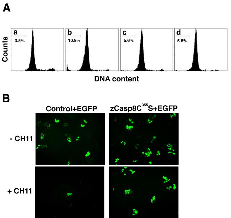
(A) The DNA content of transfectants expressing zebrafish caspase-8 or its mutant was assessed by flow cytometry. Transfectants carrying pME18S (panel a), pME18S-Flag/zCasp8 (panel b), both pME18S-Flag/zCasp8 and pCX-CrmA (panel c) or pME18S-Flag/zCasp8C355S (panel d) were analyzed for the detection of the DNA content stained with PI. Percentage indicates the cellular population detected in the sub-G1 fraction of DNA content. (B) HeLa cells were transfected with pME18S-Flag/zCasp8C355S (right panels) or control vector (left panels) together with pCX-EGFP, cultured for 48 h and treated with (lower panels) or without (upper panels) 100 ng/ml of anti-Fas antibody CH11 and 10 μg/ml of CHX for 6 h. Viable cells were detected as EGFP-positive cells under the fluorescent microscope.

