Abstract
During the last decade, alphaviruses became widely used for expression of heterologous genetic information and development of recombinant vaccines against a variety of human and animal pathogens. In this study, we compared a number of vectors based on the genome of Sindbis (SINV) and Venezuelan equine encephalitis (VEEV) viruses for their ability to express the Rift Valley fever virus (RVFV) envelope glycoprotein Gn and induce a protective immune response against RVFV infection. Our results suggest that i) application of VEEV-based expression systems appears to be advantageous, when compared to similar systems designed on the basis of the SINV genome. ii) Alphavirus-specific E3 and E2 proteins and furin-specific cleavage sites can be used for engineering secreted forms of the proteins. iii) Alphaviruses can be modified for expression of the large fragments of heterologous proteins on the surface of chimeric, infectious viral particles. Thus, alphavirus-based expression systems may have the potential for a broader application beyond their current use as replicons or double-subgenomic vectors.
Keywords: alphaviruses, recombinant vaccines, chimeric viruses
INTRODUCTION
The alphavirus genus in the Togaviridae family is a group of important and widely distributed human and animal pathogens (Griffin, 2001; Strauss and Strauss, 1994). Alphaviruses efficiently replicate in vertebrate hosts, in which they cause an acute infection characterized by high-titer viremia that is required for transmission to mosquito vectors during the blood meal. In mosquitoes, they induce a persistent, life-long infection and accumulate to high titers in salivary glands for transmission to new hosts (Weaver and Barrett, 2004).
Alphavirus virions contain icosahedral nucleocapsid surrounded by a lipid envelope with imbedded glycoprotein spikes. The genome is represented by a single-stranded RNA molecule of positive polarity, of approximately 11.7 kb in length (Kinney et al., 1989; Strauss, Rice, and Strauss, 1984; Takkinen, 1986). It mimics the structure of cellular messenger RNAs, in which it contains a 5′ methylguanylate cap and a 3′ polyadenylate tail. The genome RNA is translated into four nonstructural proteins (nsP1-4) that form, together with cellular proteins, the enzyme complex (RC) required for viral genome replication and transcription of the subgenomic RNA. The latter RNA, synthesized from the subgenomic promoter, codes for structural proteins comprising viral particles.
Since the first infectious cDNA clone of the alphavirus genome was designed (Rice et al., 1987), these viruses became an attractive system for the delivery and expression of heterologous genetic information (Bredenbeek et al., 1993; Liljeström and Garoff, 1991; Pushko et al., 1997). Their application is based on the fact that viral structural genes can be replaced by the genes of interest, and these RNAs (replicons) remain self-replicating and become capable of expressing the heterologous genetic information. Alphavirus replicons can be packaged to high titers into infectious viral particles using so-called helper RNAs (Bredenbeek et al., 1993; Fayzulin et al., 2005; Frolov et al., 1996; Liljeström and Garoff, 1991; Volkova, Gorchakov, and Frolov, 2006), which encode the cis-acting elements required for replication of helper genome and transcription of the subgenomic RNA, translated into the structural proteins. Helpers do not encode the entire set of nsPs (Volkova, Gorchakov, and Frolov, 2006), and may encode no nsPs at all (Liljeström and Garoff, 1991), and they can replicate only in the presence of replicons, which supply the replicative enzymes. Thus, they utilize a replicative machinery, produced in trans by the replicons, and synthesize structural viral proteins that package replicons into infectious viral particles to titers approaching 1010 inf.u/ml. Depending on the design, the helper RNAs are either packaged or not packaged into infectious virions (Fayzulin et al., 2005; Volkova, Gorchakov, and Frolov, 2006). At the next passage, packaged replicons infect the cells and express heterologous protein(s), encoded by the subgenomic RNA, but do not develop spreading infection, because of the inability to produce viral structural proteins. An alternative strategy of alphavirus-dependent gene expression is based on duplication of the subgenomic promoter in the viral genome and using it for expression of an additional gene (Frolov et al., 1993; Hahn et al., 1992). In this case, the RNA genome encodes all of the nonstructural and structural proteins required for virus replication and development of productive infection, and is capable of producing the heterologous protein encoded by the RNA transcribed from an additional subgenomic promoter. These two main strategies are applied in vaccine development and in the expression of genes of interest, both in vivo and in vitro.
In this study, we were interested in testing the above-described replicon expression system and developing additional alphavirus-based systems for expression of heterologous genetic information, induction of the protective immune response against lethal infection caused by Rift Valley fever virus (RVFV) and production of the recombinant viruses at a large scale.
RVFV is a representative member of the Bunyaviridae family (Shope, Peters, and Davies, 1982). This virus is continuously circulating in livestock-raising regions of Africa, and intensive farming has resulted in the emergence of mosquito-borne epidemics (Ksiazek et al., 1989). Death rates are very high for domestic animals, and, in particular, the fatality rate for fetuses in pregnant livestock can approach 100%, while for newborn lambs it is close to 90%. RVFV is spreading to new geographic areas, and the recent outbreaks include an extensive epidemics in Egypt in 1977 (Arthur et al., 1993) and in sub-Saharan Africa in 1997–98, and spread across the Red Sea to Saudi Arabia and Yemen. RVFV has the potential for use in bioterrorism, and its projected effect on U.S. agricultural interests would likely be akin to the economic disruption seen in cases of foot-and-mouth disease virus (FAMDV). However, unlike FAMDV, RVFV can be transmitted by many different species of mosquitoes (Tesh, 1988), and thus might cause outbreaks in the areas where large mosquito populations exist close to farms. RVFV is also an important human pathogen; it causes a self-limited febrile, influenza-like illness that, in ~2–10% cases, results in hemorrhagic fever, liver necrosis, encephalitis, and eye damage. Based on data from epidemics, the estimated fatality rate in humans is close to 1% (Meegan, Hoogstraal, and Moussa, 1979; Meegan, Niklasson, and Bengtsson, 1979).
The RVFV genome contains three, single-stranded RNA segments (L, M and S) (Ihara, Akashi, and Bishop, 1984; Pettersson et al., 1977). The M segment encodes the NSm and viral envelope glycoproteins Gn and Gc that are translated from a single mRNA complementary to the M segment (Collett et al., 1985). The latter RNA appears to employ a traditional strategy of viral RNA templates, in which it encodes polyprotein precursors for two mature structural proteins that are co- and posttranslationally processed. The carboxy terminal parts of NSm and Gn contain signal peptides that likely function in translocation of the following Gn and Gc, respectively, into the ER followed by transport into the Golgi compartment.
Virions of the Bunyaviridae members are formed mainly intracellularly by budding into the Golgi vesicles. However, RVFV was found to bud through the external cellular membrane as well (Anderson and Smith, 1987), which indicates that significant glycoprotein fraction can be present on the external surface of the infected cells. Immunization of mice with recombinant Gc and Gn glycoproteins of RVFV, expressed by baculoviruses or by recombinant vaccinia virus, has been shown to protect the animals from viral disease (Keegan and Collett, 1986; Schmaljohn et al., 1989). Moreover, there are two important neutralizing and protective antigenic epitopes on the Gn glycoprotein of RVFV, and they are conserved among virus strains from all over Africa (Battles and Dalrymple, 1988).
We tested a number of expression systems designed on the basis of Sindbis (SINV) and Venezuelan equine encephalitis virus (VEEV) genomes, and a variety of expression strategies for RVFV Gn protein. The accumulated data suggest that VEEV replicons expressing RVFV glycoproteins and VEE virus with a chimeric envelope elicit the most efficient immune response against RVFV infection, and such strategies might be applicable in expressing the antigens of other viruses, particularly of these having intracellular budding.
RESULTS
SINV replicons expressing RVFV Gn
The initial constructs aimed at expression of RVFV-specific antigen were designed on the basis of the SINV genome (Fig. 1A). The advantage of using SINV-based vectors is in their high safety record, efficient expression of heterologous proteins in vitro and the possibility of passaging of packaged SINV replicons at an escalating scale as a tri-component genome virus (Fayzulin et al., 2005). The SINrep/NSm+Gn+Gc replicon encoded the entire ORF of RVFV M segment (a.a. 1-1197) (Fig. 1B); SINrep/NSm+G2 encoded a.a. 1-674 with the stop codon engineered instead of the Gc-specific signal peptide; SINrep/spGn encoded a.a. 131-674 and the latter included the signal peptide of Gn and the entire sequence of this protein up to the Gc signal peptide. SINrep/spGn(opt) encoded exactly the same protein as did SINrep/spGn, but this was a synthetic gene, in which the codon frequency was the same as in the most efficiently translated human mRNAs (Haas, Park, and Seed, 1996). SINrep/spGn(secr) encoded a.a. 131-581, representing a signal peptide of Gn and the Gn itself having transmembrane and cytosol domains deleted.
FIG. 1.
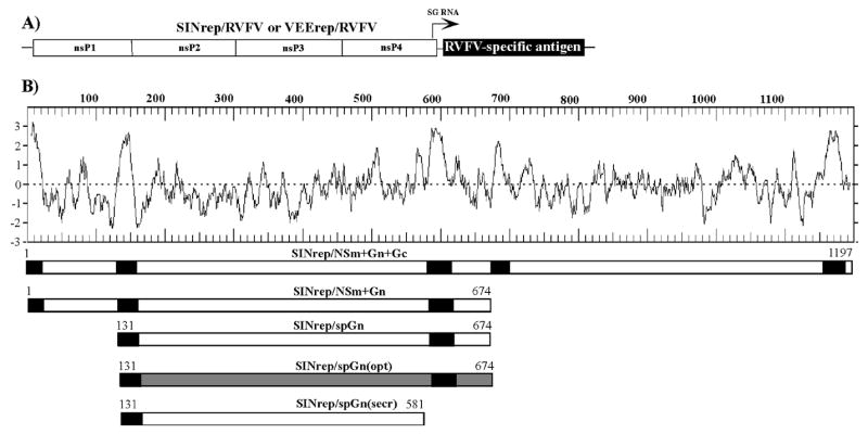
The expression cassettes designed for expression of RVFV-specific antigen from SINV and VEEV replicons. (A) The schematic representation of SINV and VEEV replicons used in the study. The arrow indicates the position of the subgenomic promoter. (B) The hydrophobicity profile (Kyte-Doolitle) of the entire polyprotein encoded by the M segment of RVFV genome, and the fragments cloned into the subgenomic RNA of SINV replicon. The positions of signal peptides and transmembrane domains are indicated by black boxes. The spGn(opt) synthetic gene, encoding exactly the same protein as does spGn, is indicated in grey color. The starting and the last a.a. of each cassette are indicated.
All of the replicons (Fig. 2A) were packaged into viral particles using two helper RNAs encoding either capsid or the glycoproteins that either packaged only replicon genomes (Bredenbeek et al., 1993) or were capable of self-packaging (Fayzulin et al., 2005). Electroporation of the replicons and the latter helpers generated tri-component genome viruses, and such viruses could be passaged in tissue culture at an escalating scale (one of the representative results is shown in Fig. 2B).
FIG. 2.
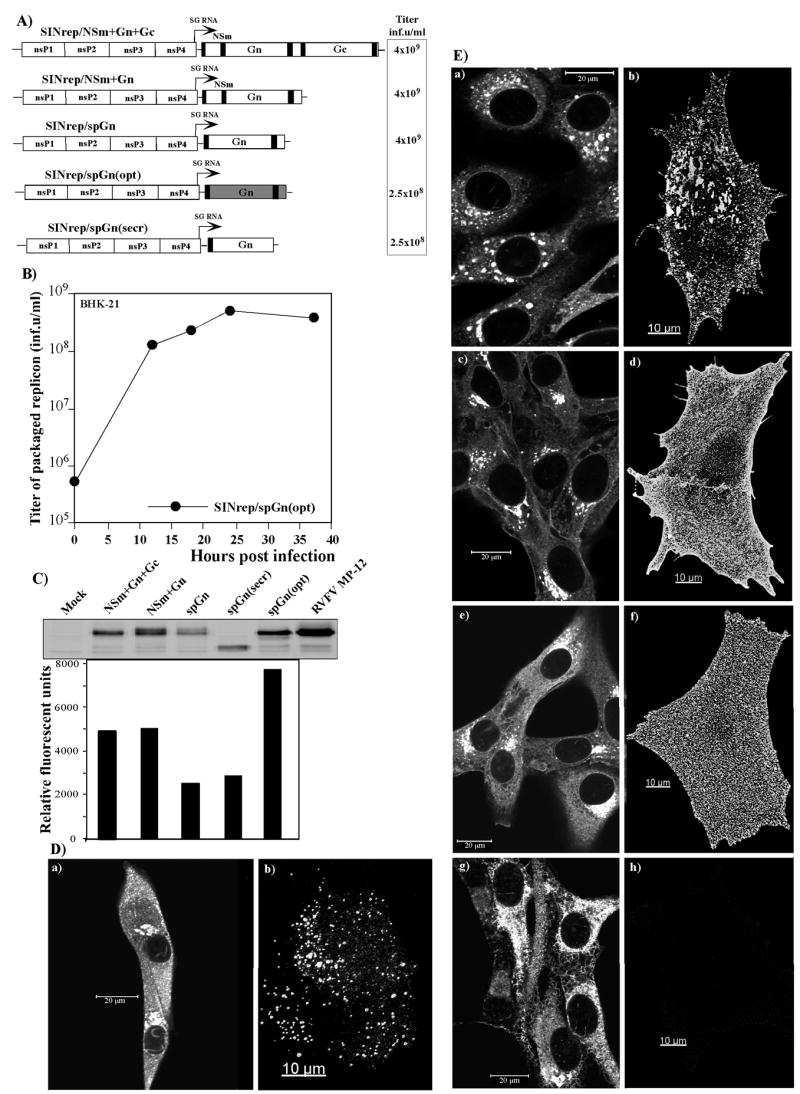
SINV-based replicons used in the study and analysis of the expression of the RVFV-specific glycoproteins by the designed constructs. (A) The schematic representation of the replicons and the titers of the packaged replicons obtained after co-transfection of the replicon and helper RNAs into BHK-21 cells. One of multiple reproducible experiments is presented. The spGn(opt) synthetic gene is indicated in grey color (B) Replication of the tri-component genome virus, having a SINrep/spGn(opt) replicon genome and the genomes of capsid- and glycoprotein-coding helpers, in BHK-21 cells. Cells were infected at an MOI of 8 inf.u/cell, and, at 12 h post infection, the FBS-containing medium was replaced by serum-free VP-SFM medium. Samples of medium were taken at the indicated time points and titers of packaged replicons were determined as described in Materials and Methods. (C) Analysis of RVFV-specific protein expression in cells infected with SINV replicons. BHK-21 cells were infected with packaged replicons at an MOI of 20 inf.u/cells or RVFV MP12 at an MOI of 2.5 PFU/cell and incubated in complete medium at 37°C for 12 h. Equal amounts of cell lysates were analyzed by electrophoresis in SDS-10% polyacryamide gel, followed by Western blotting. Membranes were processed by mouse anti-RVFV antibodies and IRdye 800CW-labeled secondary antibodies. Images were acquired on a Odyssey Infrared Imager (LI-COR). (D) Distribution of virus-specific proteins in the cells infected with RVFV. BHK-21 cells were infected with RVFV MP12 at an MOI of 50 PFU/cell and, at 13 h post infection, stained with mouse anti-RVFV antibodies and AlexaFluor 546-labeled goat anti-mouse secondary antibodies. Panel (a) presents staining of the Triton X-100-permeabilized cells; panel (b) presents surface staining of a non-permeabilized cell. Gn and Gc develop well-distinguishable patches on the cell surface. E) Distribution of the RVFV-specific proteins in the cells infected with packaged SINV replicons that express different fragments of the polyprotein encoded by the M segment of the RVFV genome. Panels (a) and (b) present cells infected with SINrep/NSm+Gn+Gc replicon, panels (c) and (d) present cells infected with SINrep/NSm+Gn replicon, panels (e) and (f) present cells infected with SINrep/spGn, replicon, panels (g) and (h) present cells infected with SINrep/spGn(secr) replicon. Panels (a), (c), (e) and (g) represent images of the cells that were permeabilized with Triton X-100 prior to immunostaining. Panels (b), (d), (f) and (h) present images of the cells stained with antibodies without permeabilization. Images were acquired on a Zeiss LSM510 META confocal microscope using a 63X 1.4NA oil immersion planapochromal lens, as described in Materials and Methods.
The designed constructs were tested for the level of RVFV antigen expression, its distribution in the cellular compartments, and accumulation on the cell surface. All of the SINV replicons efficiently expressed Gn (Fig. 2C). The NSm-containing constructs produced higher levels of Gn than did similar constructs having only the carboxy terminal NSm fragment, representing a signal peptide required for translocation of the Gn protein into the endoplasmic reticulum. However, we increased the level of signal peptide-containing Gn expression by almost 4-fold by using a codon-optimized version of the protein, instead of its natural, RVFV M segment-derived gene (Fig. 2C).
Next, the expression of Gn was further analyzed by staining the cells infected with RVFV MP12 and the packaged replicons with RVFV-specific antibodies. Prior to antibody treatment, cells were either fixed with 1% formaldehyde and permeabelized with Triton X-100 (to monitor the intracellular distribution of Gn), or only fixed (to evaluate the presence of Gn on the cell surface). In the BHK-21 cells infected with attenuated MP12 strain of RVFV, the virus-specific glycoproteins accumulated in the Golgi compartment, and a significant fraction was transported to the cell surface (Fig. 2D). The detection of antigen throughout the cytoplasm and in the nucleus was, most likely, due to N and NSs distribution, because both proteins were recognized by the used mouse antiserum on Western blotting (data not shown).
Fig. 2C presents the results of the analysis of the Gn distribution in the BHK-21 cells infected by different replicons. The Gn, having a transmembrane domain and expressed by SINrep/NSm+Gn+Gc, SINrep/NSm+Gn and SINrep/spGn, accumulated in the Golgi compartment (panels a, c and e), but was also readily detected on the cell surface (panels b, d and f), where it formed discrete subdomains reminiscent of lipid rafts. The size and distribution of these Gn-containing patches differed for cassettes expressing Gn only and Gn+Gc. The latter two proteins were distributed on the cellular membrane in a manner similar to that found in RVFV MP12-infected cells: Gn+Gc formed larger antigen-containing areas (compare Figs. 2D, panel b, and 2E, panel b). The Gn protein expressed by SINrep/spGn(secr), having the signal peptide, but lacking the transmembrane domain, was also efficiently expressed in BHK-21 cells (Fig. 2E panel g). However, in contrast to the transmembrane domain-containing versions, it did not accumulate in the Golgi compartment, but was distributed throughout the cell, most likely in the ER. This truncated protein was not detected at the cell surface (Fig. 2E panel h), and it was not secreted from the cell (data not shown). The replacement of the RVFV-specific signal peptide by a SINV-derived E3 protein that functions as a signal peptide for SINV E2 did not noticeably improve the secretion, and the Gn was not detected in the media either by immunoprecipitation or Western blotting (data not shown).
The Gn-encoding, packaged SINV replicons were tested for their ability to induce a protective immune response in mice. We were interested in identifying a construct that could protect mice with an efficiency comparable to that of live attenuated strain RVFV MP12, that is one of the presently available vaccine candidates. Therefore, mice were immunized once s.c. with 0.5–1×10⁷ inf.u of packaged replicons and 105 PFU of MP12. After 28 days they were challenged i.p. with 105 PFU of RVFV ZH501. All of the MP12-immunized animals survived the ZH501 infection without any sign of the disease; however, none of the packaged SINV replicons protected mice in the challenge experiments. In later experiments, some of the mice were challenged s.c., as described in the Materials and Methods, and were not protected against the disease as well. The PRNT80 titers detected in the immunized animals were always below 1:40. The extended survival time (data not shown) suggested that repeated vaccinations could, probably, increase the efficiency of protection. However, this is likely not an efficient means of increasing the immune response.
Application of VEEV-based replicons for RVFV Gn expression
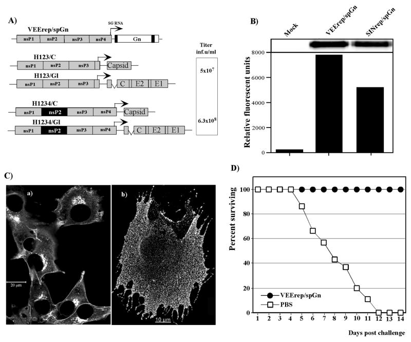
Based on our experience and published data (Perri et al., 2003), VEEV replicons are more immunogenic than are similar SINV constructs. They have a number of advantages that include the capability to more efficiently replicate in mice, large animals, primates and humans (Johnston and Peters, 1996). VEEV-based replicons can be also packaged to high titers using defective helpers and efficiently passaged in tissue culture as a virus with a tri-component genome (Volkova, Gorchakov, and Frolov, 2006). Their passaging in tissue culture at an escalating scale is achieved by using VEEV-specific helpers that strongly differ from those designed for SINV replicons. In order to package not only a replicon, but also itself, VEEV helper RNAs have to express at least nsP1-3 in cis. Thus, to compare VEEV- and SINV-based replicons as expression systems in possible vaccine applications, we designed VEErep/spGn cassette, expressing RVFV Gn, and used two different, VEEV-specific helper systems (Fig. 3A) for its packaging into infectious viral particles. This particular expression cassette was utilized, because the Gn is known to contain neutralizing and protective epitopes (Battles and Dalrymple, 1988), and expression of non-codon-optimized Gn interfered less efficiently with VEEV replicon packaging (data not shown). Helper RNAs encoded either nsP1-3 (H123/C and H123/Gl helpers), or all of the VEEV nonstructural proteins nsP1-4 (H1234/C and H1234/Gl helpers), in which nsP2 contained an attenuating P773→S mutation that had a strong negative effect on RNA and virus replication (Volkova, Gorchakov, and Frolov, 2006). All of these packaging systems required a supply of the replicative enzymes in trans by the VEEV replicons for efficient helper RNA replication and production of the structural proteins. In our previous studies, both pairs of helpers packaged not only replicons, but also their own RNAs and, thus, VEEV replicon and two helper RNAs could be efficiently passaged in the form of a tri-component genome virus (Volkova, Gorchakov, and Frolov, 2006).
FIG. 3.
VEEV replicon and helpers used in the animal studies. (A) The schematic representation of the RVFV Gn-expressing VEEV replicon and two pairs of helpers encoding VEEV capsid and glycoproteins. Representative titers, obtained in one of multiple, reproducible experiments, are indicated. (B) Analysis of RVFVGn expression in cells infected with SINrep/spGn and VEErep/spGn replicons. BHK-21 cells were infected with packaged replicons at an MOI of 20 inf.u/cells and incubated in complete medium at 37°C for 12 h. Equal amounts of cell lysates were analyzed by SDS-10% polyacryamide gel electrophoresis, followed by Western blotting. Membranes were processed by mouse anti-RVFV antibodies and IRdye 800CW-labeled secondary antibodies. Images were acquired on a Odyssey Infrared Imager (LI-COR). (C) Distribution of RVFV Gn in the cells infected with packaged VEErep/spGn replicon. BHK-21 cells were infected with the packaged replicon at an MOI of 50 inf.u/cell and, at 12 h post infection, stained with mouse anti-RVFV antibodies and AlexaFluor 546 goat anti-mouse secondary antibodies. Panel (a) presents staining of the Triton X-100-permeabilized cells; panel (b) presents cell stained with antibodies without permeabilization. Images were acquired on a Zeiss LSM510 META confocal microscope using a 63X 1.4NA oil immersion planapochromal lens, as described in Materials and Methods. (C) Survival of mice immunized with 5×10⁶ inf.u of packaged VEErep/spGn replicon and challenged in 42 days with 5×10³ PFU s.c. of RVFV ZH501. The control group was injected with PBS and challenged with the same dose of RVFV.
VEEV replicon expressing RVFV spGn was packaged into viral particles (Fig. 3A) that were used for analysis of protein expression and immunization of mice. VEErep/spGn expressed Gn noticeably more efficiently than did SINrep/spGn (Fig. 3B), and the protein was localized not only in the ER and Golgi compartment (Fig. 3C, panel a), but was also present in high concentrations on the external membrane (Fig. 3C, panel b) Its distribution was similar to that found in the cells infected with the SINrep/spGn replicon (compare Figs. 2E, panel f, and 3C, panel b). In mice, one immunization with packaged VEErep/spGn did not induce neutralizing antibodies to titers higher than those detected in mice immunized with similar SINV-based replicons (data not shown). The PRNT80 titers were always lower than 1:80. However, even one immunization efficiently protected animals against an ensuing infection with RVFV ZH501, a finding consistent with the idea that VEEV replicons are more immunogenic and, hence, more appropriate for development of recombinant vaccines. However, the negative feature of the VEErep/spGn replicon was in the strong effect of expressed Gn on the assembly and/or release of the viral particles. In contrast to our success with SINV replicons in terms of their passaging in tissue culture, we were incapable of passaging the Gn-expressing VEErep in cell culture, regardless of the helpers used. Even after electroporation, the titers of the infectious particles were always lower than in the experiments with replicons expressing other heterologous genes (data not shown). Thus, the possible problem with the large-scale production of packaged Gn-encoding VEEV replicons should be considered in the future studies.
Expression of RVFV Gn in VEEV structural polyprotein
The above-described experiments suggested a need for additional approaches to enable the expression of RVFV antigens by alphaviruses and the induction of protective immune response against RVFV infection. The rationale was to develop Gn expression systems that would be capable of producing secreted forms of Gn, either presented on the surface of alphavirus particles or secreted from the cells as an individual protein.
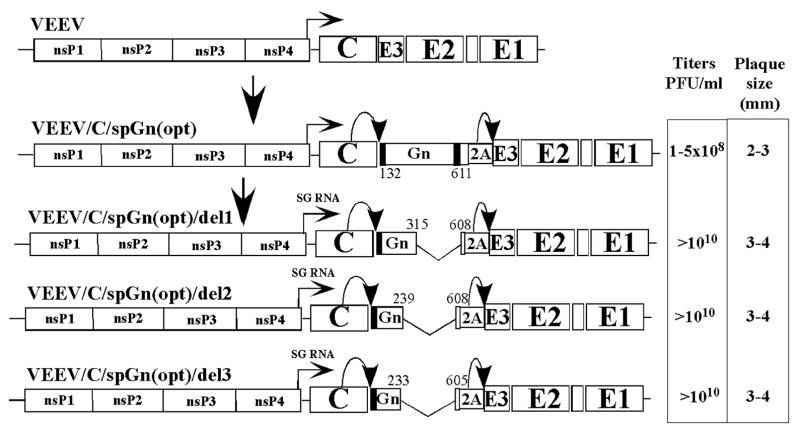
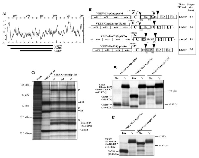
First, we attempted to express a secreted form of RVFV Gn as a part of the alphavirus structural polyprotein. The Gn-coding sequence, containing the signal peptide and the transmembrane domain, was cloned between the VEEV capsid- and E3-coding genes. The FAMDV-derived 2A protease was fused with the Gn cytosol domain to promote the processing of Gn and following E3. VEEV capsid was expected to drive the cleavage of capsid/Gn fusion by its intrinsic protease activity. The recombinant virus was viable and capable of replicating to titers higher than 108 PFU/ml. However, it demonstrated a detectable level of instability and evolution to a large plaque phenotype. Sequencing of the structural genes of the randomly selected, plaque-purified variants revealed extended deletions of the carboxy terminal fragment in the Gn-coding sequence. These variants retained 109, 112 and 188 amino terminal and very few carboxy terminal amino acids of Gn and the 2A protease gene (Fig. 4). Their common feature was a deletion of the transmembrane domain in the Gn, suggesting a negative effect of this sequence on recombinant virus replication. However, these mutants indicated that the selected expression approach might be feasible. Therefore, we designed a set of expression cassettes encoding RVFV Gn that lacked the transmembrane domain (Figs. 5A and B). In the VEEV/C/spGn(opt)/td−, this protein was expected to use its own, the NSm-derived signal peptide for translocation into the ER, and VEEV/Gn318(opt)/fur and VEEV/Gn239(opt)/fur were designed in such a manner as to use VEEV E3 for the same function. In the latter constructs, the VEEV-specific furin cleavage site was generated upstream and downstream from the Gn sequence to promote processing of the polyprotein and secretion of the recombinant Gn. All three viruses were viable (Fig. 5B), and two of them, VEEV/C/spGn(opt)/td− and VEEV/Gn239(opt)/fur, were stable during successive passages. Fig. 5C presents a pattern of protein synthesis in the cell infected by VEEV/C/spGn(opt)/td− and harvested after different passages on BHK-21 cells.
FIG. 4.
The schematic representation of the VEEV genomes encoding the RVFV Gn protein in the structural polyprotein and the genomes of the selected deletion mutants. The positions of found in-frame deletions in the Gn-coding sequence are indicated.
FIG. 5.
The expression cassettes designed for expression of secreted forms of RVFV Gn and analysis of the Gn expression. (A) The hydrophobicity profile (Kyte-Doolitle) of the Gn protein. Fragments cloned into the VEEV structural polyprotein are indicated by black lines. (B) The schematic representation of the recombinant VEEV genomes, containing the insertions of the Gn-encoding sequences in the subgenomic RNA. The numbers correspond to the positions of the first and the last a.a. of the insertion in the polyprotein encoded by the RVFV M segment. The triangles indicate positions of engineered and natural furin-specific cleavage sites. Indicated viral titers and plaque size were derived from one of multiple, highly reproducible experiments. (C) Analysis of the stability of VEEV/C/spGn(opt)td− during serial passaging in tissue culture. Virus was passaged three times in BHK-21 by infecting the monolayer of BHK-21 cells in 100-mm dishes with 100 μl of the stock harvested at the previous passage. For analysis of protein expression, BHK-21 cells were infected with harvested viruses and VEEV TC-83 at an MOI of 20 PFU/cell. At 16 h post infection, cells were labeled with [35S]methionine, as described in Materials and Methods, and equal aliquots of proteins were analyzed by electrophoresis in SDS-10% polyacrylamide gels. The positions of VEEV-specific proteins and Gn-2A are indicated. Asterisks indicate the positions of incompletely processed products. (D) and (E) Analysis of proteins expressed by the recombinant viruses and secreted to the medium. Metabolic labeling of the proteins was performed as described in Materials and Methods. Viral particles (V) were pelleted by ultracentrifugation and the RVFV Gn proteins were isolated by immunoprecipitation. Samples were analyzed by electrophoresis in SDS-10% polyacrylamide gels, followed by autoradiography. The positions of proteins and molecular weight markers are indicated. The molecular weights of the expressed Gn protein fragments and its fusion forms, predicted based on the a.a. sequence, are indicated in the brackets. Gn239 and Gn318 contain one potential glucosylation site, Gn340-2A-E3, and Gn340-E3 have two potential sites.
All three Gn proteins, Gn340, Gn318 and Gn239, were secreted, and only the VEEV E1 and E2 were detected in the viral particles (Fig. 5D), indicating the complete processing of the VEEV structural proteins. However, if the secreted Gn318 and Gn239 corresponded to the expected sizes, the secreted Gn340 had a significantly higher molecular weight than expected, and, on the gel, this protein was seen to run slower than did the Gn340-2A fusion, detected in the protein samples isolated from the infected cells (compare Figs. 5C and D). That difference was unlikely to result from additional glycosylation, because the Gn318 and the Gn340 had the same predicted glycosylation sites. The most plausible explanation for finding the protein of higher molecular weight was that, in its secreted form, the Gn remained fused with E3. Thus, the completely processed Gn340-2A appeared to be retained in the cells, and the Gn340-2A-E3 was, most likely, secreted. To test this hypothesis, we designed a VEEV/C/spGn(opt)E3/td− recombinant virus that differed from the above-described VEEV/C/spGn(opt)/td− by the lack of FAMDV 2A protease. This deletion made processing of Gn340/E3 fusion an impossible event. This virus was also viable, stable, formed large plaques and secreted Gn to the media during replication. The molecular weight of the latter protein was almost the same as that of secreted during VEEV/C/spGn/td- replication (Fig. 5E); however, a change in protein size, caused by 2A sequence deletion, was readily detectable (Figs. 5D and E). The RVFV-specific antibodies precipitated from the media proteins of lower molecular weight than that of the major secreted proteins (Figs. 5D and E). The exact nature of these products remains unclear. They could be either a result of partial degradation of the secreted Gn or incomplete glycosylation of Gn proteins. These additional polypeptides were not further investigated in this study.
Thus, fusion with E3 at least during the early stages of protein transport was required for protein secretion. Taken together, the data indicated that i) alphavirus-derived E3 can function in the transport of heterologous proteins from infected cells; ii) alphavirus spike glycoproteins are required for this efficient transport as well; ii) both carboxy and amino terminal fusions of E3 can be utilized for transport of heterologous polypeptides; iii) the furin-specific cleavage sites can efficiently function in a heterologous context to produce the proteins of interest in an unfused form.
Alphaviruses with chimeric envelopes
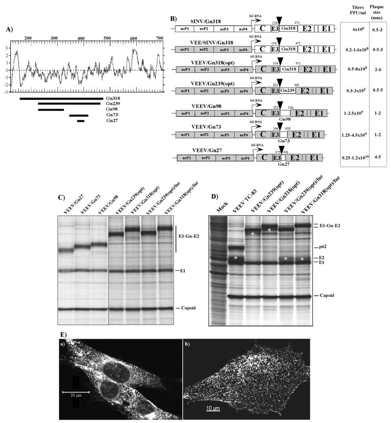
The previously published data strongly indicated that alphavirus E2 glycoprotein could tolerate insertion of extended heterologous protein sequences into the very amino terminal fragment without a deleterious effect on virus replication (Klimstra et al., 2005; London et al., 1992). To test the possibility of designing infectious alphaviruses having chimeric envelopes, which contain large RVFV Gn fragments, we developed a variety of VEEV genome-based constructs with Gn-specific insertions in the E2 gene (Fig. 6A). The selection of these sequences was based on an analysis of Gn’s hydrophobicity profile and the available data about the antigenic structure of the protein (London et al., 1992). These Gn-specific fragments were cloned into the amino terminus of E2 in such a manner as to save a furin-specific cleavage site downstream from E3, and no cleavage sites for proteolytic processing between the Gn sequence and E2 were created. The recombinant, viable viruses were rescued from the cDNA, and it was not surprising that their replication efficiency decreased with the increase in the length of a Gn-specific insertion (Fig. 6B). The peptides’ insertions were readily detectable in the pulse-labeling experiments (Fig. 6C), and, in the pulse-chase experiments, we found a processed Gn/E2 (lacking E3), but no E2 (Fig. 6D). The latter protein was present only in the samples prepared from cells infected with VEEV/Gn239(opt)/fur and VEEV/Gn318(opt)/fur viruses (described in the previous section) encoding the same Gn fragments separated from E2 by engineered furin-specific cleavage sites (see Figs. 5B and 6D). Even the longest peptide, Gn318, was efficiently transported as Gn/E2 chimeric protein to the cell surface (Fig. 6E, panel b) and secreted in the form of infectious viral particles (data not shown) that had E2 replaced by Gn/E2 fusion.
FIG. 6.
The expression cassettes designed for expression of RVFV Gn in viral particles and analysis of the protein expression. (A) The hydrophobicity profile (Kyte-Doolitle) of the Gn protein. Fragments cloned into VEEV structural polyprotein are indicated by black lines. (B) The schematic representation of the recombinant VEEV genomes containing the insertions of the Gn-encoding sequences in the subgenomic RNA. The numbers correspond to the positions of the first and the last a.a. of the insertion in the polyprotein encoded by the RVFV M segment. The triangles indicate positions of engineered furin-specific cleavage sites. VEEV-specific sequences are indicated in grey color. Indicated viral titers and plaque size were derived from multiple experiments. (C and D) The results of metabolic labeling of the proteins expressed by recombinant viruses. The pulse labeling (C) and pulse-chase labeling (D) with [35S]methionine, followed by SDS-10% polyacrylamide gel electrophoresis, were performed at 16 h post infection as described in the Materials and Methods. Final processing products of furin protease are indicated by asterisks. (E) Distribution of Gn-E2 fusion protein in the VEEV/Gn318(opt)-infected cells. Panels (a) represent images of the cells that were permeabilized with Triton X-100 prior to immunostaining with RVFV-specific antibodies. Panels (b) present images of the cells stained with the same antibodies without permeabilization. Images were acquired on a Zeiss LSM510 META confocal microscope using a 63X 1.4NA oil immersion planapochromal lens, as described in Materials and Methods.
Taken together, these data indicated that the application of alphaviruses for expression of RVFV Gn is not limited by using them in a replicon form that encodes heterologous proteins under control of the additional subgenomic promoter. The structural protein-specific signal sequences and the structural proteins themselves can be used for secretion of heterologous proteins in form of infectious viral particles having chimeric envelope.
Protection experiments
Some of the designed recombinant viruses were tested in mice for their ability to induce a protective immune response against RVFV infection. The results of these experiments are presented in Figs. 7A and B. Mice were immunized once or twice with the recombinant viruses that expressed either a secreted form of RVFV Gn (VEEV/Gn239(opt)/fur and VEEV/C/spGn(opt)/td−) or a fusion of E2 with the Gn fragment [VEE/SINV/Gn318 and VEEV/Gn318(opt)]. After immunization, mice were challenged s.c. with 5×10³ PFU of RVFV ZH501 in 42 days (or 21 days, if two immunizations were used). The results of these experiments (Figs. 7A and B) suggested that all of the constructs induced at least partial protection against the challenging virus, and two immunizations were detectably more efficient than a single one. However, the most important result was that replicating viruses with chimeric envelope, having VEEV or SINV E2 fused in the amino terminus with the 318-a.a. long fragment of RVFV Gn, protected mice more efficiently. One immunization with VEEV/Gn318(opt) protected against the dose of ZH501 used, and mice developed no clinical sign of the disease.
FIG. 7.
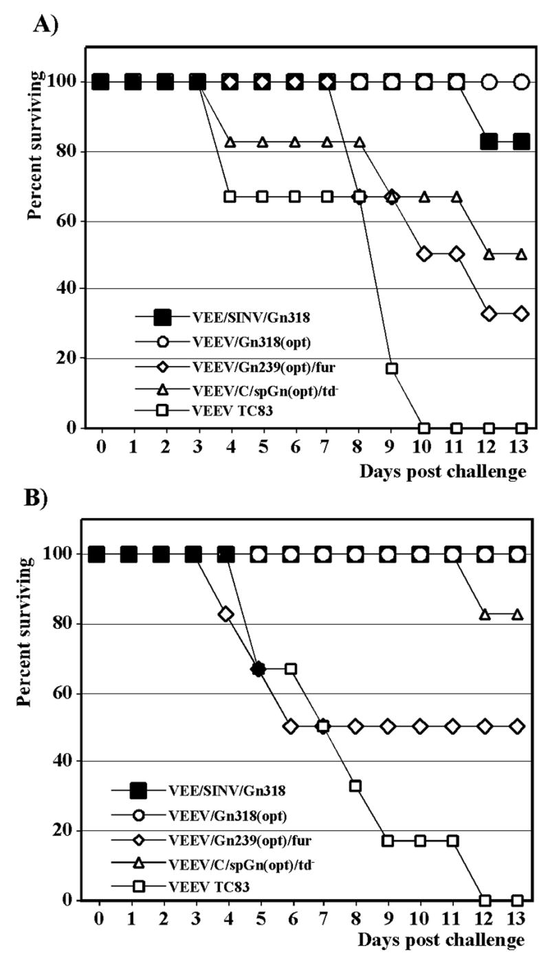
Survival of mice immunized with the indicated recombinant viruses after s.c. challenge with 5×10³ PFU of RVFV ZH501. Mice were immunized either once (A) or twice (B) and challenged as described in the Materials and Methods.
DISCUSSION
Within the last several years, alphavirus expression systems have become widely used for the delivery and expression of heterologous genetic information both in vitro and in vivo. SINV and VEEV expression systems are mainly based on the application of so-called replicons, self-replicating RNAs, in which the structural genes, encoded by the subgenomic RNAs, are replaced by the genes of interest. These recombinant virus-specific RNAs can be packaged into infectious viral particles by the structural proteins, supplied in trans by replication-competent helpers or other expression systems, and infect the cells as do normal, unmodified alphaviruses. However, replicons are not the only means of heterologous gene expression. Alphaviruses have also developed unique mechanisms of structural protein transport and processing. For example, their E3 proteins are highly unusual signal peptides that do not remain in the cell-specific membranes after translocation of the following E2 into the ER, but are rather secreted from the cells in free form or as a component of mature viral particles (Mayne et al., 1984; Strauss and Strauss, 1994). Therefore, in the present study, in addition to evaluating the antigen presentation capacity of the alphavirus replicons, we made an attempt to use VEEV- and SINV-specific intracellular protein transport machinery for delivery of the RVFV envelope glycoprotein Gn outside of the cells for its presentation to the immune system.
Both VEEV- and SINV-based replicons were capable of efficient Gn expression, and surprisingly high concentration of this protein accumulated not only in the ER and trans-Golgi compartment, but was present at the cell surface at a high concentration, regardless of its expression in natural NSm+Gn+Gc cassette or as a single protein having its signal peptide. However, after one immunization, only VEEV-specific, packaged replicons were capable of inducing the immune response that protected mice against the RVFV ZH501 challenge. This can be explained by the higher levels of VEEV RNA replication in vivo and/or the stronger resistance of this virus replication to the autocrine action of IFN-α/β (White et al., 2001). However, VEEV replicons appear to be imperfect in vaccine applications against RVFV infection, because of the strong interference of Gn with VEEV replicon packaging. This makes technically difficult passaging of replicons with Gn-specific insertions for large-scale production of infectious viral particles.
The deletion of the Gn transmembrane domain did not cause the latter protein to be secreted from the cells, and it no longer accumulated in the trans-Golgi compartment, but was mostly present in the ER. The replacement of a Gn signal peptide with E3 did not cause improved secretion of this protein from the cells. In order to be secreted that fusion protein required other VEEV or SINV structural proteins to be involved in transport. Consequently, the truncated Gn was transported outside of the cell when it was fused with the carboxy or amino terminus of E3 and synthesized as an E2-containing polyprotein. Thus, both E3 and E2 proteins were required for efficient intracellular transport and, ultimately, for secretion. This was particularly noticeable in the study of Gn340 synthesis and transport in VEEV/C/spGn(opt)/td−-infected cells, in which the Gn340-2A remained in the cells, and the uncleaved Gn340-2A-E3 (that remained fused with E2 until the processing with furin protease at the very late stages of transport) was secreted (Fig. 5). Thus, the use of alphavirus-specific E3 and E2, and engineering of furin-specific cleavage sites in the carboxy and/or amino termini of the expressed protein provides an efficient means for secretion of the proteins that have a strong potential for the intracellular accumulation. In this study, we succeeded in secretion of large Gn fragments both as free proteins and on the surface of the alphavirus particles. The secreted forms of Gn, expressed by VEEV/Gn239(opt)/fur, VEEV/C/spGn(opt)/td− and VEEV/C/spGn(opt)E3/td− (Fig. 5B) demonstrated poor immunogenicity (Fig. 7 and data not shown). However, the Gn fragments, expressed as Gn/E2 fusions on the cell surface and in the envelope of viral particles, produced by VEE/SINV/Gn318 or VEEV/Gn318(opt) (Figs. 6B and E) during virus replication in vivo, induced a protective immune response against RVFV ZH501 even after a single immunization (Fig. 7). It should be noted that after one immunization, the titers of neutralizing antibodies (PRNT80) did not exceed 1:60 (data not shown). Previous studies of the immune response induced by the nonsecreted VEEV glycoproteins expressed on the cell surface by the recombinant vaccinia virus suggested that protection against very high doses of VEEV TRD, those approaching 108 LD50, could be achieved even when the neutralizing antibodies titers were at very low levels, similar to those detected in the present experiments (Kinney et al., 1988a; Kinney et al., 1988b; Sviatchenko et al., 1993). In the above-described studies, we could not distinguish between the possibilities of whether the RVFV-specific antigen expressed on the surface of the cells during virus replication (Fig. 6E) was sufficient for eliciting an immune response, or the Gn in the viral particles made an additional contribution to protection. However, the accumulated data about the high level of immunogenicity of HBs (Fomsgaard et al., 1998; Woo et al., 2006) or HBc (Fehr et al., 1998; Mihailova et al., 2006) particles, expressing additional antigens, suggest the latter possibility as a possible event.
Taken together, the results of this study indicate that i) application of VEEV-based replicons and other VEEV-based expression systems for induction of protective immune response against RVFV infection appears to be advantageous, when compared to similar systems designed on the basis of the SINV genome. ii) Alphavirus-specific E3 protein, viral structural protein E2 and furin-specific cleavage sites can be used for engineering secreted forms of the proteins that are normally transported to the cell surface and released from the cells very inefficiently. However, as we demonstrated for RVFV Gn, the secretion of these proteins does not necessarily lead to the induction of an efficient immune response. iii) Alphaviruses can be modified for expression of the large fragments of heterologous proteins as fusions with E2 glycoproteins, and such fusions can be used in the context of replicating virus for presentation of the protein both on the cell surface and on the surface of the chimeric, infectious viral particles. Thus, alphavirus-based expression systems may have the potential for a broader application beyond their current use as replicons or double-subgenomic vectors.
MATERIALS AND METHODS
Cell cultures and viruses
BHK-21 cells were kindly provided by Dr. Sondra Schlesinger (Washington University, St. Louis, MO). Cells were maintained at 37°C in alpha minimum essential medium (αMEM) supplemented with 10% fetal bovine serum (FBS) and vitamins. VEEV TC-83 was derived from an infectious cDNA clone of the viral genome. The attenuated strain of RVFV MP12 (Morrill, Mebus, and Peters, 1997) was obtained from Dr. Clarence Peters (UTMB).
Plasmid constructs
The plasmid encoding the entire ORF of RVFV ZH501 M segment was provided by Dr. Connie S. Schmaljohn (USAMRIID). pSINrep/NSm+Gn+Gc encoded SINV replicon (Bredenbeek et al., 1993), cloned under the control of SP6 RNA polymerase promoter and containing the entire ORF of the M segment (a.a. 1-1197) under the control of the subgenomic promoter. pSINrep/NSm+Gn encoded the entire NSm and Gn (a.a. 1-674) of the polyprotein, while pSINrep/spGn encoded the Gn only, including the amino terminal signal peptide. pSINrep/spGn(secr) contained in the subgenomic RNA the ORF that covers a.a. 131-581 of Gn. pSINrep/spGn(opt) encoded the same RVFV-specific protein sequence as did pSINrep/spGn, but the nucleotide sequence was designed based on the codon frequency of human genes that demonstrate a high translation efficiency (Haas, Park, and Seed, 1996). The spGn(opt) ORF was synthesized from the oligonucleotides using the PCR-based technique. A schematic representation of the Gn fragments and replicons is shown in Figs. 1B and 2A, respectively. pVEErep/spGn contained VEEV TC-83-based replicon (Petrakova et al., 2005) having the described above spGn cassette cloned under the control of the subgenomic promoter. Helpers used for SINV replicon packaging are described elsewhere (Fayzulin et al., 2005), and VEEV helpers encoding nsP1-3, H123/C and H123/Gl, were described in our early publication (Volkova, Gorchakov, and Frolov, 2006). VEEV-specific helpers H1234/C and H1234/Gl had a very similar design, in which they encoded the same subgenomic RNAs as did H123/C and H123/Gl. However, in their genomes, all of the nonstructural genes were present and the nsP2 contained an attenuating mutation P773 to S, that strongly reduced their cytopathogenicity (Petrakova et al., 2005). The schematic representation of the VEEV replicon and helper genomes is shown in Fig. 3A. pVEEV/C/spGn(opt) contained a promoter of SP6 RNA polymerase, followed by a cDNA copy of the VEEV TC-83 genome, in which the spGn(opt) (fused with FAMDV 2A protease) cassette was cloned in frame between a capsid-coding sequence and E3. In addition, 9 nucleotides, coding 3 amino terminal amino acids of VEEV E3, were left upstream of the signal peptide-coding sequence of codon-optimized Gn and an extra proline-coding codon was added to the 5′ terminus of the E3 gene. These two modifications were required for polyprotein processing by capsid and 2A proteases. The schematic representation of recombinant viral genome is shown in Fig. 4. pVEEV/C/spGn(opt)/td− had a design similar to that of pVEEV/C/spGn(opt), but in the Gn-coding fragment, a sequence encoding a carboxy terminal part of the protein, including the transmembrane domain, was deleted, and the ectodomain fragment of Gn was fused with 2A protease (Fig. 5). In pVEEV/C/spGn(opt)E3/td−, a 2A protease sequence was deleted, and the Gn ectodomain-coding gene (a.a. 132-471) was fused with E3 (Fig. 5B). pVEEV/Gn318(opt)/fur and pVEEV/Gn239(opt)/fur contained VEEV TC-83 genomes, in which the insertions encoding 318 and 239 a.a. of Gn, respectively, were made between E3 and E2. The Gn-specific sequences did not contain both the signal peptide- and trasmembrane domain-coding fragments. The furin-specific cleavage sites were engineered between VEEV E3 and Gn, and between Gn and VEEV E2. The schematic representation of viral genomes is presented in Fig. 5B. pVEEV/Gn318(opt), VEEV/Gn239(opt), pVEEV/Gn98, pVEEV/Gn73 and pVEEV/Gn27 contained VEEV TC-83 genomes having the insertions of codon-optimized or wt RVFV Gn-coding sequence of different length, fused with the VEEV E2 gene (see Fig. 6B for details). The furin-specific cleavage site sequence was engineered in the junction between VEEV E3- and RVFV Gn-coding genes. pSINV/Gn318 and pVEE/SINV/Gn318 had a design similar to that of pVEEV/Gn318(opt). Insertions of the Gn fragment (having a wt, but not codon-optimized sequence) encoding 318 a.a. of the protein were cloned between E3 and E2 genes into an infectious cDNA clone of the SINV genome (SINV/Gn318) or chimeric VEEV genome, in which the VEEV structural genes were replaced by those of SINV (Garmashova et al., 2007).
All of the described replicons and viruses encoding the RVFV-specific sequences were engineered using standard PCR-based cloning techniques. All of the PCR products were sequenced to avoid any accumulation of spontaneous mutations in the recombinant DNAs. The details of the cloning procedures and sequences of the plasmids can be provided upon request.
RNA transcriptions
Plasmids were purified by centrifugation in CsCl gradients. Before the transcription reaction, SINV genome-based plasmids were linearized using the XhoI restriction site located downstream of the poly(A) sequence. Plasmids containing recombinant VEEV genomes were linearized by NotI or MluI. RNAs were synthesized by SP6 RNA polymerase in the presence of cap analog by previously described conditions (Rice et al., 1987). The yield and integrity of the transcripts were analyzed by gel electrophoresis under non-denaturing conditions. Transcription reactions were used for electroporation without additional purification.
RNA transfections
BHK-21 cells were electroporated by using previously described conditions (Liljeström et al., 1991). Packaged replicons were harvested at 19–30 h post transfection. Titers were determined by infecting BHK-21 cells with serial dilutions of the stocks. After 16 h incubation at 30°C in a CO2 incubator, cells were fixed with 1% formaldehyde, permeabilized with Triton X-100 and stained with RVFV-specific antibodies, obtained from Dr. Robert Tesh (UTMB), and secondary antibodies labeled with AlexaFluor 546. Cell samples electroporated with the in vitro-synthesized viral genomes were tested in the infectious center as previously described (Gorchakov, Frolova, and Frolov, 2005) to assess the accumulation of deletion mutants having higher rates of replication and forming larger plaques. The residual electroporated cells were seeded into 100-mm dishes, and viruses were harvested after development of profound CPE. Virus titers were determined using a standard plaque assay on BHK-21 cells (Lemm et al., 1990).
Analysis of protein synthesis
BHK-21 cells were seeded into six-well Costar plates at a concentration of 5×10⁵ cells/well. After 4 h incubation at 37°C in 5% CO2 they were infected at an MOI of 20 PFU/cell or 20 infectious units (inf.u)/cell by viruses or packaged replicons, respectively, in 200 μl of αMEM supplemented with 1% FBS at 37°C for 1 h with shaking. The medium was then replaced by a corresponding complete medium, and incubation continued at 37°C. At 16 h post infection, the cells were washed three times with phosphate-buffered saline (PBS) and then incubated for 30 minutes at 37°C in 0.8 ml of DMEM lacking methionine, supplemented with 0.1% FBS and 20 μCi/ml of [35S]methionine. After this incubation, cells were scraped into the media, pelleted by centrifugation and dissolved in 300 μl of standard protein gel loading buffer. Equal amounts of proteins were loaded onto sodium dodecyl sulfate-10% polyacrylamide gels. After electrophoresis, gels were dried and autoradiographed. In the pulse-chase protein labeling experiments, the [35S]methionine-containing media were replaced by complete media, and incubation was continued for 2 h in the CO2 incubator at 37°C. Then cells were collected, and proteins were analyzed by electrophoresis in sodium dodecyl sulfate-10% polyacrylamide gels.
Immunoprecipitation experiments
BHK-21 cells were seeded at a concentration of 5 × 10⁵ cells/35-mm dish. After 4 h incubation at 37°C, the subconfluent monolayers that formed were infected with different viruses at an MOI of 20 PFU/cell for 1 h, and incubated in complete medium in a CO2 incubator at 37°C. At 20 h post infection, the cells were washed three times with PBS and then incubated for 6 h at 37°C in 0.8 ml of DMEM lacking methionine, supplemented with 0.1% FBS and 20 μCi/ml of [35S]methionine. By that time, cells did not demonstrate profound CPE, characterized by destruction of the membrane and release of the cytoplasmic content. Harvested media were additionally centrifugated at 13,500 rpm for 5 min at room temperature and used for analysis of both VEEV particles and secreted RVFV-specific proteins. Virions were pelleted by centrifugation through the cushion of 20% sucrose in an MLS-50 rotor in Beckman Optima MAX ultracentrifuge at 48,000 rpm for 1 h at 4°C, then dissolved in standard protein gel loading buffer and analyzed on sodium dodecyl sulfate-10% polyacrylamide gels. The supernatants, harvested after ultracentrifugation, were incubated with mouse anti-RVFV antibodies and protein A, linked to magnetic beads (Miltenyi Biotec). The 35S-labeled RVFV Gn was eluted from the μColumns (Miltenyi Biotec) by SDS-containing protein gel loading buffer and analyzed on sodium dodecyl sulfate-10% polyacrylamide gels.
Microscopy
For microscopy, BHK-21 cells were seeded on the glass chamber slides (Nunc) and infected with recombinant viruses or packaged replicons at an MOI of 50 PFU or inf.u per cell. Then glass chambers were incubated in a CO2 incubator at 37°C for 12 h. Next, they were fixed in 1% formaldehyde in PBS and either directly stained with RVFV-specific mouse antibodies and goat anti-mouse, AlexaFluor 546-labeled secondary antibodies (for surface staining). Alternatively, the same staining was done after their permeabilization with 0.5% Triton X-100 (for staining of the intracellular proteins). Further analysis was performed on a Zeiss LSM510 META confocal microscope using a 63X 1.4NA oil immersion planapochromal lense. For three-dimensional analysis of surface staining, the image stacks were further processed using Huygens Essential v2.7 deconvolution software (Scientific Volume Imaging) and 3D rendering software Imaris v 4.2 (Bitplane AG).
Animals
Balb/c mice, 6-week-old females, were purchased from Harlan and housed for at least 7 days in a specific pathogen-free environment until they were immunized. Immunization studies were carried out under ABSL-2 conditions, and all of challenge studies were conducted under ABSL-4 conditions in the Robert E. Shope, BSL4 Laboratory, UTMB. All of the animal studies were approved by the Institutional Animal Care and Use Committee and carried out according to NIH guidelines.
Immunization and challenge with virulent RVFV
Mice were inoculated on day 0 subcutaneously (s.c.) into the medial thigh with tested viruses or packaged replicons at a dose of 5×10⁶ PFU or inf.u, respectively, in a total volume of 100 μl of PBS. Some animals received an additional booster on day 21, which was performed in the same way as the initial immunization. Mice were challenged either s.c. (5×10³ PFU) or i.p. (1×10⁵ PFU) with virulent RVFV ZH501.
Acknowledgments
We thank Dr. Robert Tesh, UTMB, for supplying RVFV-specific antibodies, and Dr. Connie S. Schmaljohn, USAMRIID, for supplying the plasmid encoding the entire ORF of the M segment of RVFV ZH501. We also thank Mardelle Susman, technical editor, for critical reading and editing of the manuscript. This work was supported by Public Health Service grant AI053135.
Footnotes
Publisher's Disclaimer: This is a PDF file of an unedited manuscript that has been accepted for publication. As a service to our customers we are providing this early version of the manuscript. The manuscript will undergo copyediting, typesetting, and review of the resulting proof before it is published in its final citable form. Please note that during the production process errors may be discovered which could affect the content, and all legal disclaimers that apply to the journal pertain.
References
- Anderson GW, Jr, Smith JF. Immunoelectron microscopy of Rift Valley fever viral morphogenesis in primary rat hepatocytes. Virology. 1987;161(1):91–100. doi: 10.1016/0042-6822(87)90174-7. [DOI] [PubMed] [Google Scholar]
- Arthur RR, el-Sharkawy MS, Cope SE, Botros BA, Oun S, Morrill JC, Shope RE, Hibbs RG, Darwish MA, Imam IZ. Recurrence of Rift Valley fever in Egypt. Lancet. 1993;342(8880):1149–50. doi: 10.1016/0140-6736(93)92128-g. [DOI] [PubMed] [Google Scholar]
- Battles JK, Dalrymple JM. Genetic variation among geographic isolates of Rift Valley fever virus. Am J Trop Med Hyg. 1988;39:617–631. doi: 10.4269/ajtmh.1988.39.617. [DOI] [PubMed] [Google Scholar]
- Bredenbeek PJ, Frolov I, Rice CM, Schlesinger S. Sindbis virus expression vectors: Packaging of RNA replicons by using defective helper RNAs. J Virol. 1993;67:6439–6446. doi: 10.1128/jvi.67.11.6439-6446.1993. [DOI] [PMC free article] [PubMed] [Google Scholar]
- Collett MS, Purchio AF, Keegan K, Frazier S, Hays W, Anderson DK, Parker MD, Schmaljohn C, Schmidt J, Dalrymple JM. Complete nucleotide sequence of the M RNA segment of Rift Valley fever virus. Virol. 1985;144:228–245. doi: 10.1016/0042-6822(85)90320-4. [DOI] [PubMed] [Google Scholar]
- Fayzulin R, Gorchakov R, Petrakova O, Volkova E, Frolov I. Sindbis virus with a tricomponent genome. J Virol. 2005;79(1):637–43. doi: 10.1128/JVI.79.1.637-643.2005. [DOI] [PMC free article] [PubMed] [Google Scholar]
- Fehr T, Skrastina D, Pumpens P, Zinkernagel RM. T cell-independent type I antibody response against B cell epitopes expressed repetitively on recombinant virus particles. Proc Natl Acad Sci U S A. 1998;95(16):9477–81. doi: 10.1073/pnas.95.16.9477. [DOI] [PMC free article] [PubMed] [Google Scholar]
- Fomsgaard A, Nielsen HV, Bryder K, Nielsen C, Machuca R, Bruun L, Hansen J, Buus S. Improved humoral and cellular immune responses against the gp120 V3 loop of HIV-1 following genetic immunization with a chimeric DNA vaccine encoding the V3 inserted into the hepatitis B surface antigen. Scand J Immunol. 1998;47(4):289–95. doi: 10.1046/j.1365-3083.1998.00323.x. [DOI] [PubMed] [Google Scholar]
- Frolov I, Hoffman TA, Prágai BM, Dryga SA, Huang HV, Schlesinger S, Rice CM. Alphavirus-based expression systems: strategies and applications. Proc Natl Acad Sci USA. 1996;93:11371–11377. doi: 10.1073/pnas.93.21.11371. [DOI] [PMC free article] [PubMed] [Google Scholar]
- Frolov IV, Dryga SA, Kolykhalov AA, Mikriukova TP, Agapov EV, Netesov SV. [Recombinant equine Venezuelan encephalomyelitis virus, expressing HBsAg] Dokl Akad Nauk. 1993;332(6):789–91. [PubMed] [Google Scholar]
- Garmashova N, Gorchakov R, Volkova E, Paessler S, Frolova E, Frolov I. The Old World and New World alphaviruses use different virus-specific proteins for induction of transcriptional shutoff. J Virol. 2007;81(5):2472–84. doi: 10.1128/JVI.02073-06. [DOI] [PMC free article] [PubMed] [Google Scholar]
- Gorchakov R, Frolova E, Frolov I. Inhibition of transcription and translation in Sindbis virus-infected cells. J Virol. 2005;79(15):9397–409. doi: 10.1128/JVI.79.15.9397-9409.2005. [DOI] [PMC free article] [PubMed] [Google Scholar]
- Griffin DE. Alphaviruses. In: Knipe DM, Howley PM, editors. “Fields’ Virology. 4. Lippincott, Williams and Wilkins; New York: 2001. pp. 917–962. [Google Scholar]
- Haas J, Park EU, Seed B. Codon usage limitation in the expression of HIV-1 envelope glycoprotein. Current Biology. 1996;6(3):315–324. doi: 10.1016/s0960-9822(02)00482-7. [DOI] [PubMed] [Google Scholar]
- Hahn CS, Hahn YS, Braciale TJ, Rice CM. Infectious Sindbis virus transient expression vectors for studying antigen processing and presentation. Proc Natl Acad Sci USA. 1992;89:2679–2683. doi: 10.1073/pnas.89.7.2679. [DOI] [PMC free article] [PubMed] [Google Scholar]
- Ihara T, Akashi H, Bishop DH. Novel coding strategy (ambisense genomic RNA) revealed by sequence analyses of Punta Toro Phlebovirus S RNA. Virology. 1984;136(2):293–306. doi: 10.1016/0042-6822(84)90166-1. [DOI] [PubMed] [Google Scholar]
- Johnston RE, Peters CJ. Alphaviruses. In: Fields BN, Knipe DM, Howley PM, editors. Virology. 3. Lippincott-Raven; New York: 1996. pp. 843–898. [Google Scholar]
- Keegan K, Collett MS. Use of bacterial expression cloning to define the amino acid sequences of antigenic determinants on the G2 glycoprotein of Rift Valley fever virus. J Virol. 1986;58(2):263–70. doi: 10.1128/jvi.58.2.263-270.1986. [DOI] [PMC free article] [PubMed] [Google Scholar]
- Kinney RM, Esposito JJ, Johnson BJ, Roehrig JT, Mathews JH, Barrett AD, Trent DW. Recombinant vaccinia/Venezuelan equine encephalitis (VEE) virus expresses VEE structural proteins. J Gen Virol. 1988a;69 ( Pt 12):3005–13. doi: 10.1099/0022-1317-69-12-3005. [DOI] [PubMed] [Google Scholar]
- Kinney RM, Esposito JJ, Mathews JH, Johnson BJ, Roehrig JT, Barrett AD, Trent DW. Recombinant vaccinia virus/Venezuelan equine encephalitis (VEE) virus protects mice from peripheral VEE virus challenge. J Virol. 1988b;62(12):4697–702. doi: 10.1128/jvi.62.12.4697-4702.1988. [DOI] [PMC free article] [PubMed] [Google Scholar]
- Kinney RM, Johnson BJB, Welch JB, Tsuchiya KR, Trent DW. The full-length nucleotide sequences of the virulent Trinidad donkey strain of Venezuelan equine encephalitis virus and its attenuated vaccine derivative, strain TC-83. Virology. 1989;170:19–30. doi: 10.1016/0042-6822(89)90347-4. [DOI] [PubMed] [Google Scholar]
- Klimstra WB, Williams JC, Ryman KD, Heidner HW. Targeting Sindbis virus-based vectors to Fc receptor-positive cell types. Virology. 2005;338(1):9–21. doi: 10.1016/j.virol.2005.04.039. [DOI] [PubMed] [Google Scholar]
- Ksiazek TG, Jouan A, Meegan JM, Le Guenno B, Wilson ML, Peters CJ, Digoutte JP, Guillaud M, Merzoug NO, Touray EM. Rift Valley fever among domestic animals in the recent West African outbreak. Res Virol. 1989;140(1):67–77. doi: 10.1016/s0923-2516(89)80086-x. [DOI] [PubMed] [Google Scholar]
- Lemm JA, Durbin RK, Stollar V, Rice CM. Mutations which alter the level or structure of nsP4 can affect the efficiency of Sindbis virus replication in a host-dependent manner. J Virol. 1990;64:3001–3011. doi: 10.1128/jvi.64.6.3001-3011.1990. [DOI] [PMC free article] [PubMed] [Google Scholar]
- Liljeström P, Garoff H. A new generation of animal cell expression vectors based on the Semliki Forest virus replicon. BioTechnology. 1991;9:1356–1361. doi: 10.1038/nbt1291-1356. [DOI] [PubMed] [Google Scholar]
- Liljeström P, Lusa S, Huylebroeck D, Garoff H. In vitro mutagenesis of a full-length cDNA clone of Semliki Forest virus: the small 6,000-molecular-weight membrane protein modulates virus release. J Virol. 1991;65:4107–4113. doi: 10.1128/jvi.65.8.4107-4113.1991. [DOI] [PMC free article] [PubMed] [Google Scholar]
- London SD, Schmaljohn AL, Dalrymple JM, Rice CM. Infectious enveloped RNA virus antigenic chimeras. Proc Natl Acad Sci USA. 1992;89:207–211. doi: 10.1073/pnas.89.1.207. [DOI] [PMC free article] [PubMed] [Google Scholar]
- Mayne JT, Rice CM, Strauss EG, Hunkapiller MW, Strauss JH. Biochemical studies of the maturation of the small Sindbis virus glycoprotein E3. Virology. 1984;134:338–357. doi: 10.1016/0042-6822(84)90302-7. [DOI] [PubMed] [Google Scholar]
- Meegan JM, Hoogstraal H, Moussa MI. An epizootic of Rift Valley fever in Egypt in 1977. Vet Rec. 1979;105(6):124–5. doi: 10.1136/vr.105.6.124. [DOI] [PubMed] [Google Scholar]
- Meegan JM, Niklasson B, Bengtsson E. Spread of Rift Valley fever virus from continental Africa. Lancet. 1979;2(8153):1184–5. doi: 10.1016/s0140-6736(79)92406-1. [DOI] [PubMed] [Google Scholar]
- Mihailova M, Boos M, Petrovskis I, Ose V, Skrastina D, Fiedler M, Sominskaya I, Ross S, Pumpens P, Roggendorf M, Viazov S. Recombinant virus-like particles as a carrier of B- and T-cell epitopes of hepatitis C virus (HCV) Vaccine. 2006;24(20):4369–77. doi: 10.1016/j.vaccine.2006.02.051. [DOI] [PubMed] [Google Scholar]
- Morrill JC, Mebus CA, Peters CJ. Safety and efficacy of a mutagen-attenuated Rift Valley fever virus vaccine in cattle. Am J Vet Res. 1997;58(10):1104–9. [PubMed] [Google Scholar]
- Perri S, Greer CE, Thudium K, Doe B, Legg H, Liu H, Romero RE, Tang Z, Bin Q, Dubensky TW, Jr, Vajdy M, Otten GR, Polo JM. An alphavirus replicon particle chimera derived from venezuelan equine encephalitis and sindbis viruses is a potent gene-based vaccine delivery vector. J Virol. 2003;77(19):10394–403. doi: 10.1128/JVI.77.19.10394-10403.2003. [DOI] [PMC free article] [PubMed] [Google Scholar]
- Petrakova O, Volkova E, Gorchakov R, Paessler S, Kinney RM, Frolov I. Noncytopathic replication of Venezuelan equine encephalitis virus and eastern equine encephalitis virus replicons in Mammalian cells. J Virol. 2005;79(12):7597–608. doi: 10.1128/JVI.79.12.7597-7608.2005. [DOI] [PMC free article] [PubMed] [Google Scholar]
- Pettersson RF, Hewlett MJ, Baltimore D, Coffin JM. The genome of Uukuniemi virus consists of three unique RNA segments. Cell. 1977;11(1):51–63. doi: 10.1016/0092-8674(77)90316-6. [DOI] [PubMed] [Google Scholar]
- Pushko P, Parker M, Ludwig GV, Davis NL, Johnston RE, Smith JF. Replicon-helper systems from attenuated Venezuelan equine encephalitis virus: expression of heterologous genes in vitro and immunization against heterologous pathogens in vivo. Virology. 1997;239(2):389–401. doi: 10.1006/viro.1997.8878. [DOI] [PubMed] [Google Scholar]
- Rice CM, Levis R, Strauss JH, Huang HV. Production of infectious RNA transcripts from Sindbis virus cDNA clones: Mapping of lethal mutations, rescue of a temperature-sensitive marker, and in vitro mutagenesis to generate defined mutants. J Virol. 1987;61(12):3809–3819. doi: 10.1128/jvi.61.12.3809-3819.1987. [DOI] [PMC free article] [PubMed] [Google Scholar]
- Schmaljohn CS, Parker MD, Ennis WH, Dalrymple JM, Collett MS, Suzich JA, Schmaljohn AL. Baculovirus expression of the M genome segment of Rift Valley fever virus and examination of antigenic and immunogenic properties of the expressed proteins. Virology. 1989;170(1):184–92. doi: 10.1016/0042-6822(89)90365-6. [DOI] [PubMed] [Google Scholar]
- Shope RE, Peters CJ, Davies FG. The spread of Rift Valley fever and approaches to its control. Bull World Health Organ. 1982;60(3):299–304. [PMC free article] [PubMed] [Google Scholar]
- Strauss EG, Rice CM, Strauss JH. Complete nucleotide sequence of the genomic RNA of Sindbis virus. Virology. 1984;133:92–110. doi: 10.1016/0042-6822(84)90428-8. [DOI] [PubMed] [Google Scholar]
- Strauss JH, Strauss EG. The alphaviruses: gene expression, replication, evolution. Microbiol Rev. 1994;58:491–562. doi: 10.1128/mr.58.3.491-562.1994. [DOI] [PMC free article] [PubMed] [Google Scholar]
- Sviatchenko VA, Agapov EV, Urmanov I, Serpinskii OI, Frolov IV, Kolykhalov AA, Ryzhikov AB, Netesov SV. [The immunogenic properties of a recombinant vaccinia virus with an incorporated DNA copy of the 26S RNA of the Venezuelan equine encephalomyelitis virus] Vopr Virusol. 1993;38(5):222–6. [PubMed] [Google Scholar]
- Takkinen K. Complete nucleotide sequence of the non-structural protein genes of Semliki Forest virus. Nucleic Acids Res. 1986;14:5667–5682. doi: 10.1093/nar/14.14.5667. [DOI] [PMC free article] [PubMed] [Google Scholar]
- Tesh RB. The genus Phlebovirus and its vectors. Annu Rev Entomol. 1988;33:169–81. doi: 10.1146/annurev.en.33.010188.001125. [DOI] [PubMed] [Google Scholar]
- Volkova E, Gorchakov R, Frolov I. The efficient packaging of Venezuelan equine encephalitis virus-specific RNAs into viral particles is determined by nsP1-3 synthesis. Virology. 2006;344(2):315–27. doi: 10.1016/j.virol.2005.09.010. [DOI] [PMC free article] [PubMed] [Google Scholar]
- Weaver SC, Barrett AD. Transmission cycles, host range, evolution and emergence of arboviral disease. Nat Rev Microbiol. 2004;2(10):789–801. doi: 10.1038/nrmicro1006. [DOI] [PMC free article] [PubMed] [Google Scholar]
- White LJ, Wang JG, Davis NL, Johnston RE. Role of Alpha/Beta Interferon in Venezuelan Equine Encephalitis Virus Pathogenesis: Effect of an Attenuating Mutation in the 5′ Untranslated Region. J Virol. 2001;75(8):3706–18. doi: 10.1128/JVI.75.8.3706-3718.2001. [DOI] [PMC free article] [PubMed] [Google Scholar]
- Woo WP, Doan T, Herd KA, Netter HJ, Tindle RW. Hepatitis B surface antigen vector delivers protective cytotoxic T-lymphocyte responses to disease-relevant foreign epitopes. J Virol. 2006;80(8):3975–84. doi: 10.1128/JVI.80.8.3975-3984.2006. [DOI] [PMC free article] [PubMed] [Google Scholar]


