

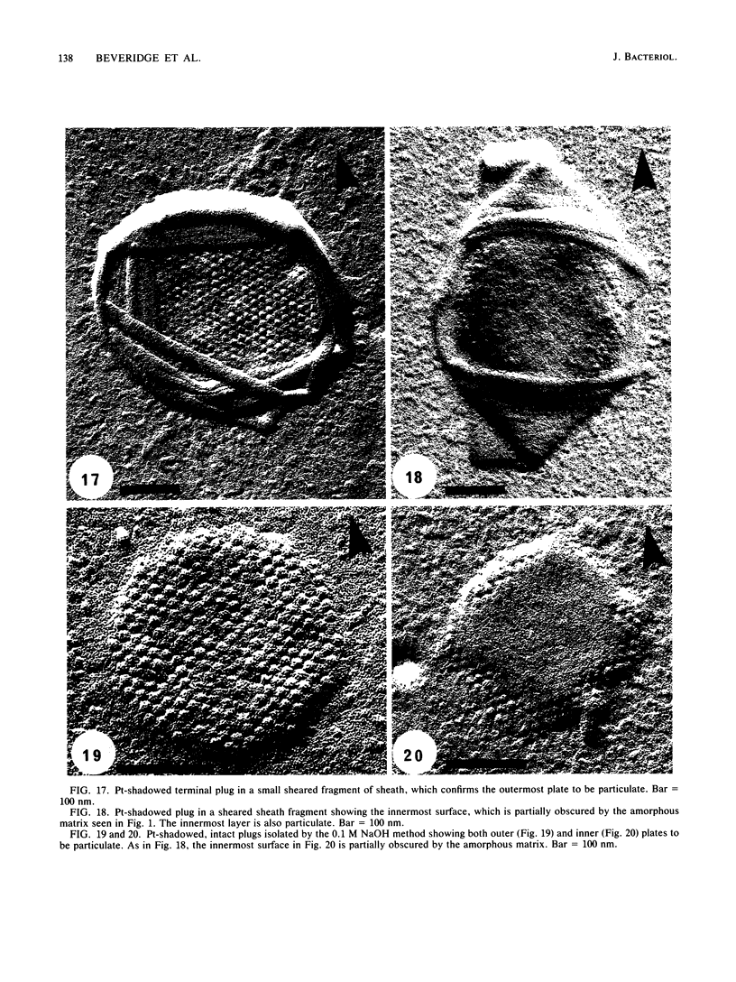
Abstract
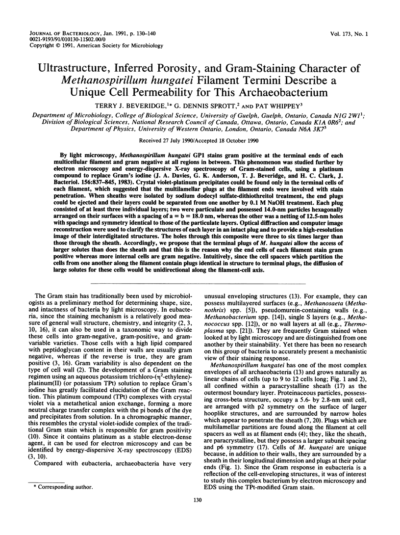
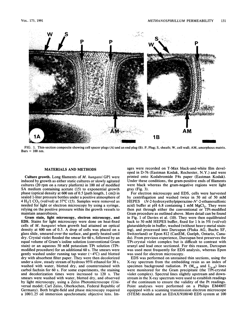
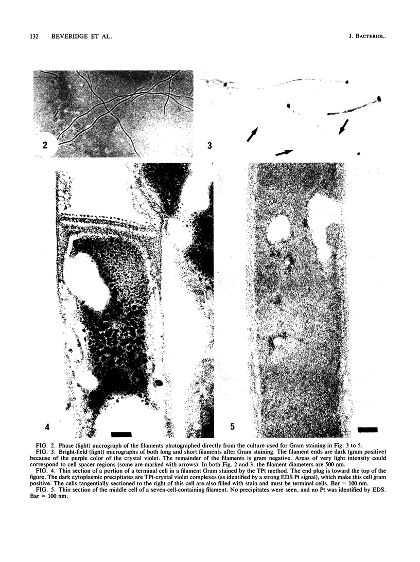
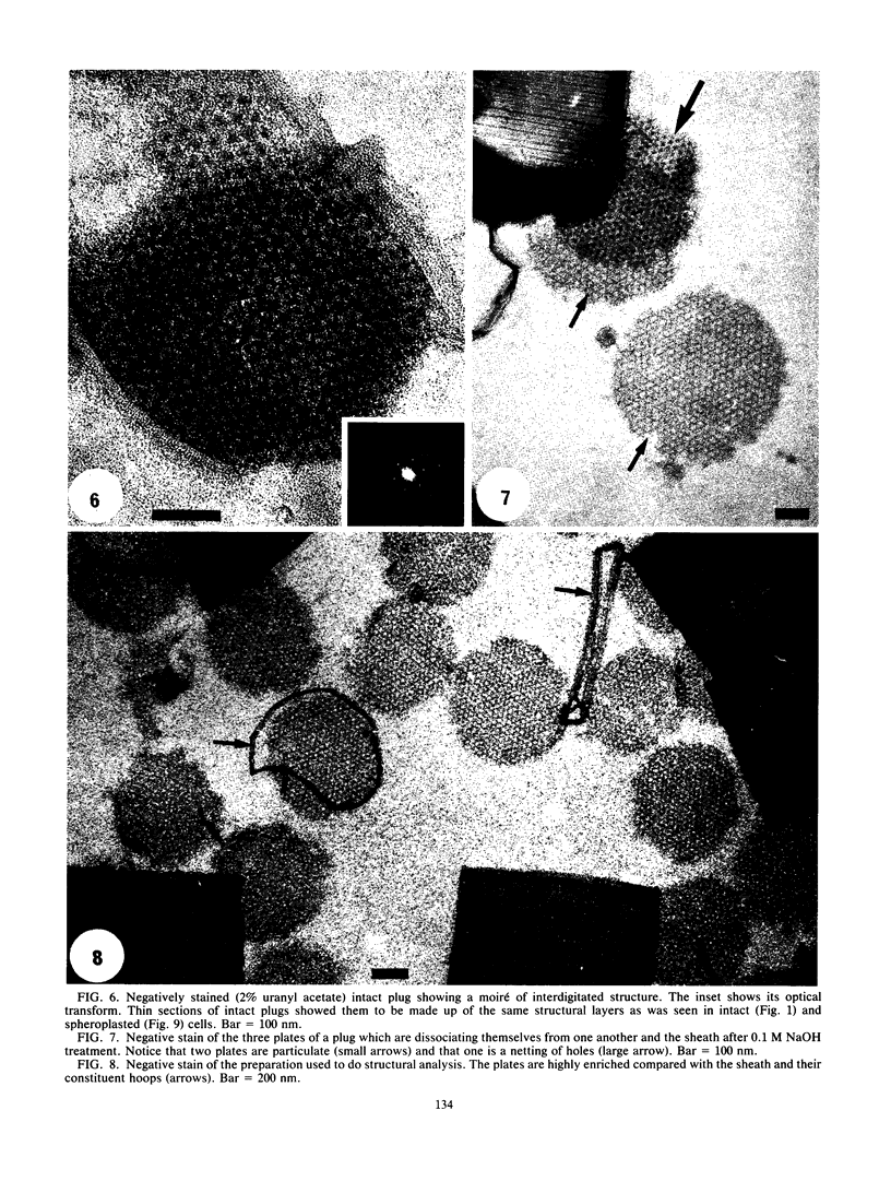
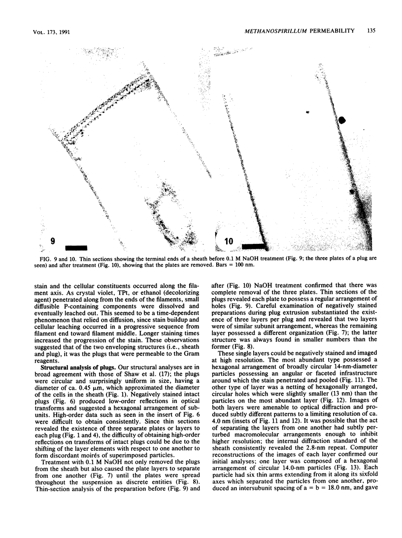
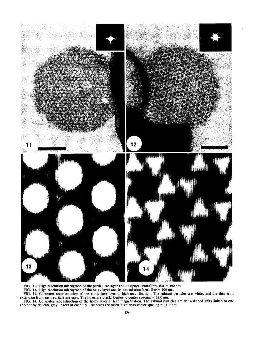
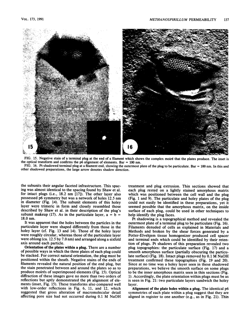
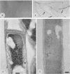
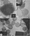
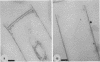
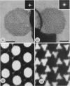
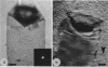
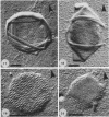
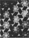
By light microscopy, Methanospirillum hungatei GP1 stains gram positive at the terminal ends of each multicellular filament and gram negative at all regions in between. This phenomenon was studied further by electron microscopy and energy-dispersive X-ray spectroscopy of Gram-stained cells, using a platinum compound to replace Gram's iodine (J. A. Davies, G. K. Anderson, T. J. Beveridge, and H. C. Clark, J. Bacteriol. 156:837-845, 1983). Crystal violet-platinum precipitates could be found only in the terminal cells of each filament, which suggested that the multilamellar plugs at the filament ends were involved with stain penetration. When sheaths were isolated by sodium dodecyl sulfate-dithiothreitol treatment, the end plugs could be ejected and their layers could be separated from one another by 0.1 M NaOH treatment. Each plug consisted of at least three individual layers; two were particulate and possessed 14.0-nm particles hexagonally arranged on their surfaces with a spacing of a = b = 18.0 nm, whereas the other was a netting of 12.5-nm holes with spacings and symmetry identical to those of the particulate layers. Optical diffraction and computer image reconstruction were used to clarify the structures of each layer in an intact plug and to provide a high-resolution image of their interdigitated structures. The holes through this composite were three to six times larger than those through the sheath. Accordingly, we propose that the terminal plugs of M. hungatei allow the access of larger solutes than does the sheath and that this is the reason why the end cells of each filament stain gram positive whereas more internal cells are gram negative. Intuitively, since the cell spacers which partition the cells from one another along the filament contain plugs identical in structure to terminal plugs, the diffusion of large solutes for these cells would be unidirectional along the filament-cell axis.
Full text
PDF
Images in this article
Selected References
These references are in PubMed. This may not be the complete list of references from this article.
- Beveridge T. J., Davies J. A. Cellular responses of Bacillus subtilis and Escherichia coli to the Gram stain. J Bacteriol. 1983 Nov;156(2):846–858. doi: 10.1128/jb.156.2.846-858.1983. [DOI] [PMC free article] [PubMed] [Google Scholar]
- Beveridge T. J. Mechanism of gram variability in select bacteria. J Bacteriol. 1990 Mar;172(3):1609–1620. doi: 10.1128/jb.172.3.1609-1620.1990. [DOI] [PMC free article] [PubMed] [Google Scholar]
- Beveridge T. J., Southam G., Jericho M. H., Blackford B. L. High-resolution topography of the S-layer sheath of the archaebacterium Methanospirillum hungatei provided by scanning tunneling microscopy. J Bacteriol. 1990 Nov;172(11):6589–6595. doi: 10.1128/jb.172.11.6589-6595.1990. [DOI] [PMC free article] [PubMed] [Google Scholar]
- Beveridge T. J., Stewart M., Doyle R. J., Sprott G. D. Unusual stability of the Methanospirillum hungatei sheath. J Bacteriol. 1985 May;162(2):728–737. doi: 10.1128/jb.162.2.728-737.1985. [DOI] [PMC free article] [PubMed] [Google Scholar]
- Beveridge T. J. The bacterial surface: general considerations towards design and function. Can J Microbiol. 1988 Apr;34(4):363–372. doi: 10.1139/m88-067. [DOI] [PubMed] [Google Scholar]
- Davies J. A., Anderson G. K., Beveridge T. J., Clark H. C. Chemical mechanism of the Gram stain and synthesis of a new electron-opaque marker for electron microscopy which replaces the iodine mordant of the stain. J Bacteriol. 1983 Nov;156(2):837–845. doi: 10.1128/jb.156.2.837-845.1983. [DOI] [PMC free article] [PubMed] [Google Scholar]
- Helenius A., McCaslin D. R., Fries E., Tanford C. Properties of detergents. Methods Enzymol. 1979;56:734–749. doi: 10.1016/0076-6879(79)56066-2. [DOI] [PubMed] [Google Scholar]
- Jarrell K. F., Koval S. F. Ultrastructure and biochemistry of Methanococcus voltae. Crit Rev Microbiol. 1989;17(1):53–87. doi: 10.3109/10408418909105722. [DOI] [PubMed] [Google Scholar]
- SALTON M. R. The relationship between the nature of the cell wall and the Gram stain. J Gen Microbiol. 1963 Feb;30:223–235. doi: 10.1099/00221287-30-2-223. [DOI] [PubMed] [Google Scholar]
- Shaw P. J., Hills G. J., Henwood J. A., Harris J. E., Archer D. B. Three-dimensional architecture of the cell sheath and septa of Methanospirillum hungatei. J Bacteriol. 1985 Feb;161(2):750–757. doi: 10.1128/jb.161.2.750-757.1985. [DOI] [PMC free article] [PubMed] [Google Scholar]
- Southam G., Kalmokoff M. L., Jarrell K. F., Koval S. F., Beveridge T. J. Isolation, characterization, and cellular insertion of the flagella from two strains of the archaebacterium Methanospirillum hungatei. J Bacteriol. 1990 Jun;172(6):3221–3228. doi: 10.1128/jb.172.6.3221-3228.1990. [DOI] [PMC free article] [PubMed] [Google Scholar]
- Sprott G. D., Shaw K. M., Jarrell K. F. Isolation and chemical composition of the cytoplasmic membrane of the archaebacterium Methanospirillum hungatei. J Biol Chem. 1983 Mar 25;258(6):4026–4031. [PubMed] [Google Scholar]
- Stewart M., Beveridge T. J., Sprott G. D. Crystalline order to high resolution in the sheath of Methanospirillum hungatei: a cross-beta structure. J Mol Biol. 1985 Jun 5;183(3):509–515. doi: 10.1016/0022-2836(85)90019-1. [DOI] [PubMed] [Google Scholar]
- Yang L. L., Haug A. Purification and partial characterization of a procaryotic glycoprotein from the plasma membrane of Thermoplasma acidophilum. Biochim Biophys Acta. 1979 Sep 21;556(2):265–277. doi: 10.1016/0005-2736(79)90047-6. [DOI] [PubMed] [Google Scholar]