

Abstract
Connective tissue cells were cultured from buffy coat after contamination had been reduced to an absolute minimum by cannulating a large vessel and using special diffusion chambers.
The cells were identified by the presence of connective tissue in the diffusion chambers and not by cell morphology.
There can be few explanations for their presence. One is that they represent cells of low differentiation with a multipotentiality which have passed into the circulating blood from the vessel wall or some other source. Another is that they are transformed leucocytes.
The implications of their presence for wound healing, the pathogenesis of atherosclerosis and organisation of thrombi is indicated.
Full text
PDF
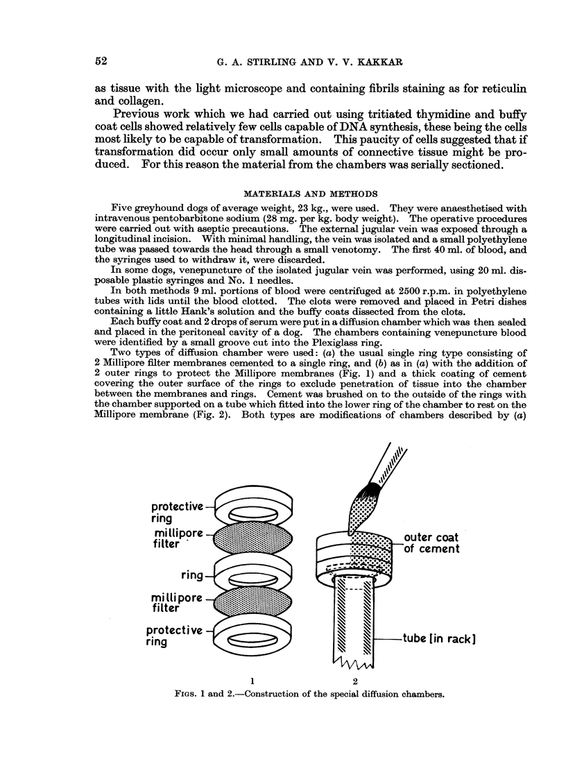
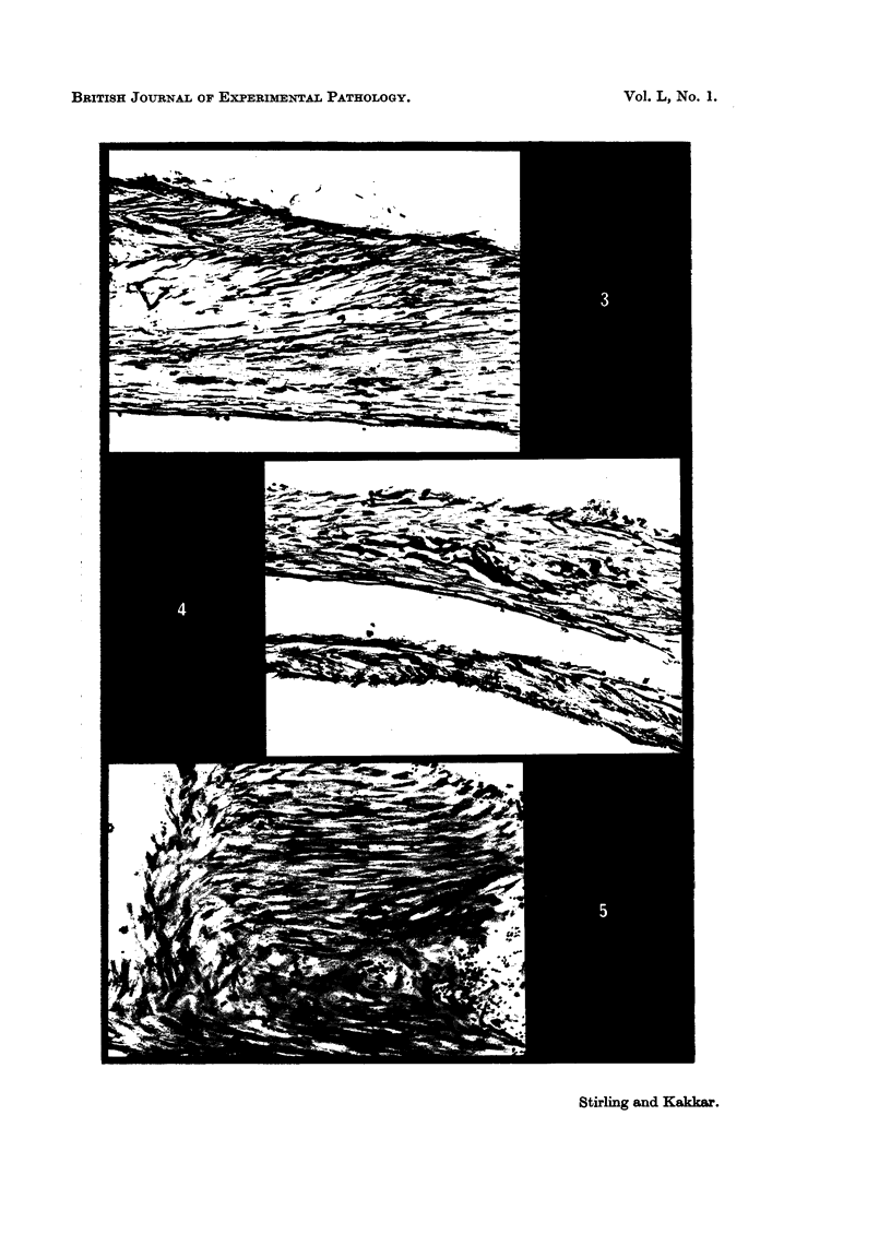

Images in this article


Selected References
These references are in PubMed. This may not be the complete list of references from this article.
- ALGIRE G. H., WEAVER J. M., PREHN R. T. Growth of cells in vivo in diffusion chambers. I. Survival of homografts in immunized mice. J Natl Cancer Inst. 1954 Dec;15(3):493–507. [PubMed] [Google Scholar]
- HULLIGER L. Uber die unterschiedlichen Entwicklungsfähigkeiten der Zellen des Blutes und der Lymphe in vitro. Virchows Arch Pathol Anat Physiol Klin Med. 1956;329(3):289–318. doi: 10.1007/BF00955132. [DOI] [PubMed] [Google Scholar]
- LENDRUM A. C., FRASER D. S., SLIDDERS W., HENDERSON R. Studies on the character and staining of fibrin. J Clin Pathol. 1962 Sep;15:401–413. doi: 10.1136/jcp.15.5.401. [DOI] [PMC free article] [PubMed] [Google Scholar]
- PAUL J. Establishment of permanent cell strains from human adult peripheral blood. Nature. 1958 Sep 20;182(4638):808–808. doi: 10.1038/182808a0. [DOI] [PubMed] [Google Scholar]
- PETRAKIS N. L., DAVIS M., LUCIA S. P. The in vivo differentiation of human leukocytes into histiocytes, fibroblasts and fat cells in subcutaneous diffusion chambers. Blood. 1961 Jan;17:109–118. [PubMed] [Google Scholar]
- Rangan S. R. Origin of the fibroblastic growths in chicken buffy coat macrophage cultures. Exp Cell Res. 1967 Jun;46(3):477–487. doi: 10.1016/0014-4827(67)90374-6. [DOI] [PubMed] [Google Scholar]
- Ross R., Lillywhite J. W. The fate of buffy coat cells grown in subcutaneously implanted diffusion chambers. A light and electron microscopic study. Lab Invest. 1965 Sep;14(9):1568–1585. [PubMed] [Google Scholar]
- SANFORD K. K., LIKELY G. D., BRYAN W. R., EARLE W. R. The infection of cells in tissue culture with Rous sarcoma virus. J Natl Cancer Inst. 1952 Jun;12(6):1317–1343. [PubMed] [Google Scholar]