

Abstract
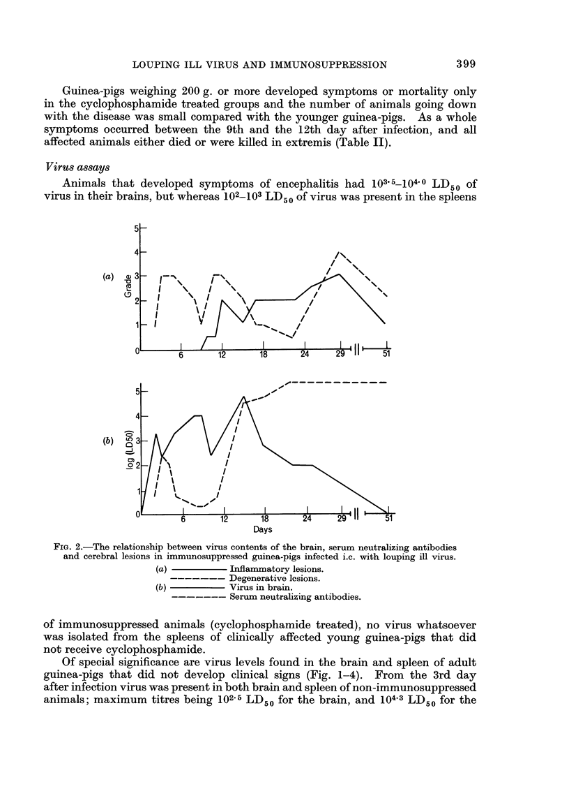
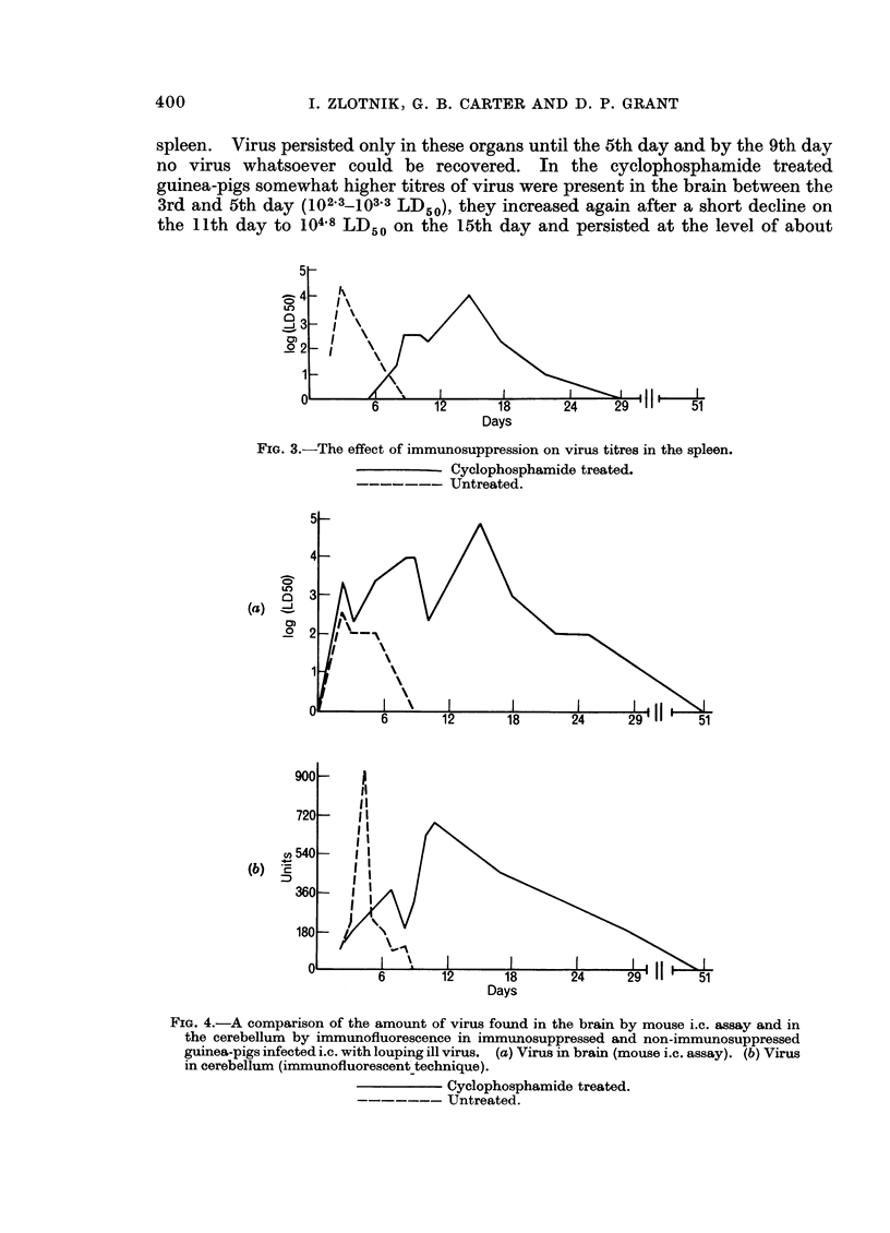
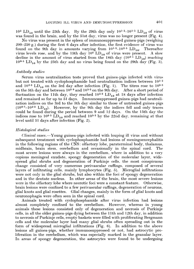
Louping ill virus proved to be pathogenic for young guinea-pigs, about 20 days old weighing about 150-180 g. In older guinea-pigs there was inapparent infection with virus replication in the brain and spleen, inflammatory lesions of encephalitis but without clinical signs or mortality. In non-immunosuppressed guinea-pigs virus replication was noted in the brain and spleen only during the first 5 to 7 days after infection. Serum antibodies appeared on the 3rd day, increased quickly and reached the maximum after 9 days, remaining at the same level for up to the 51st day. In immunosuppressed animals replication of virus in the brain was similar during the first 7 days to that of untreated guinea-pigs, but thereafter, it increased considerably reaching 105 LD50 of virus by the 15th day and later, although in diminished quantities, persisted for at least 29 days after infection. Replication of virus was not demonstrated in the spleen of immunosuppressed animals during the first 7 days, but virus appeared on the 9th day, attained a peak by the 15th day; after a slow decline it could still be detected by the 25th day. Serum neutralizing antibodies after a very short appearance between the 3rd and 5th day, declined quickly and disappeared almost completely until the 12th day. From the 15th day the amount of antibody increased reaching the top level by the 22nd day, and remained so at least until the 51st day after infection.
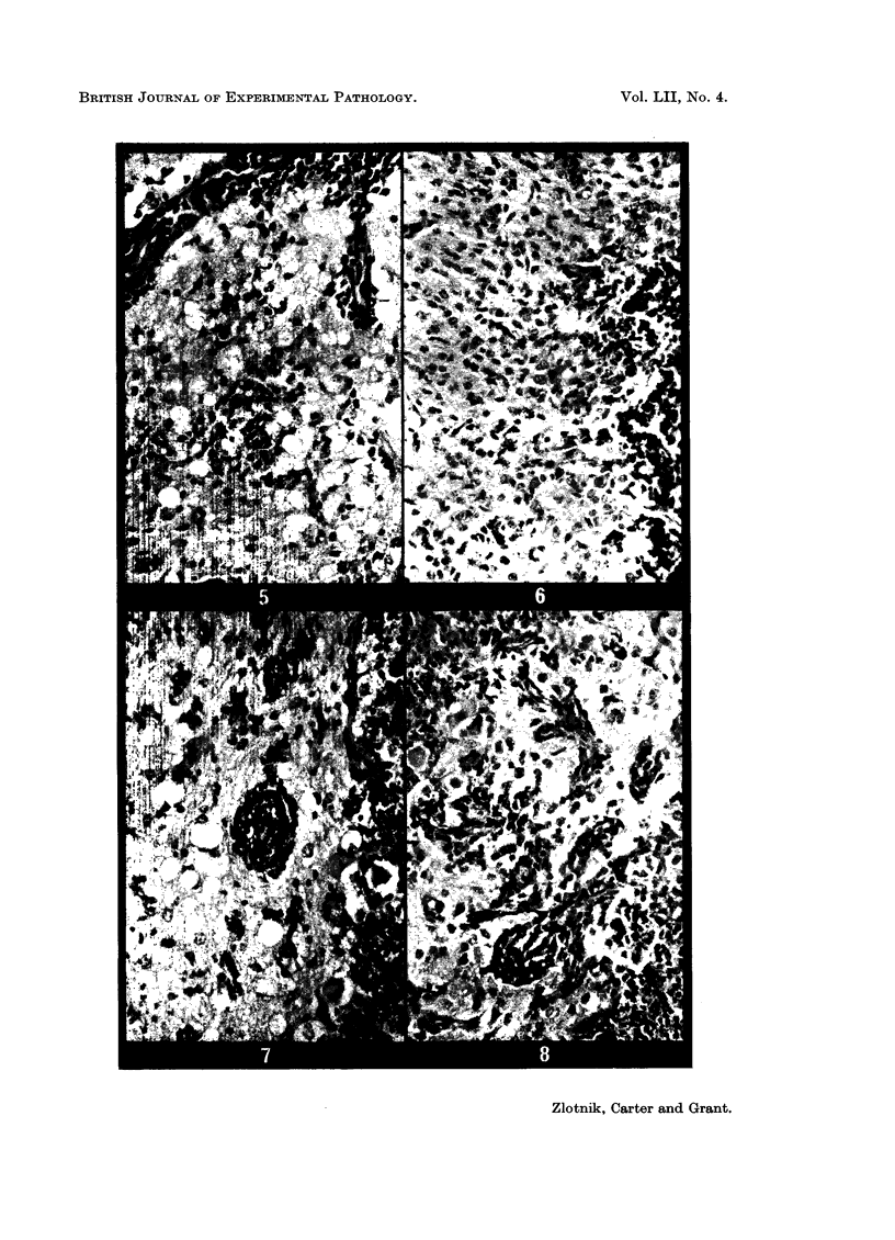
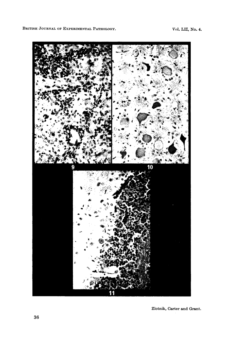
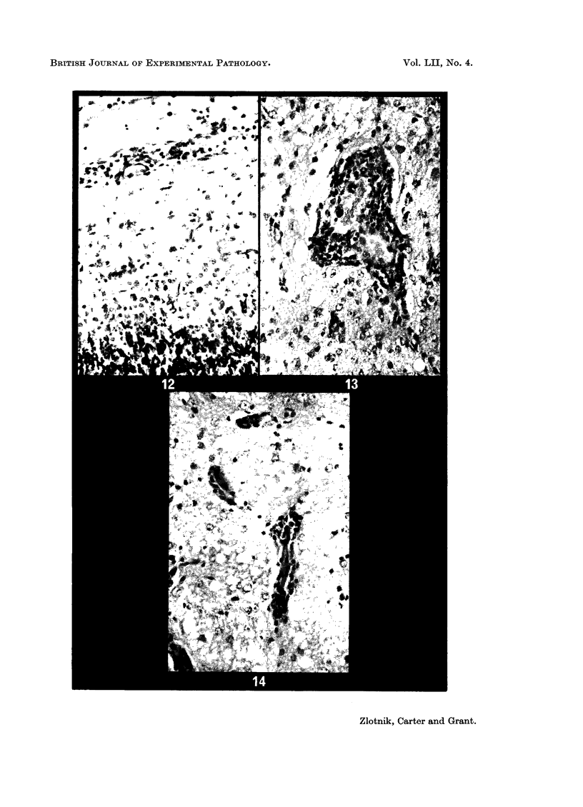
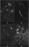
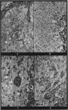
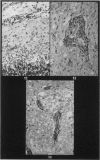
Inflammatory and degenerative brain lesions were noted in both immunosuppressed and non-immunosuppressed animals, but whereas the curve representing inflammatory lesions followed the pattern of serum antibodies, the degenerative changes appeared to follow the pattern of virus replication. While inflammatory changes were present in non-immunosuppressed animals beginning from the 3rd day, in immunosuppressed guinea-pigs no inflammatory lesions were detected until about 12-15 days after infection. The absence of inflammatory lesions at the beginning of infection in immunosuppressed animals, coincided with the critical decline in the presence of serum antibodies. Although lesions were present in most parts of the brain the most severe and constant lesions were found invariably in the cerebellum.
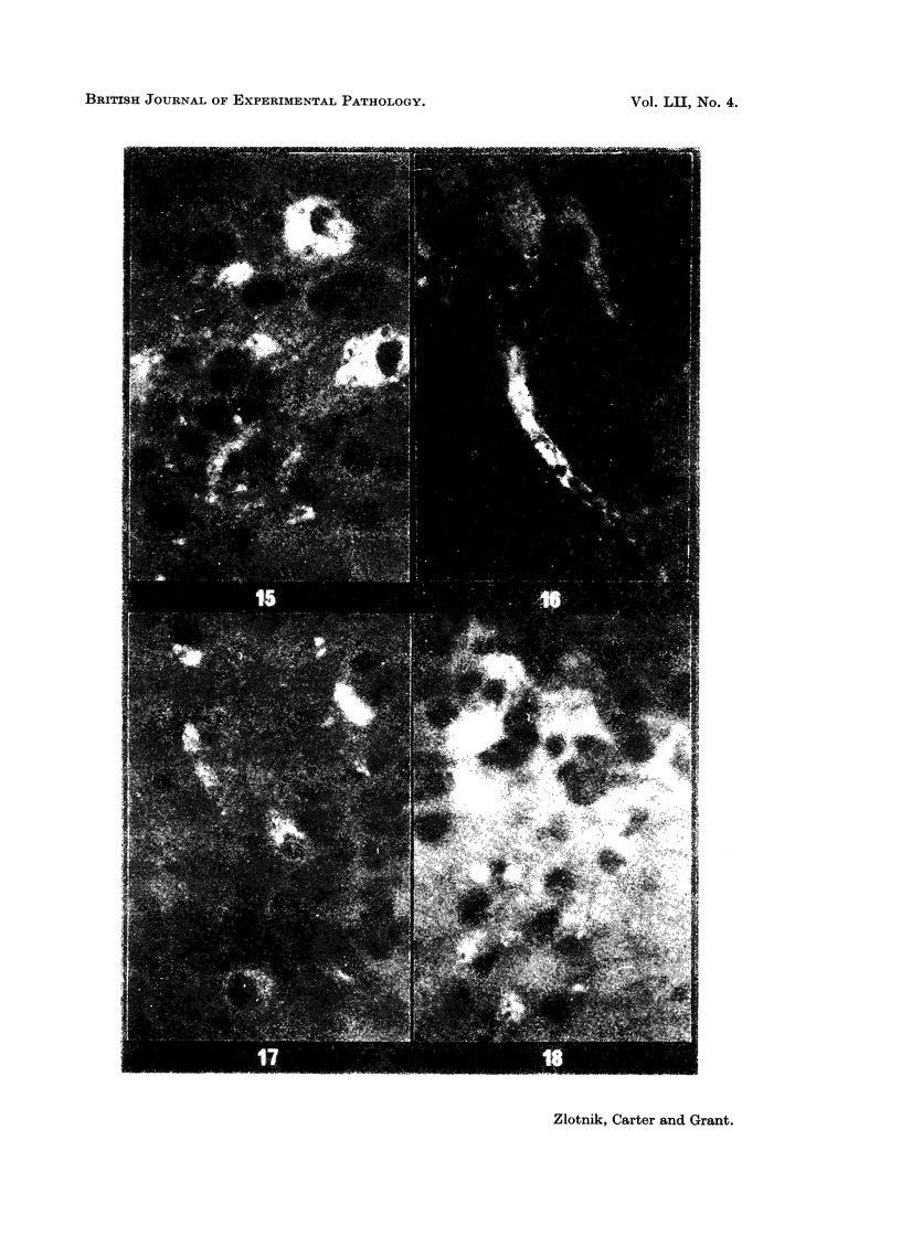
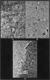
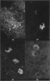
The successful tracing of louping ill virus by immunofluorescence proved that this method is highly sensitive and that it could be adapted for reasonably accurate quantitation of virus.
Full text
PDF
Images in this article
Selected References
These references are in PubMed. This may not be the complete list of references from this article.
- Carter G. B. The rapid titration of Semliki forest virus in cell monolayers by immunofluorescence. J Gen Virol. 1969 Jan;4(1):139–143. doi: 10.1099/0022-1317-4-1-139. [DOI] [PubMed] [Google Scholar]
- Cole G. A., Nathanson N. Potentiation of experimental arbovirus encephalitis by immunosuppressive doses of cyclophosphamide. Nature. 1968 Oct 26;220(5165):399–401. doi: 10.1038/220399a0. [DOI] [PubMed] [Google Scholar]
- HITCHINGS G. H., ELION G. B. Chemical suppression of the immune response. Pharmacol Rev. 1963 Jun;15:365–405. [PubMed] [Google Scholar]
- Nathanson N., Cole G. A. Fatal Japanese encephalitis virus infection in immunosuppresed spider monkeys. Clin Exp Immunol. 1970 Jan;6(1):161–166. [PMC free article] [PubMed] [Google Scholar]
- Paterson P. Y., Hanson M. A. Cyclophosphamide inhibition of experimental allergic encephalomyelitis and cellular transfer of the disease in Lewis rats. J Immunol. 1969 Dec;103(6):1311–1316. [PubMed] [Google Scholar]
- Reid H. W., Doherty P. C. Experimental louping-ill in sheep and lambs. I. Viraemia and the antibody response. J Comp Pathol. 1971 Apr;81(2):291–298. doi: 10.1016/0021-9975(71)90103-4. [DOI] [PubMed] [Google Scholar]
- Robinson T. W., Cureton R. J., Heath R. B. The effect of cyclophosphamide on Sendai virus infection of mice. J Med Microbiol. 1969 May;2(2):137–145. doi: 10.1099/00222615-2-2-137. [DOI] [PubMed] [Google Scholar]
- Thind I. S., Price W. H. The effect of cyclophosphamide treatment on experimental arbovirus infections. Am J Epidemiol. 1969 Jul;90(1):62–68. doi: 10.1093/oxfordjournals.aje.a121050. [DOI] [PubMed] [Google Scholar]
- Webb H. E., Wight D. G., Platt G. S., Smith C. E. Langat virus encephalitis in mice. I. The effect of the administration of specific antiserum. J Hyg (Lond) 1968 Sep;66(3):343–354. doi: 10.1017/s0022172400041218. [DOI] [PMC free article] [PubMed] [Google Scholar]
- Zlotnik I., Keppie J., Grant D. P. A method of testing the efficacy of louping-ill vaccines in sheep. Vet Rec. 1970 May 30;86(22):659–660. doi: 10.1136/vr.86.22.659. [DOI] [PubMed] [Google Scholar]
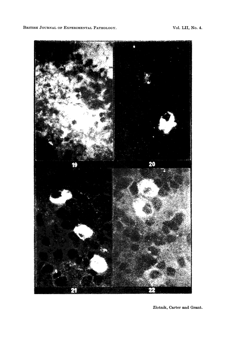
- Zlotnik I., Smith C. E., Grant D. P., Peacock S. The effect of immunosuppression on viral encephalitis, with special reference to cyclophosphamide. Br J Exp Pathol. 1970 Aug;51(4):434–439. [PMC free article] [PubMed] [Google Scholar]