Abstract
A group of guinea-pigs and another of rats were successively placed for 400 hours in an atmosphere containing a high concentration of North-west Cape (NWC) crocidolite fibres. A further group of guinea-pigs inhaled Transvaal (TVL) crocidolite for an equal time.
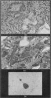
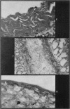
Guinea-pigs that inhaled NWC crocidolite were substantially outlived by animals of the other 2 groups, of which the longest survivors were 2 guinea-pigs. No mesotheliomata were found but in all the lungs cellular proliferation of the septa caused a reduction in alveolar space. Giant cells were common and asbestos bodies developed in guinea-pigs, but in rats only a very few, atypical asbestos bodies were seen, and giant cells were rare. All lungs contained uncoated fibres, long fibres (occasionally exceeding 50 μm) being seen in subpleural regions in older animals and in peribronchiolar accumulations. NWC fibres reached the muscular coats of larger bronchioles of guinea-pigs sooner than TVL crocidolite. Proliferation of bronchiolar epithelium was produced in guinea-pigs and 2 rats by NWC crocidolite, but not by TVL crocidolite. Guinea-pigs dying over 3 months and rats over 18 months after inhalation of NWC crocidolite had several small emphysematous regions at the lung periphery, but only one small area was seen in each of the last 2 guinea-pigs that inhaled TVL fibres. Thus, NWC crocidolite produced greater disruption of the respiratory surfaces.
Severe lung fibrosis was not seen, although fine collagen strands were found in interalveolar septa of animals dying at over 5 months after dusting began. Dense subpleural areas of collagen produced by TVL crocidolite were, however, found in the 4 oldest guinea-pigs.
Inhalation of these 2 crocidolites results in different pathological responses, although no difference in distribution in the lung were found to explain differences in survival time, bronchiolar epithelial proliferation or subpleural collagen formation.
Full text
PDF


Images in this article
Selected References
These references are in PubMed. This may not be the complete list of references from this article.
- Botham S. K., Holt P. F. Development of asbestos bodies on amosite, chrysotile and crocidolite fibres in guinea-pig lungs. J Pathol. 1971 Nov;105(3):159–167. doi: 10.1002/path.1711050303. [DOI] [PubMed] [Google Scholar]
- HOLT P. F., MILLS J., YOUNG D. K. THE EARLY EFFECTS OF CHRYSOTILE ASBESTOS DUST ON THE RAT LUNG. J Pathol Bacteriol. 1964 Jan;87:15–23. doi: 10.1002/path.1700870103. [DOI] [PubMed] [Google Scholar]
- Henderson W. J., Harse J., Griffiths K. A replication technique for the identification of asbestos fibres in mesotheliomas. Eur J Cancer. 1969 Dec;5(6):621–624. doi: 10.1016/0014-2964(69)90011-5. [DOI] [PubMed] [Google Scholar]
- Holmes A., Morgan A., Sandalls J. Determination of iron, chromium, cobalt, nickel, and scandium in asbestos by neutron activation analysis. Am Ind Hyg Assoc J. 1971 May;32(5):281–286. doi: 10.1080/0002889718506461. [DOI] [PubMed] [Google Scholar]
- Holmes S. Developments in dust sampling and counting techniques in the asbestos industry. Ann N Y Acad Sci. 1965 Dec 31;132(1):288–297. doi: 10.1111/j.1749-6632.1965.tb41109.x. [DOI] [PubMed] [Google Scholar]
- Roberts G. H. The pathology of parietal pleural plaques. J Clin Pathol. 1971 May;24(4):348–353. doi: 10.1136/jcp.24.4.348. [DOI] [PMC free article] [PubMed] [Google Scholar]
- Roe F. J. Asbestos as a carcinogenic hazard. Food Cosmet Toxicol. 1968 Dec;6(5):565–566. doi: 10.1016/0015-6264(68)90289-7. [DOI] [PubMed] [Google Scholar]
- Roe F. J., Carter R. L., Walters M. A., Harington J. S. The pathological effects of subcutaneous injections of asbestos fibres in mice: migration of fibres to submesothelial tissues and induction of mesotheliomata. Int J Cancer. 1967 Nov 15;2(6):628–638. doi: 10.1002/ijc.2910020624. [DOI] [PubMed] [Google Scholar]
- Timbrell V., Griffiths D. M., Pooley F. D. Possible biological importance of fibre diameters of South African amphiboles. Nature. 1971 Jul 2;232(5305):55–56. doi: 10.1038/232055a0. [DOI] [PubMed] [Google Scholar]
- WAGNER J. C. Experimental production of mesothelial tumours of the pleura by implantation of dusts in laboratory animals. Nature. 1962 Oct 13;196:180–181. doi: 10.1038/196180a0. [DOI] [PubMed] [Google Scholar]
- WAGNER J. C., SLEGGS C. A., MARCHAND P. Diffuse pleural mesothelioma and asbestos exposure in the North Western Cape Province. Br J Ind Med. 1960 Oct;17:260–271. doi: 10.1136/oem.17.4.260. [DOI] [PMC free article] [PubMed] [Google Scholar]
- Wagner J. C., Berry G. Mesotheliomas in rats following inoculation with asbestos. Br J Cancer. 1969 Sep;23(3):567–581. doi: 10.1038/bjc.1969.70. [DOI] [PMC free article] [PubMed] [Google Scholar]
- Webster I. Mesotheliomatous tumors in South Africa: pathology and experimental pathology. Ann N Y Acad Sci. 1965 Dec 31;132(1):623–646. doi: 10.1111/j.1749-6632.1965.tb41142.x. [DOI] [PubMed] [Google Scholar]


