

Abstract
Viable mesenchymal cells were isolated from pronase digests of mouse liver. The majority of these cells have the ultrastructural features of macrophages, while the remainder probably include cells of the perisinusoidal type. Both protocollagen proline hydroxylase and the substrate for the enzyme have been demonstrated in these cell preparations. The substrate is increased in cells derived from animals with acute carbon tetrachloride liver injury.
Full text
PDF


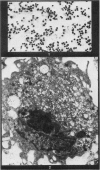
Images in this article


Selected References
These references are in PubMed. This may not be the complete list of references from this article.
- GARVEY J. S. Separation and in vitro culture of cells from liver tissue. Nature. 1961 Sep 2;191:972–974. doi: 10.1038/191972a0. [DOI] [PubMed] [Google Scholar]
- Heppleston A. G., Styles J. A. Activity of a macrophage factor in collagen formation by silica. Nature. 1967 Apr 29;214(5087):521–522. doi: 10.1038/214521a0. [DOI] [PubMed] [Google Scholar]
- ITO T., NEMOTO M. Uber die Kupfferschen Sternzellen und die Fettspeicherungszellen (fat storing cells) in der Blutkapillarenwand der memschlichen Leber. Okajimas Folia Anat Jpn. 1952 Oct;24(4):243–258. doi: 10.2535/ofaj1936.24.4_243. [DOI] [PubMed] [Google Scholar]
- McGee J. O., Patrick R. S. The role of perisinusoidal cells in hepatic fibrogenesis. An electron microscopic study of acute carbon tetrachloride liver injury. Lab Invest. 1972 Apr;26(4):429–440. [PubMed] [Google Scholar]
- McGee J. O., Patrick R. S. The synthesis of sulphated mucopolysaccharide in mouse liver following carbon tetrachloride injury. I. Autoradiographic studies. Br J Exp Pathol. 1969 Dec;50(6):521–526. [PMC free article] [PubMed] [Google Scholar]
- Mills D. M., Zucker-Franklin D. Electron microscopic study of isolated Kupffer cells. Am J Pathol. 1969 Feb;54(2):147–166. [PMC free article] [PubMed] [Google Scholar]
- Nicolescu P., Rouiller C. BEZIEHUNGEN ZWISCHEN DEN Endothelzellen der Lebersinusoide und den von Kupfferschen Sternzellen. Elektronenmikroskopische Untersuchung. Z Zellforsch Mikrosk Anat. 1967;76(3):313–338. [PubMed] [Google Scholar]
- Ohuchi K., Tsurufuji S. Protocollagen proline hydroxylase in isolated rat liver cells. Biochim Biophys Acta. 1972 Mar 8;258(3):731–740. doi: 10.1016/0005-2744(72)90174-x. [DOI] [PubMed] [Google Scholar]
- PETERKOFSKY B., UDENFRIEND S. CONVERSION OF PROLINE TO COLLAGEN HYDROXYPROLINE IN A CELL-FREE SYSTEM FROM CHICK EMBRYO. J Biol Chem. 1963 Dec;238:3966–3977. [PubMed] [Google Scholar]
- PETRAKIS N. L. In vivo cultivation of leukocytes in diffusion chambers: requirement of ascorbic acid for differentiation of mononuclear leukocytes to fibroblasts. Blood. 1961 Sep;18:310–316. [PubMed] [Google Scholar]
- Patrick R. S., McGee J. O. The utilisation of proline by the sinusoidal cells of mouse liver damaged by hepatotoxic agents. J Pathol Bacteriol. 1967 Jan;93(1):309–315. doi: 10.1002/path.1700930129. [DOI] [PubMed] [Google Scholar]
- Pisano J. C., Filkins J. P., Di Luzio N. R. Phagocytic and metabolic activities of isolated rat Kupffer cells. Proc Soc Exp Biol Med. 1968 Jul;128(3):917–922. doi: 10.3181/00379727-128-33157. [DOI] [PubMed] [Google Scholar]
- ROSS R., BENDITT E. P. Wound healing and collagen formation. I. Sequential changes in components of guinea pig skin wounds observed in the electron microscope. J Biophys Biochem Cytol. 1961 Dec;11:677–700. doi: 10.1083/jcb.11.3.677. [DOI] [PMC free article] [PubMed] [Google Scholar]
- Roser B. The distribution of intravenously injected Kupffer cellsin the mouse. J Reticuloendothel Soc. 1968 Oct;5(5):455–471. [PubMed] [Google Scholar]
- ST. GEORGE S., FRIEDMAN M., BYERS S. O. Mass separation of reticuloendothelial and parenchymal cells of rat's liver. Science. 1954 Sep 17;120(3116):463–465. doi: 10.1126/science.120.3116.463. [DOI] [PubMed] [Google Scholar]
- Schnack H., Stockinger L., Wewalka F. Adventitious connective tissue cells in the space of Disse and their relation to fibre formation. Rev Int Hepatol. 1967;17(8):855–860. [PubMed] [Google Scholar]
- Widmann J. J., Cotran R. S., Fahimi H. D. Mononuclear phagocytes (Kupffer cells) and endothelial cells. Identification of two functional cell types in rat liver sinusoids by endogenous peroxidase activity. J Cell Biol. 1972 Jan;52(1):159–170. doi: 10.1083/jcb.52.1.159. [DOI] [PMC free article] [PubMed] [Google Scholar]