

Abstract
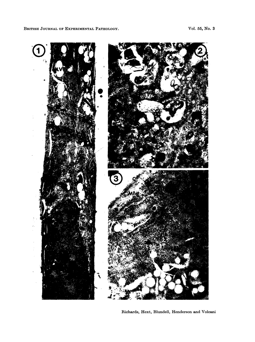
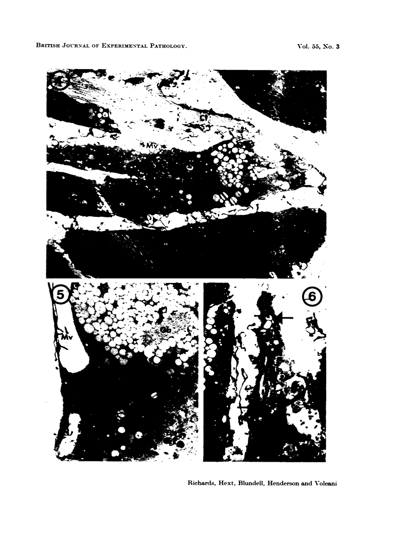
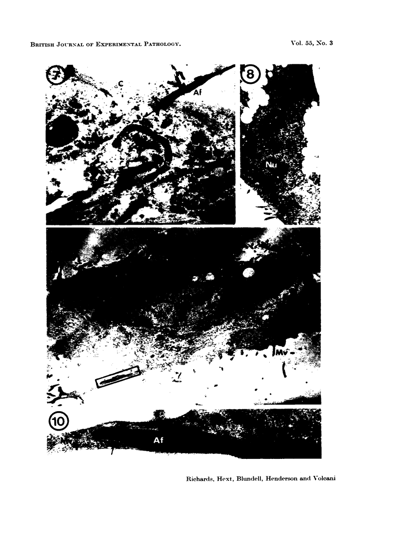
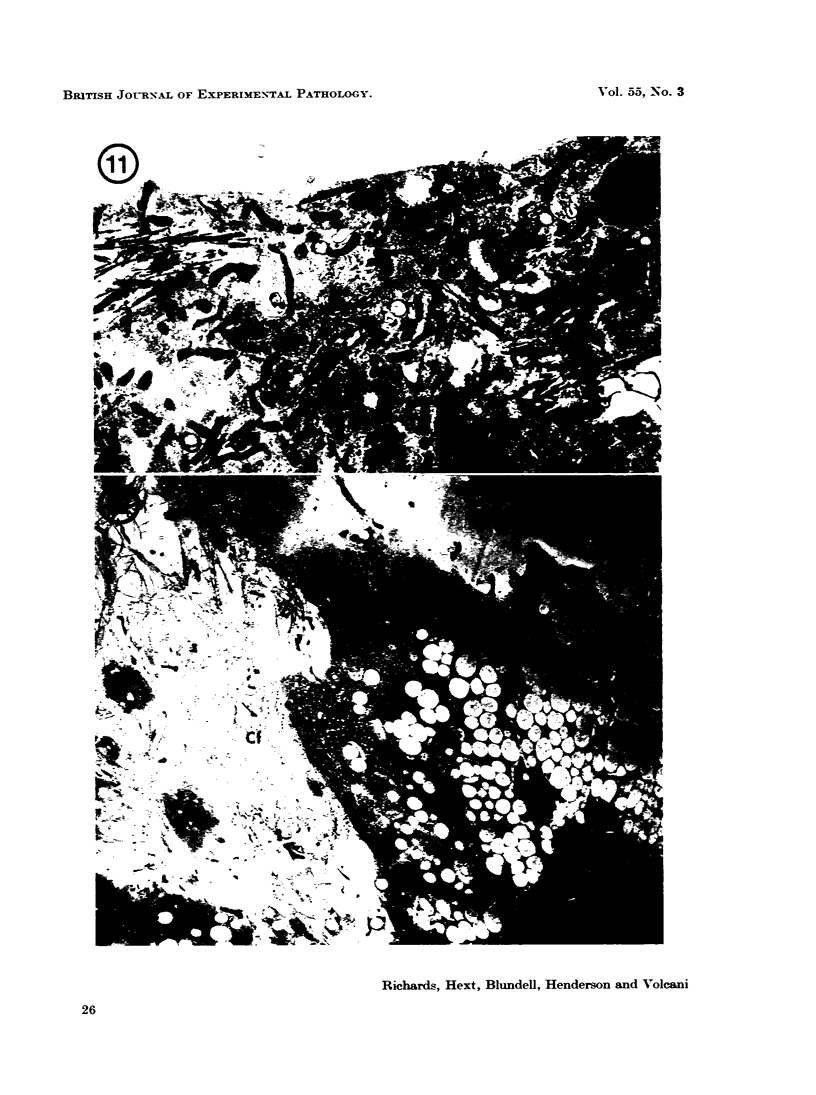
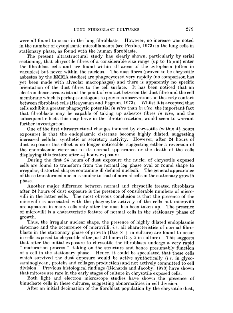
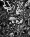
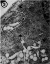
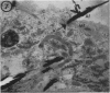
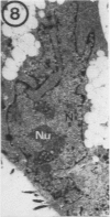
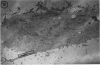
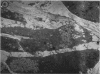
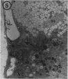
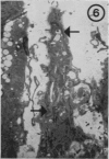
Early changes in the ultrastructure of lung fibroblasts exposed to chrysotile asbestos in vitro are described. The dust exposed cells rapidly undergo a “maturation process” reflected by highly dilated endoplasmic cisternae, irregularity in nuclear outline and composition and the appearance of membrane microvilli which are all characteristics of normal cells in the stationary phase. It is suggested the surviving cells undergoing this “maturation process” may account in part for the higher synthetic activity, leading to increased collagen levels, in chrysotile exposed cultures.
Full text
PDF
Images in this article
Selected References
These references are in PubMed. This may not be the complete list of references from this article.
- Beck E. G., Holt P. F., Nasrallah E. T. Effects of chrysotile and acid-treated chrysotile on macrophage cultures. Br J Ind Med. 1971 Apr;28(2):179–185. doi: 10.1136/oem.28.2.179. [DOI] [PMC free article] [PubMed] [Google Scholar]
- Comings D. E., Okada T. A. Electron microscopy of human fibroblasts in tissue culture during logarithmic and confluent stages of growth. Exp Cell Res. 1970 Aug;61(2):295–301. doi: 10.1016/0014-4827(70)90451-9. [DOI] [PubMed] [Google Scholar]
- GOLDBERG B., GREEN H. AN ANALYSIS OF COLLAGEN SECRETION BY ESTABLISHED MOUSE FIBROBLAST LINES. J Cell Biol. 1964 Jul;22:227–258. doi: 10.1083/jcb.22.1.227. [DOI] [PMC free article] [PubMed] [Google Scholar]
- Harington J. S., Miller K., Macnab G. Hemolysis by asbestos. Environ Res. 1971 Apr;4(2):95–117. doi: 10.1016/0013-9351(71)90038-7. [DOI] [PubMed] [Google Scholar]
- Heaysman J. E., Pegrum S. M. Early contacts between fibroblasts. An ultrastructural study. Exp Cell Res. 1973 Mar 30;78(1):71–78. doi: 10.1016/0014-4827(73)90039-6. [DOI] [PubMed] [Google Scholar]
- Miller K., Harington J. S. Some biochemical effects of asbestos on macrophages. Br J Exp Pathol. 1972 Aug;53(4):397–405. [PMC free article] [PubMed] [Google Scholar]
- Perdue J. F. The distribution, ultrastructure, and chemistry of microfilaments in cultured chick embryo fibroblasts. J Cell Biol. 1973 Aug;58(2):265–283. doi: 10.1083/jcb.58.2.265. [DOI] [PMC free article] [PubMed] [Google Scholar]
- Richards R. J., Wusteman F. S., Dodgson K. S. The direct effects of dusts on lung fibroblasts grown in vitro. Life Sci I. 1971 Oct 15;10(20):1149–1159. doi: 10.1016/0024-3205(71)90275-x. [DOI] [PubMed] [Google Scholar]
- Suzuki Y., Churg J., Ono T. Phagocytic activity of the alveolar epithelial cells in pulmonary asbestosis. Am J Pathol. 1972 Dec;69(3):373–388. [PMC free article] [PubMed] [Google Scholar]