

Abstract
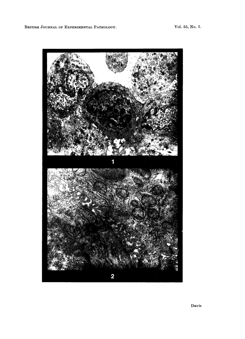
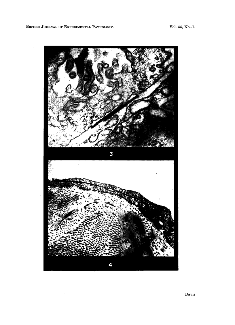
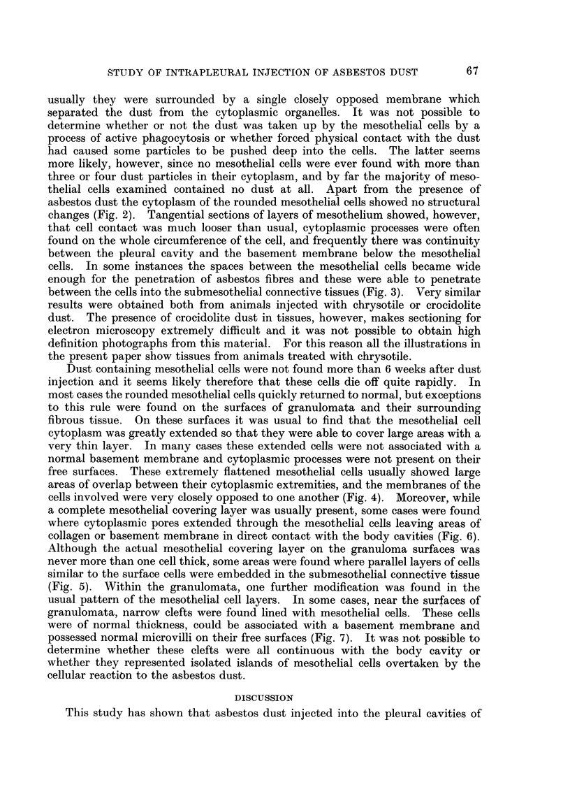
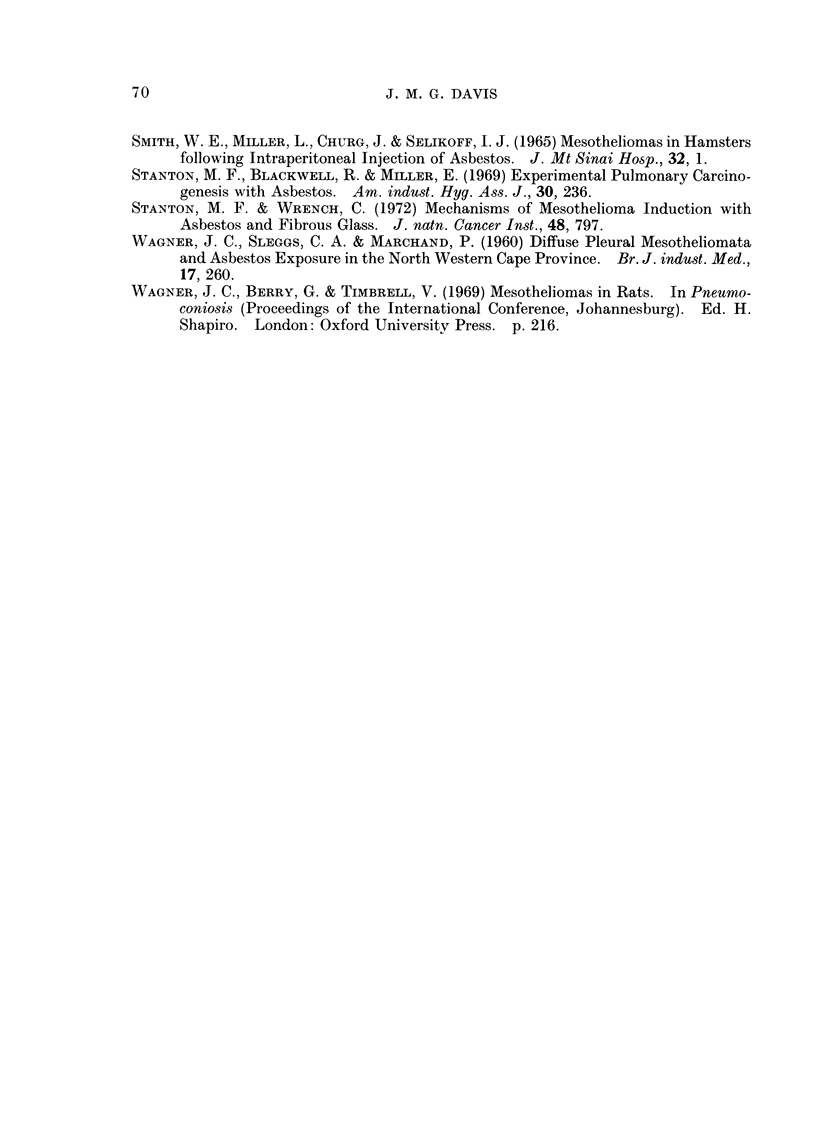
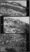
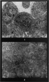
Studies of the pleural mesothelium in rats, mice, and guinea-pigs following the intrapleural injection of asbestos dust, showed that for 6 months at least this dust did not induce mesothelial hyperplasia. During the first few days after injection some areas of mesothelial cells became rounded and less clearly attached to one another, and a few were found to contain small numbers of asbestos fibres. During this period there was evidence of the penetration of asbestos fibres between the mesothelial cells, into the submesothelial connective tissues. Later in the studies the mesothelium covering most of the pleural cavity returned to normal, but where mesothelium covered asbestos granulomata, the cells were found to be extremely flattened, and without surface microvilli. Usually the mesothelial covering was complete, but in some areas pores were found penetrating the mesothelial cell cytoplasm and leaving areas of connective tissue in direct contact with the pleural cavity. In a few cases mesothelial cells were found lining clefts within the connective tissue of asbestos granulomata.
Full text
PDF

Images in this article
Selected References
These references are in PubMed. This may not be the complete list of references from this article.
- BARADI A. F., HOPE J. OBSERVATIONS ON ULTRASTRUCTURE OF RABBIT MESOTHELIUM. Exp Cell Res. 1964 Mar;34:33–44. doi: 10.1016/0014-4827(64)90180-6. [DOI] [PubMed] [Google Scholar]
- Cotran R. S., Karnovsky M. J. Ultrastructural studies on the permeability of the mesothelium to horseradish peroxidase. J Cell Biol. 1968 Apr;37(1):123–137. doi: 10.1083/jcb.37.1.123. [DOI] [PMC free article] [PubMed] [Google Scholar]
- DALTON A. J., FELIX M. D. A comparison of mesothelial cells and macrophages in mice after the intraperitoneal inoculation of melanin granules. J Biophys Biochem Cytol. 1956 Jul 25;2(4 Suppl):109–114. doi: 10.1083/jcb.2.4.109. [DOI] [PMC free article] [PubMed] [Google Scholar]
- Davis J. M. The long term fibrogenic effects of chrysotile and crocidolite asbestos dust injected into the pleural cavity of experimental animals. Br J Exp Pathol. 1970 Dec;51(6):617–627. [PMC free article] [PubMed] [Google Scholar]
- FUKATA H. ELECTRON MICROSCOPIC STUDY ON NORMAL RAT PERITONEAL MESOTHELIUM AND ITS CHANGES IN ABSORPTION OF PARTICULATE IRON DEXTRAN COMPLEX. Acta Pathol Jpn. 1963 Oct;13:309–325. doi: 10.1111/j.1440-1827.1963.tb03161.x. [DOI] [PubMed] [Google Scholar]
- HOLT P. F., YOUNG D. K. A dust-feed mechanism suitable for fibrous dust. Ann Occup Hyg. 1960 Nov;2:249–256. [PubMed] [Google Scholar]
- Kanazawa K., Birbeck M. S., Carter R. L., Roe F. J. Migration of asbestos fibres from subcutaneous injection sites in mice. Br J Cancer. 1970 Mar;24(1):96–106. doi: 10.1038/bjc.1970.13. [DOI] [PMC free article] [PubMed] [Google Scholar]
- Libánský J. The source of mononuclears at a site inflammation. Blut. 1966 Apr;13(1):20–29. doi: 10.1007/BF01631066. [DOI] [PubMed] [Google Scholar]
- ODOR D. L. Observations of the rat mesothelium with the electron and phase microscopes. Am J Anat. 1954 Nov;95(3):433–465. doi: 10.1002/aja.1000950304. [DOI] [PubMed] [Google Scholar]
- Roe F. J., Carter R. L., Walters M. A., Harington J. S. The pathological effects of subcutaneous injections of asbestos fibres in mice: migration of fibres to submesothelial tissues and induction of mesotheliomata. Int J Cancer. 1967 Nov 15;2(6):628–638. doi: 10.1002/ijc.2910020624. [DOI] [PubMed] [Google Scholar]
- Stanton M. F., Blackwell R., Miller E. Experimental pulmonary carcinogenesis with asbestos. Am Ind Hyg Assoc J. 1969 May-Jun;30(3):236–244. doi: 10.1080/00028896909343117. [DOI] [PubMed] [Google Scholar]
- Stanton M. F., Wrench C. Mechanisms of mesothelioma induction with asbestos and fibrous glass. J Natl Cancer Inst. 1972 Mar;48(3):797–821. [PubMed] [Google Scholar]
- WAGNER J. C., SLEGGS C. A., MARCHAND P. Diffuse pleural mesothelioma and asbestos exposure in the North Western Cape Province. Br J Ind Med. 1960 Oct;17:260–271. doi: 10.1136/oem.17.4.260. [DOI] [PMC free article] [PubMed] [Google Scholar]