Introduction
In the very early stages of development Intraoperative Neurophysiology (ION), somatosensory evoked potentials (SEPs) was the only available modality for monitoring functional integrity of the spinal cord and was used for many years in USA, Europe and Japan. This uni-modality approach very soon showed many disadvantages: it was nonspecific for the motor tracts, sensitive to inhalation anesthetics, needed relatively long time to update data, and patients with spinal cord pathology would sometimes have low quality of SEPs. Neuroanesthesia at that time, 30 years ago, was mainly based on inhalational anesthetics, the “deadly enemy” for SEPs when used with a higher amount of minimal alveolar concentration. An early report by Lesser et al. [10], showed that injury to the motor tracts of the spinal cord can happen with no changes within the parameters of SEPs. This first observation is later on confirmed by excellent and very well documented examples by Pelosi et al. [16], Minahan et al.[ 12], Jones et al. [9].
To avoid the influence of anesthetics and to show other advantages of direct recordings of traveling and stationary waves of SEPs, a team led by Dr. S. Jones from the UK, introduced monitoring of SEPs by the catheter-electrode in Europe placed in the epidural space of the spinal cord [7]. As highly and reliable this technique was it didn’t gain popularity as an intraoperative monitoring method of SEPs because of the relative invasiveness.
During the process of developing different methods for spinal cord monitoring, there were many trials and errors, but probably the most challenging one was when the intraoperative monitoring method of “neurogenic motor evoked potentials” (NMEPS) was introduced [13]. This method was based on the translaminar electrical stimulation of the spinal cord and recording time locked electrical activity over peripheral nerves, mainly of the low extremities. The author of this method claimed that activity recorded from limb muscles represented motor evoked potentials. Later on, collision studies [21] and unfortunate postoperative paraplegic patients with preserved NMEPS showed that this activity was mostly an antidromic stimulation of the dorsal columns. Therefore, it might be used for monitoring the functional integrity of the dorsal, but not for lateral columns [5, 12].
In search of a more specific monitoring method for the functional integrity of the spinal cord’s motor tracts, Machida et al. [11] introduced a method of electrical stimulation of the spinal cord and recording activity over the limb muscles.
The breakthrough in ION of the spinal cord took place by introducing methods of intraoperative monitoring of the motor evoked potentials (MEPs), eliciting them by applying single transcranial electrical stimulus and recording D wave from the vicinity of the spinal cord [1]. Using a technique of short train of stimuli, instead of single stimulus, Taniguchi and colleagues, elicited MEPs by direct stimulation of the exposed motor cortex [20] or transcranially and recording them from the limb muscles [15].
An important milestone in the preservation of spinal roots is the development of methodology for electrical stimulation of roots during placement of pedicle screws [2, for review see 22].
In this paper we will describe the methodological aspects of: (a) motor evoked potentials (MEPs) elicited by transcranial electrical stimulation (TES), (b) muscle activity after electrical stimulation of the spinal cord and (c) pedicle screw stimulation.
The methodological aspect of intraoperative monitoring with somato-sensory evoked potentials is described in this issue by MacDonald. The importance of combining the use of multimodal evoked potentials during spinal cord surgeries has been addressed by Sala et al., in this issue as well.
Methodological aspects of TES and recordings of D and I waves from the spinal cord and MEPs from limb muscles
Electrode montage for eliciting MEPs (for single and multipulse stimulation techniques)
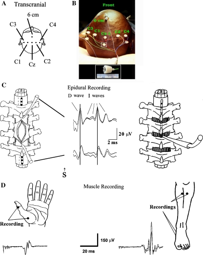
The electrode placement on the skull is based on the international 10/20 EEG system (Fig. 1). For transcranial stimulation, we prefer corkscrew-like electrodes (CS electrode, Nicolet Biomedical, Reading, WI) due to their secure placement and low impedance (usually 1 kΩ). Alternatively, an EEG needle electrode may be used. Some programs prefer standard EEG cup electrodes that are also effective and have an excellent safety profile. Their secure and low-impedance application with collodion requires skill and consumes more time. However, it can be done before the patient’s call to the operating room, saving time once there. They should be routine for young children in whom the fontanel still exists, since CS or needle electrodes could penetrate the fontanel during placement.
Fig. 1.
Schematic drawing of intraoperative methodology for eliciting and recording motor evoked potentials from the spinal cord and limb muscles. Schematic (a) and actual (b) illustrations of electrode positions for transcranial electrical stimulation of the motor cortex according to the International 10–20 EEG system. Front row: C3, C1, Cz, C2, and 6 cm (Cz + 6 cm). Note, instead of CZ, the CZ electrode is placed 1 cm behind the typical CZ point. Electrode positioned posterior are for recording somatosensory evoked potentials. Insert: Corkscrew electrode. Enlarged with a scale in mm. cto the left: schematic diagram of the positions of the catheter electrodes (each with three recording surfaces) placed cranial to the tumor (control electrode) and caudal to the tumor to monitor the incoming signal passing through the site of surgery. In the middle: D and I waves recorded rostral and caudal to the tumor site. Please note the peak latency difference between cranial and caudal recordings of the D and I waves are marked with vertical lines. To the right: placement of catheter electrode through ligamentum flavum. d Recording of muscle motor evoked potentials from the thenar, tibialis anterior and abductor hallucis muscles after eliciting them with a short train of electrical pulses applied transcranially. (Modified from Deletis V, Sala F. The role of Intraoperative Neurophysiology in the protection and documentation of surgically induced injury to the spinal cord. Ann NY Acad Sci 2001; 939:137–144)
The skull presents a barrier of high impedance to the electrode current applied transcranially, therefore we can not completely control the spread of electrical current when it is applied. Therefore, various combinations of electrode montages may need to be explored to obtain an optimal response. The standard montage is C3/C4 for eliciting MEPs in the upper extremities and C1/2 for eliciting MEPs in the lower extremities. With sufficient intensity of stimulation, C1/2 preferentially elicits MEPs on the right limb muscles while C2/1 elicits MEPs in the left limb muscles [17]. The first electrode in the montage represents an anode, while the second represents the cathode.
With stronger electrical stimulation, the current will penetrate the brain more deeply, stimulating the corticospinal tract (CT) at a different depth from the motor cortex. On the basis of measurements of the D wave latency it has been postulated that there are three favorable points which are susceptible to depolarization of the CT: cortex/ subcortex (weak electrical stimulation), internal capsula (moderate electrical stimulation), and brainstem/foramen magnum (strong electrical stimulation). Selectivity is only possible at the level of the cortex (subcortex). Therefore, only the application of relatively weak electrical stimuli to the cortex is selective, and it activates only a small portion of the CT fibers (e.g., activating only one extremity) or only one CT. It is important to remember that during electrical stimulation of the motor cortex, the anode is preferentially the stimulating electrode. With increasing intensity of the current, the cathode becomes the stimulating electrode as well.
As an example, stimulation with the C3+/C4− will selectively activate muscles of the right arm. When stimulation intensity is increased, the cathode (C4−) becomes the stimulating electrode as well, resulting in the stimulation of the left arm. Finally, when current intensity becomes strong enough to penetrate to the internal capsule more caudally, all four-extremity muscles can be activated. For anatomical reasons (deep position of the leg motor area in the interhemispheric fissure), more intense current is usually needed to obtain MEPs in the lower extremities. It is especially difficult to obtain them separately without also activating the upper extremities. Our observation has been that it can be done in certain patients, especially when using the “CZ/6 cm in front” montage.
The neurophysiological mechanism for eliciting MEPs by stimulating the motor cortex in patients under the influence of anesthetics is different from the mechanism in the awake subject. In the latter, electrical current stimulates the body of the motor neuron transynaptically over the chain of vertically oriented excitatory neurons resulting in I waves (indirect activation of the motoneurons). At the same time, electrical current activates axons of the cortical motoneurons, directly generating D waves [14]. In anesthetized patients, anesthetics block the synapses of the vertically oriented excitatory chains of neurons terminating on the cortical motoneurons body. Therefore, in most patients only the D wave is generated after electrical stimulation of the motor cortex [8, 14]. Patients with idiopathic scoliosis are an exception. In this group, abundant I waves can be recorded. We believe that this is one of the neurogenic markers of the disease present in these patients [4].
Recording of MEPs over the spinal cord (epidural or subdural space) as D and I waves using single pulse stimulating technique. D wave recording technique through an epidurally or subdurally inserted electrode
This method is a direct clinical application of Patton and Amassian’s discovery in the 1950s that electrically stimulated motor cortex in monkeys generates a series of well synchronized descending volleys in the pyramidal tract. This knowledge of CT neurophysiology, which was collected in primates, can be applied to humans in most cases.
We have to be aware that even small methodological aspects of recording D waves are of utmost importance and should be followed in order to achieve reliable results.
Choice of electrode
Practically any type of catheter type electrode, designed for electrical stimulation of the spinal cord epidurally, can be used for recording D and I wave [6].
Most epidural electrodes are disposable. If one uses a non-disposable type, extreme care should be taken to assure the electrode is clean before sterilization in order to improve its electrical properties. To clean the electrode, we recommend one of the following procedures: First, immerse the electrode tip in saline and pass a 9 Volt DC current (regardless of polarity) through it until a bubble of gas cleans the contact surface for a period of a few minutes. Second, you can use an ultrasound cleaner (Branson 1210, Branson Ultrasonics Corporation, Danbury, CT) by submersing the electrode in the cleaner for 5 min. Both techniques will remove any film or biological material remaining on the electrode, from the contact surfaces, and decrease their impedance. This maneuver will diminish the stimulus artifact, which usually appears when contact surfaces have high impedance. Because of the short latency of the D wave, a large stimulus artifact in an uncleaned electrode can pose an insurmountable obstacle for D wave recording.
Proper placement of the epidural electrodes
Depending on the surgical procedure, there are two methods of electrode placement: (a) percutaneously or (b) after laminectomy/ laminotomy, or flavotomy/flavectomy.
Percutaneous placement of the catheter electrode
This technique is rather popular in Japan [19]. The technique used in Japan is slightly different from the one we employ, since the neurosurgeon may ask for a subdural placement of the catheter electrode. Today, due to the increasing popularity of muscle MEPs monitoring during procedures involving the spinal cord and brainstem, the demand (indications) for percutaneous placement of this type of electrode has diminished. Confirmation of appropriate electrode placement can be done either by X-ray or by recording epidural SEPs from the same electrode after stimulation of nerves for upper or lower extremities.
Placement of electrode after laminectomy/laminotomy or flavectomy/flavotomy
These procedures include surgery for the removal of spinal cord tumors and different surgical interventions on the spinal cord. The surgeon places two catheter electrodes in the epi- or subdural space at the rostral and caudal edge of the laminectomy. The rostral electrode is the control electrode for non-surgically induced changes in the D wave, while the caudal one monitors the surgically induced changes to the CT (see Fig. 1). The amplitude of the D wave recorded over the cervical spinal cord could be 60 μV or more, while over thoracic segments it may be only 10 μV. With a stimulating rate of 2 Hz, it takes 2–4 averaged responses to get a reliable D wave. These results give in an update every second.
In surgical procedures in which the spine is exposed but a laminectomy is not performed (e.g., dorsal approach to spine stabilization), the catheter electrode may be inserted through a flavotomy/flavectomy.
Factors influencing the D and I wave recordings
D waves represent a neurogram of the CT, which is not significantly influenced by non-surgically induced factors. Stimulation of the CT takes place intracranially, distal to the cortical motoneuron body while recording is done caudal to the surgical site, but above the synapses of the CT at the α-motoneuron. Since no synapses are involved between the stimulating site and the recording site, the D wave is very stable and reliable. Therefore, we consider them to be the “gold standard” for measuring the functional integrity of the CT.
Massive dural adhesions, usually from previous surgery or after spinal cord radiation, can prevent the placement of the catheter electrode. Also, placement below the T10 bony level can not record a D wave of sufficient amplitude due to a lack of enough numbers of CT fibers at his level. The control (rostral) electrode can not be placed in patients with high cervical spinal cord pathology, due to the lack of space. Furthermore, plurisegmental intramedullary spinal cord tumors can desynchronize the D wave to such an extent that it becomes not recordable with an epidural catheter electrode. In this case MEPs from limb muscle are still recordable [6]. It has recently been shown that D wave monitoring during scoliosis surgery can give false positive and false negative data due to the new anatomical relationship between corrected scoliotic curvature and recording catheter electrode; therefore, it should not be used for monitoring these patients [23].
Recording of MEPs in limb muscles elicited by a multipulse stimulating technique
The selection of appropriate muscles to record MEP is an important issue in the monitoring of MEPs. In certain patients having deep paresis, not choosing the optimal muscles can result in “non-monitorable” patients. The small hand muscles e.g. abductor pollicis brevis (APB), or first dorsal interosseus muscle, are the optimal muscles to monitor the CT for the upper extremities. It has been shown that a good alternative is the long forearm flexors or even the forearm extensors. The spinal motoneurons for these muscle groups have rich CT innervation and are therefore suitable for monitoring the functional integrity of the CT. This is not the case with the proximal muscle of the arm or of the shoulder (biceps, triceps, or deltoid muscles)
For the lower extremities, abductor hallucis brevis (AHB) is the optimal muscle because of its dominant CT innervation. An alternative to this muscle is the tibial anterior muscle (TA). Our standard electrode montage for recording muscle MEPs in the upper and lower extremities are the AHB and TA for the lower, and the APB for the upper extremities.
Understanding the mechanism involved in the generation of muscle MEPs is essential for describing their appropriate use, explaining their behavior, understanding their value and knowing their limits during the monitoring of the CT. Generation of muscle MEPs is more complex in nature than the generation of the D and I waves. Therefore, their interpretation, especially during anesthesia, is rather complex. Generation of muscle MEPs and their propagation to the end organ (muscle) depends on: (a) the excitability of the motor cortex and the CT tract, (b) the conductivity of CT axons, (c) the excitability level of α-motoneuron pools, (d) the role played by the supportive system of the spinal cord (helping to increase the excitability of α-motoneurons), and (e) the integrity of motor nerves, the motor endplates and muscles.
Methodology of recording muscle activity after electrical stimulation of the spinal cord
This technique operates with non selective stimulation of the spinal cord, usually by inserting epidural catheter, or by translaminar needle electrodes and recordings from the limb muscles [11]. Later on, Taylor and colleagues found out that the optimal amplitude of compound action muscle potentials can be obtained after delivering two stimuli over the spinal cord 2 ms apart [18]. This technique is based on presumption that after electrical stimulation of the spinal cord, α-motoneurons are activated only by the CT tract. Therefore, compound muscle action potentials in the limb muscles or electrical activity in the peripheral nerves are generated by selective CT stimulation. Unfortunately, electrical stimulation of the spinal cord is not selective and therefore α-motoneurons can also be activated by any of the multiple descending tracts within the spinal cord, after strong electrical stimulation of the spinal cord, and/or by antidromically activated dorsal columns and their segmental branches that mediate the H-reflex [3]. So far no paraplegic patient has been reported having preserved muscle activity after electrical stimulation of the spinal cord.
Furthermore, these techniques (for methodological reasons) cannot evaluate the functional integrity of the CT from the motor cortex to the upper cervical spinal cord. Therefore, supratentorial, brainstem, foramen magnum and upper cervical spinal cord surgeries cannot be monitored using these techniques.
Methodology of pedicle screw stimulation
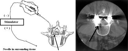
This technique is based on the previous measurements that relative distance between the pedicle screw and the neighboring root can be estimated by the intensity of the current required to activate the root and appropriate muscle. It has been shown that if the current which activates the root by stimulating it either through inserted pedicle screw or through the hole before screw insertion, is not lower than 5 mA, then the screw is a safe distance from the root (Fig. 2). Furthermore, if the pedicle breaks during the pedicle screw placement or the drilling of the hole in the pedicle, the current for root activation will be lower than 5 mA. This simple and straightforward technique is the standard of care during pedicle screw placement. Some variation in the results has been reported in some diabetic patients because they have a higher threshold. Conversely, some patients with osteoporosis have a lower threshold. (for review see 22].
Fig. 2.
To the left: The stimulation technique used to assess pedicle screw placements. The same technique can be used to test markers, taps, and pedicle holes. In this case, the pedicle screw has broken through the wall of the pedicle and is situated very close to an emerging nerve root (marked by arrow). As a result, electrical current, following the path of least resistance through the pedicle screw and the breach in the pedicle wall, is expected to excite the nerve root. Excitation typically occurs at very low stimulation intensities and results in triggered myogenic responses from muscles innervated by the nerve root. To the right: The CT scan showed the pedicle screw has entered the spinal canal and is in a position where L5 spinal nerve root (marked by arrow), was found on surgical inspection to be significantly compromised. (Combined and modified from: Toleikis, JR et al. The usefulness of electrical stimulation for assessing pedicle screw placements. J. Spin. Disord. 2000, 13(4), 283–289 and Toleikis, JR et al. The use of dermatomal evoked responses during surgical procedures that use intrapedicular fixation of the lumbosacral spine. Spine 1993, 18(16), 2401–2407)
Acknowledgments
Conflict of interest statement None of the authors has any potential conflict of interest.
References
- 1.Boyd SG, Rothwell JC, Cowan JMA, Webb PJ, Morley T, Asselman P, Marsden CD. A method of monitoring function in cortical pathways during scoliosis surgery with a note on motor conduction velocities. J Neurol Neurosurg Psych. 1986;49:251–257. doi: 10.1136/jnnp.49.3.251. [DOI] [PMC free article] [PubMed] [Google Scholar]
- 2.Calancie B, Madsen P, Lebwohl N. Stimulus-evoked EMG monitoring during transpedicular lumbosacral spine instrumentation: Initial clinical results. Spine. 1994;19:2780–2786. doi: 10.1097/00007632-199412150-00008. [DOI] [PubMed] [Google Scholar]
- 3.Deletis V. Intraoperative monitoring of the functional integrity of the motor pathways. In: Devinsky O, Beric A, Dogali M, editors. Advances in neurology: electrical and magnetic stimulation of the brain. New York: Raven; 1993. pp. 201–214. [PubMed] [Google Scholar]
- 4.Deletis V, Kiprovski K, Neuwirth M, Engler G. Do neurogenic lesions of the spinal cord generate distinctive features of the epidurally recorded motor evoked potentials? In: Jones SJ, Boyd SM, Smith NJ, editors. Handbook of spinal cord monitoring. Dordrecht: Kluwer; 1994. pp. 266–271. [Google Scholar]
- 5.Deletis V. The “motor” inaccuracy in neurogenic motor evoked potentials (Editorial) Clin Neurophys. 2001;112:1365–1366. doi: 10.1016/S1388-2457(01)00596-X. [DOI] [PubMed] [Google Scholar]
- 6.Deletis V. Intraoperative neurophysiology and methodology used to monitor the functional integrity of the motor system. In: Deletis, editor. Neurophysiology in neurosurgery. A modern intraoperative approach. Dublin: Academic; 2002. pp. 25–51. [Google Scholar]
- 7.Halonen JP, Jones SJ, Edgar MA, Ransford AO. Conduction properties of epidurally recorded spinal cord potentials following lower limb stimulation in man. EEG Clin Neurophysiol. 1989;74:161–174. doi: 10.1016/0013-4694(89)90002-3. [DOI] [PubMed] [Google Scholar]
- 8.Hicks R, Burke D, Stephen J, Woodforth I, Crawford M. Corticospinal volleys evoked by electrical stimulation of human motor cortex after withdrawal of volatile anesthetics. J Physiol (London) 1992;456:393–404. doi: 10.1113/jphysiol.1992.sp019342. [DOI] [PMC free article] [PubMed] [Google Scholar]
- 9.Jones SJ, Buonamassa S, Crockard HA. Two cases of quadriparesis following anterior cervical discectomy, with normal perioperative somatosensory evoked potentials. J Neurol Neurosurg Psych. 2003;44:273–276. doi: 10.1136/jnnp.74.2.273. [DOI] [PMC free article] [PubMed] [Google Scholar]
- 10.Lesser RP, Raudzens P, Lüders H, Nuwer MR, Goldie WD, Morris HH, Dinner DS, Klem G, Hahn JF, Shetter AG, Ginsburg HH, Gurd AR. Postoperative neurological deficits may occur despite unchanged intraoperative somatosensory evoked potentials. Ann Neurol. 1986;19:22–25. doi: 10.1002/ana.410190105. [DOI] [PubMed] [Google Scholar]
- 11.Machida M, Weinstein SL, Yamada T, Kimura J. Spinal cord monitoring––electrophysiological measures of sensory and motor function during spinal surgery. Spine. 1985;10:407–413. doi: 10.1097/00007632-198506000-00001. [DOI] [PubMed] [Google Scholar]
- 12.Minahan RE, Sepkuty JP, Lesser RP, Sponseller PD, Kostuik JP. Anterior spinal cord injury with preserved neurogenic ‘motor’ evoked potentials. Clin Neurophysiol. 2001;112:1442–1450. doi: 10.1016/S1388-2457(01)00567-3. [DOI] [PubMed] [Google Scholar]
- 13.Owen JH, Laschinger J, Bridwell K, Shiman S, Nielsen C, Dunlap J, Kain C. Sensitivity and specificity of somatosensory and neurogenic-motor evoked potentials in animals and humans. Spine. 1988;19(10):1111–1117. doi: 10.1097/00007632-198810000-00010. [DOI] [PubMed] [Google Scholar]
- 14.Patton HD, Amassian VE. Single and multiple unit analysis of cortical state of pyramidal tract activation. J Neurophysiol. 1954;17:345–363. doi: 10.1152/jn.1954.17.4.345. [DOI] [PubMed] [Google Scholar]
- 15.Pechstein U, Cedzich C, Nadstawek J, Schramm J. Transcranial high-frequency repetitive electrical stimulation of recording myogenic motor evoked potential with the patient under general anesthesia. Neurosurgery. 1996;39:335–344. doi: 10.1097/00006123-199608000-00020. [DOI] [PubMed] [Google Scholar]
- 16.Pelosi L, Jardine A, Webb JK. Neurological complications of anterior spinal surgery for kyphosis with normal somatosensory evoked potentials (SEPs) J Neurol Neurosurg Psych. 1999;66:662–664. doi: 10.1136/jnnp.66.5.662. [DOI] [PMC free article] [PubMed] [Google Scholar]
- 17.Szelenyi A, Kothbauer K, Deletis V. Transcranial electric stimulation for intraoperative motor evoked potential monitoring: stimulation parameters and electrode montages. Clinical Neurophysiol. 2007;118:1586–1595. doi: 10.1016/j.clinph.2007.04.008. [DOI] [PubMed] [Google Scholar]
- 18.Taylor BA, Taylor A, Farrell J. Temporal summation––the key to motor evoked potential spinal cord monitoring in humans. J Neurol Neurosurg Psychiatry. 1993;56:104–106. doi: 10.1136/jnnp.56.1.104. [DOI] [PMC free article] [PubMed] [Google Scholar]
- 19.Tamaki T, Takano H, Takakuwa K. Spinal cord monitoring: Basic principles and experimental aspects. Cent Nerv Syst Trauma. 1985;2:137–149. doi: 10.1089/cns.1985.2.137. [DOI] [PubMed] [Google Scholar]
- 20.Taniguchi M, Cedzich C, Schramm J. Modification of cortical stimulation for motor evoked potentials under general anesthesia: technical description. Neurosurgery. 1993;32(2):219–226. doi: 10.1097/00006123-199302000-00011. [DOI] [PubMed] [Google Scholar]
- 21.Toleikis JR, Skelly JP, Carlvin AO, Burkus JK. Spinally elicited peripheral nerve responses are sensory rather than motor. Clin Neurophysiol. 2000;111:736–742. doi: 10.1016/S1388-2457(99)00317-X. [DOI] [PubMed] [Google Scholar]
- 22.Toleikis R. Neurophysiological monitoring during pedicle screw placement. In: Deletis, editor. Neurophysiology in neurosurgery. A modern intraoperative approach. Dublin: Academic; 2002. pp. 231–259. [Google Scholar]
- 23.Ulkatan S, Neuwirth M, Bitan F, Minardi C, Kokoszka A, Deletis V. Monitoring of scoliosis surgery with epidurally recorded motor evoked potentials (D Wave) revealed false results. Clin Neurophysiol. 2006;117:2093–2101. doi: 10.1016/j.clinph.2006.05.021. [DOI] [PubMed] [Google Scholar]


