Abstract
Rickettsia prowazekii, the causative agent of epidemic typhus, is an obligate intracellular bacterium that grows directly within the cytoplasm of its host cell, unbounded by a vacuolar membrane. The obligate intracytoplasmic nature of rickettsial growth places severe restrictions on the genetic analysis of this distinctive human pathogen. In order to expand the repertoire of genetic tools available for the study of this pathogen, we have employed the versatile mariner-based, Himar1 transposon system to generate insertional mutants of R. prowazekii. A transposon containing the R. prowazekii arr-2 rifampin resistance gene and a gene coding for a green fluorescent protein (GFPUV) was constructed and placed on a plasmid expressing the Himar1 transposase. Electroporation of this plasmid into R. prowazekii resulted in numerous transpositions into the rickettsial genome. Transposon insertion sites were identified by rescue cloning, followed by DNA sequencing. Random transpositions integrating at TA sites in both gene coding and intergenic regions were identified. Individual rickettsial clones were isolated by the limiting-dilution technique. Using both fixed and live-cell techniques, R. prowazekii transformants expressing GFPUV were easily visible by fluorescence microscopy. Thus, a mariner-based system provides an additional mechanism for generating rickettsial mutants that can be screened using GFPUV fluorescence.
Rickettsia prowazekii is the etiologic agent of epidemic typhus, a historically significant human disease that continues to threaten populations subjected to nonhygienic, vector-laden conditions. Tragically, due to the continuing human legacy of war and extreme poverty, such conditions are not rare and recent outbreaks of this disease have been reported (13). In addition, R. prowazekii is a category B select agent and thus is classified as a potential agent of bioterrorism.
R. prowazekii is an obligate intracellular bacterium that grows directly within the cytoplasm of eukaryotic host cells, unbounded by a vacuolar membrane. The pathogenicity of R. prowazekii is dependent on its ability to grow to high numbers within the rich cytoplasmic environment, leading to lysis of the host cell and the release of rickettsiae that can then invade and destroy additional host cells. It accomplishes this feat, in organisms as diverse as humans and arthropods, with a small genome of approximately 106 base pairs predicted to encode 835 proteins (1, 18).
Defining the importance of specific rickettsial genes in intracellular invasion and growth has been hampered by the lack of genetic tools. However, progress in developing such tools has been made. Rickettsial genome manipulation has been performed using both homologous recombination mechanisms and transposon-based systems (3, 10-12, 14, 17). Recently, Felsheim et al. (7) described the use of a Himar1-based system to generate transposon insertions in the genome of another obligate intracellular pathogen, Anaplasma phagocytophilum. In this study, we report the use of the mariner-based Himar1 transposition system to obtain transposon insertions into the genome of R. prowazekii. We have successfully identified multiple insertional mutants, some of which are within predicted protein-encoding regions. This system provides an additional mechanism for identifying rickettsial proteins that are not essential for intracellular growth under selected growth conditions. The accumulation of such information is essential for identifying genes that play a critical role in rickettsial intracellular growth, pathogenicity, and vector transmission.
MATERIALS AND METHODS
Bacterial strains and culture conditions.
R. prowazekii Madrid E strain (6) rickettsiae were purified from the yolk sacs of embryonated chicken eggs as described previously (19). Rickettsiae purified from yolk sacs were frozen in a sucrose-phosphate-glutamate-magnesium solution (0.218 M sucrose, 3.76 mM KH2PO4, 7.1 mM K2HPO4, 4.9 mM potassium glutamate, and 10 mM MgCl2) and subsequently used to infect mouse L929 cells. Rickettsia-infected L929 cells were grown in modified Eagle medium (Mediatech, Inc., Herndon, VA) supplemented with 10% newborn calf serum and 2 mM l-glutamine (HyClone, Logan, UT; and Mediatech).
Escherichia coli strains XL1-Blue (Stratagene, La Jolla, CA) (5) and Top10 (Invitrogen, Carlsbad, CA) were grown in Luria-Bertani medium (2). For the selection of antibiotic-resistant transformants, ampicillin and rifampin were added to a final concentration of 50 μg/ml for each and kanamycin was added to a final concentration of 25 μg/ml.
R. prowazekii transformation.
R. prowazekii Madrid E strain rickettsiae were harvested from mouse L929 cells, prepared for electroporation, and electroporated in the presence of plasmid DNA as previously described (10, 12). Briefly, rickettsia-infected L929 cells (∼6 × 10⁷ L929 cells containing ∼200 rickettsiae per cell) were disrupted using minibeads and a minibead mixer. Isolated rickettsiae were electroporated in the presence of 20 μg of pMW1650, and then L929 cells were infected with the rickettsiae. Twenty-four hours after infection of the L929 cells by the electroporated rickettsiae, rifampin was added to a final concentration of 200 ng/ml. Rickettsial growth was monitored by microscopic examination of infected cells on coverslips stained by the method of Gimenez (8). Typically, the presence of rifampin clears the culture of detectable rickettsiae within several days. Rifampin-resistant rickettsiae are subsequently detected at approximately day 12 following the addition of rifampin. Once the rickettsiae were visually detected, the rickettsial culture was maintained under continued selection for approximately 7 days to amplify the resistant population. Following growth, infected L929 cells were diluted into 96-well plates for clonal isolation by limiting dilution as described previously (10) or with a slight variation as described in Results. After 1 week, the 96-well plates were screened for green fluorescent protein (GFPUV) expression using a Nikon Eclipse TE200-U fluorescence microscope and/or for the presence of the R. prowazekii arr-2 (arr-2Rp) rifampin resistance gene by PCR. Chromosomal DNA was isolated with the Puregene DNA purification kit (Gentra Systems, Inc., Minneapolis, MN), while plasmid DNA was isolated using the QIAPrep spin miniprep kit (QIAGEN, Valencia, CA). DNA sequencing was performed by the DNA Sequencing and Synthesis Facility, Iowa State University, Ames. Probes used in Southern hybridizations (15) were 32P labeled using the Multiprime DNA labeling system (Amersham, Piscataway, NJ) and [α-32P]dATP (ICN, Irvine, CA).
Construction of the mariner delivery vector.
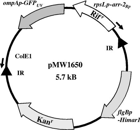
The 4.95-kb pGKT plasmid containing the Himar1 A7 transposase and transposon was kindly provided by Philip E. Stewart, Rocky Mountain Laboratories, NIAID. This plasmid is a derivative of the pMarGent plasmid (16) and contains a kanamycin resistance gene cassette inserted adjacent to the Himar1 transposase gene. A 0.8-kb fragment of pGKT containing the gentamicin resistance gene was removed by digestion with AvrII and BsaAI and replaced with a 1.6-kb PCR product containing the arr-2Rp rifampin resistance cassette and a gene encoding GFPUV under the control of the spotted fever group rickettsial ompA gene promoter (3). Plasmid pMODompACAT\GFPUV658, containing GFPUV under the control of the ompA promoter, was kindly provided by Gerald Baldridge and Ulrike Munderloh, University of Minnesota. The ompA promoter-GFPUV cassette was removed from plasmid pMODompACAT\GFPUV658 by XbaI digestion and inserted downstream of the arr-2Rp cassette contained in plasmid pMW1409 (10), generating a plasmid designated pMW1630. Plasmid pMW1630 was used as a template for PCR amplification using primers DW1011 (5′-GGCGGCCCTAGGAACATACTTGCTTTTATAGG-3′) and DW1013 (5′-CGGCGGTACGTGTCTAGAGTCGACCTGCAGGC-3′) to generate the 1.6-kb cassette fragment containing compatible restriction sites for cloning into the AvrII- and BsaAI-digested pGKT plasmid. Amplification was performed using Vent polymerase (New England Biolabs, Ipswich, MA) for 35 cycles of 94°C for 30 s, 54°C for 1 min, and 72°C for 1.5 min. The recombinant plasmid containing the arr-2Rp-GFPUV gene cassette was designated pMW1650 (Fig. 1). The sequence of pMW1650 is included in the supplementary material.
FIG. 1.
Physical map of pMW1650. The transposase, Himar1, is under the control of the borrelial promoter, flgBp (16). The region including rpsLp-arr-2Rp (Rifr), ompAp-GFPUV, and ColE1 represents the transposable element and is bounded by inverted repeats (IR), denoted by triangles. The small arrows indicate oligonucleotides (DW317 and DW1100) used for subsequent sequencing after rescue in E. coli. ColE1, an E. coli origin of replication; Kanr, the kanamycin resistance gene.
Rescue cloning.
The identification of transposon insertion sites was accomplished using rescue cloning as previously described (10). Briefly, chromosomal DNA (4 μg) was isolated from rifampin-resistant rickettsiae that were positive for the arr-2Rp gene by PCR and digested with BclI or HindIII, enzymes that do not cleave within the transposon. The resulting fragments were self-ligated in a 500-μl volume of ligation buffer containing 24 units of T4 DNA ligase (New England Biolabs) and incubated overnight at 16°C. Large-volume ligations were performed to favor intramolecular interactions. Following ligation, the DNA was precipitated with 95% ethanol; the precipitated DNA was washed twice with 70% ethanol, dried, and suspended in 16 μl of water. This DNA preparation was used to transform chemically competent E. coli Top10 cells. Transformants were selected on LB medium plates containing rifampin. Plasmids from individual clones were sequenced using primers DW317 (5′-ATGTCGGCATATGAAAACACTCC-3′) and DW1100 (5′-CGCCACCTCTGACTTGAGCGTCG-3′) that sequence out of the transposon into adjacent rickettsial DNA (Fig. 1).
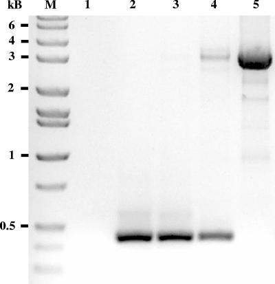
PCR detection of R. prowazekii gene RP401 and the insertion within this gene were accomplished using RP401-specific primers DW1151 (5′-ATGCAACAAATAGAGAAGTAC-3′) and DW1152 (5′-TACATCACTTGTTTTAGTGC-3′) that would amplify a 420-bp fragment of the wild-type open reading frame (ORF) or a 3,011-bp fragment of the Himar1 insertion within this gene. Amplification was performed using the FastStart high-fidelity PCR system (Roche, Indianapolis, IN) for 35 cycles of 94°C for 30 s, 56°C for 30 s, and 72°C for 3 min.
Imaging of fluorescent rickettsiae.
For fluorescent imaging, mouse L929 cells infected with rifampin-resistant rickettsiae, PCR positive for the arr-2Rp gene, were planted (3 × 10⁵ cells per 35-mm coverslip dish) on 22-mm glass coverslips and allowed to adhere for at least 24 h. The coverslips were then washed three times with phosphate-buffered saline (PBS) prior to fixation with ice-cold acetone for 2 min. The cells were then washed three times with PBS and mounted with VectaShield antifade mounting medium containing DAPI (4′,6′-diamidino-2-phenylindole) to counterstain the DNA (Vector Laboratories, Burlingame, CA). Fluorescent rickettsiae were visualized using a Nikon Eclipse TE200-U microscope and MetaMorph Imaging System software (Universal Imaging Corporation/Molecular Devices Corporation, Sunnyvale, CA).
For visualization of viable rickettsiae, L929 cells were planted as described above, washed with PBS, and visualized on an Olympus BX41 microscope with an Olympus FLN oil immersion iris 40× lens and a CytoViva 150 with Exfo X-Cite 120 fluorescent light source with the dual-mode fluorescence attachment that allows for the observation of both fluorescent and nonfluorescent sample portions simultaneously and in real time (CytoViva, Inc., Auburn, AL). Fluorescent rickettsiae were visualized using a universal DAPI-fluorescence isothiocyanate (FITC)-Texas Red emitter and excitation filter set. Images were captured using an Olympus Q Color 3 charge-coupled-device camera with QCapture V2.81.0 software.
RESULTS
Himar1 transposon mutagenesis of R. prowazekii.
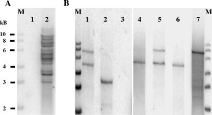
By the use of previously established protocols (10, 12), rickettsiae were purified from host cells and washed with and eventually suspended in 250 mM sucrose, and the electrocompetent rickettsiae were electroporated in the presence of plasmid pMW1650 containing the Himar1 rifampin resistance transposon. Selection for rifampin resistance resulted in a population of rickettsiae that at day 18 following electroporation was positive by PCR for the presence of the arr-2Rp rifampin resistance gene (data not shown). Southern blotting of DNA extracted from this population revealed that it contained multiple rickettsial populations exhibiting numerous transposon insertion sites (Fig. 2A). In order to determine whether any of these insertions involved recombination of the entire plasmid into the rickettsial genome, a Southern blot using sequences outside of the transposon as a probe was also performed. No plasmid DNA was detected in the rifampin-resistant population (see Fig. S1 in the supplemental material).
FIG. 2.
Southern blot analysis of transformed rickettsiae. (A) Lane 1, a mixture of chromosomal DNA isolated from R. prowazekii Madrid E (1 μg) and from L929 host cells (1.5 μg); lane 2, DNA (2 μg) isolated from a population of rifampin-resistant rickettsiae following transformation. (B) Lanes 1, 2, and 4 to 7, rickettsial chromosomal DNA (2 μg) from putative clones isolated by limiting dilution; lane 3, a mixture of chromosomal DNA isolated from R. prowazekii Madrid E (2 μg) and from L929 host cells (0.5 μg). In all analyses, chromosomal DNA was digested with HindIII and membranes were probed with a 32P-labeled GFPUV ORF. Lanes M, molecular size markers.
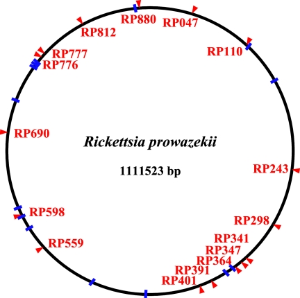
Identification of the insertion sites within the R. prowazekii genome was accomplished through rescue cloning. The Himar1 transposon used in these studies contains an E. coli ColE1 origin (16). Digestion of the DNA with an enzyme, BclI or HindIII, which does not cut within the transposon but which cuts often within the rickettsial genome, results in fragments that can be self-ligated and transformed into an E. coli recipient with selection for transposon-associated rifampin resistance. The resulting colonies were fluorescent when screened under UV light. Plasmids isolated from these clones were sequenced using primers DW317 and DW1100 to identify the rickettsial DNA linked to the transposon. Transposon insertion sites identified within the R. prowazekii genome are shown in Fig. 3. Of the 30 sites currently identified from three independent transformations, 16 are within predicted coding sequences. Transposon integration occurred at TA sites in a random fashion (see Table S1 in the supplemental material).
FIG. 3.
Schematic of the R. prowazekii chromosome showing the locations of Himar1 insertions. Insertions into coding regions are indicated with red arrowheads and the respective ORF number. Intergenic insertions are indicated with blue lines. Specific insertion sites are listed in Table S1 in the supplementary material.
Isolation of R. prowazekii transposon insertion clones and visualization of fluorescent rickettsiae.
Due to the obligate intracellular existence of R. prowazekii, a situation that precludes the ability to form clonal bacterial colonies on agar surfaces, it is impossible to determine frequencies of transformation or the spontaneous mutation rate using methods familiar to investigators of free-living or facultative bacteria. A typical experiment starts with approximately 1 × 10⁹ rickettsiae and generates approximately 10 identified insertions, indicating a transformation frequency of <1 × 10⁻⁸. Likewise, calculating the spontaneous mutation rate is problematic. Previous studies have shown the emergence of spontaneously occurring rifampin resistance as a result of single point mutations in the rpoB gene (12). During the extensive growth period under selective conditions, both transformants and spontaneous mutants continue to divide as a single, mixed population, making it impossible to identify distinct mutants from siblings.
Limiting-dilution techniques, as previously described (10), were used to isolate specific rickettsial clones. Briefly, this entailed the growth of the electroporated rickettsiae in mouse L929 host cells until a rifampin-resistant population could be detected microscopically. Dilution of this population in a 96-well format permits the isolation of individual clones (Fig. 2). To detect potential GFPUV-expressing clones, the 96-well plates were screened using a fluorescence microscope. Wells exhibiting fluorescence above background were expanded into six-well plates and eventually into tissue culture flasks. Southern blot analysis of DNA extracted from these expanded clones revealed both mixed populations (Fig. 2B, lanes 1, 5, and 7) and single, pure clones (Fig. 2B, lanes 2, 4, and 6), several of which were later shown to be siblings (lanes 1 and 5; lanes 4 and 6). However, the presence of a high percentage of spontaneously occurring, rifampin-resistant mutants within this population greatly increases the number of plates that must be screened and the number of dilutions that must be generated in order to isolate pure clones. To minimize this problem, we initiated limiting-dilution cloning at an earlier time point, 11 days postelectroporation, a time when the initial rifampin-sensitive population is no longer detectable but before rifampin-resistant rickettsiae can be visualized microscopically. This variation also resulted in the isolation of rickettsial clones, thus demonstrating a more efficient method for overcoming a major barrier of rickettsial genetic manipulation, namely, the isolation of clonal rickettsial mutant populations.
To demonstrate the low frequency of transformation involved in these studies, we examined DNA isolated at different points of an experiment by PCR for the presence of a specific gene insertion that had previously been identified in the population by rescue cloning. As shown in Fig. 4, lane 3, only wild-type sequence is detected in the rifampin-resistant population isolated at day 18, confirming the low numbers of the RP401 insertional mutant present in this population. A mixed population of spontaneous, rifampin-resistant mutants and the specific insertion could be identified in the first limiting-dilution cloning on day 49 (Fig. 4, lane 4). A pure clone, with no detectable wild-type sequence, is finally identified on day 83, following an additional round of limiting-dilution cloning (Fig. 4, lane 5).
FIG. 4.
PCR analysis of a limiting-dilution clone. DNA, isolated from rifampin-resistant rickettsiae at selected time points, was used as a template in a PCR designed to detect the RP401 gene. The presence of wild-type sequence results in the amplification of a 420-bp fragment. A transposon insertion within the RP401 gene results in a 3,011-bp fragment. Lane 1, negative control; lane 2, wild-type Madrid E chromosomal DNA; lane 3, DNA isolated from a population of rifampin-resistant rickettsiae harvested at day 18; lane 4, DNA isolated from rickettsiae harvested on day 49 following an initial round of limiting dilution; lane 5, DNA isolated on day 83 from a cloned population following a second round of limiting dilution. Lane M, molecular size markers.
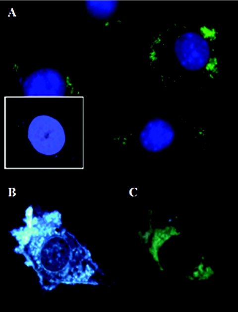
The transposon used in this study contained the GFPUV gene, thus providing an additional marker for screening rickettsial clones by their fluorescence. When the rifampin-resistant population was distributed in 96-well plates during limiting-dilution cloning, individual wells exhibiting greater-than-background fluorescence could be detected during low-power microscopic screening. Subsequent expansion of the rickettsiae within these wells revealed rickettsial populations that exhibited intense fluorescence when the cells were fixed and observed by standard fluorescence microscopy (Fig. 5A). The existence of fluorescent rickettsiae also permitted the visualization of viable rickettsiae within a living host cell. This was accomplished by observing a rickettsial clone with the CytoViva system, a system that permits high-resolution microscopy of living cells. As shown in Fig. 5B, GFPUV-expressing rickettsiae are difficult to visualize under visible light within the host cell, due to the obstructing characteristics of the eukaryotic host cell. However, the rickettsiae were clearly distinguishable by fluorescence microcopy under these conditions (Fig. 5C).
FIG. 5.
Fluorescence microscopy of rickettsial transformants. (A) The image is an overlay of intracellular rickettsiae expressing the GFPUV protein (green) visualized with an FITC filter set and the nuclear counterstain DAPI visualized with a DAPI filter set (blue) on acetone-fixed cells. As a negative control, the inset in panel A shows an L929 cell infected with wild-type R. prowazekii viewed with the same FITC and DAPI filter sets. An increase in gain was used to visualize the nonfluorescent rickettsiae counterstained with DAPI. (B) Live mouse L929 cell infected with viable rickettsiae expressing GFPUV protein visualized with CytoViva 150 Exfo X-Cite 120 fluorescent light source. (C) The same cell visualized in panel B with fluorescent rickettsiae (green) viewed with the CytoViva and a universal fluorescent filter set.
DISCUSSION
Due to the obligate intracellular nature of their existence, genetic manipulation of rickettsial species is difficult. Transformation of these organisms currently requires isolation of the rickettsiae from the host cell, electroporation, and reinfection of additional host cells. Following selection for rickettsiae that have been transformed comes a step that for free-living bacteria is simple but for rickettsiae is a major obstacle, namely, the isolation of individual clones. While limiting dilution followed by PCR detection of rickettsiae containing transposon insertions is a proven method of obtaining rickettsial mutant clones (10), it is laborious and expensive. The modification of this technique instituted in this study, in which we diluted out transformed populations early in the growth and selection phase, was an improvement that reduced the time and effort required for the isolation of rickettsial clones. However, even with this improvement, limiting dilution remains a labor-intensive method of clonal isolation. The expression of GFPUV by R. prowazekii mutants provides an additional mechanism for screening large numbers of putative clones. In this study it was used to screen 96-well plates for positive wells during cloning by limiting dilution. In addition, it is anticipated that cells infected with fluorescent rickettsial mutants could be separated from spontaneous resistance mutants by flow cytometry, greatly reducing the number of 96-well plates that would need to be planted in order to obtain single mutants. Single-cell techniques using micromanipulation, a protocol that is being used successfully for other obligate intracellular bacteria like Coxiella burnetii (4), can also be considered. The feasibility of this approach is greatly increased by the existence of fluorescent rickettsiae. Because the rickettsiae do not grow within vacuoles, they are difficult to visualize in unstained cells. The ability to identify live, fluorescent rickettsiae opens the door for applying single-cell techniques. The combination of live-cell imaging using the CytoViva system and fluorescent rickettsial clones also provides the means for observing rickettsial growth and intracellular distribution within their intracytoplasmic environment.
Due to the inherent difficulties in manipulating obligate intracellular bacteria, efficient methods for generating mutants are critical. Previously, it had been demonstrated that the transposome system marketed by Epicentre Technologies could be used to generate insertional mutants of rickettsial species (3, 10). While this system was appealing, due to the fact that the transposase entered the bacterium in complex with the transposon and did not have to be expressed by the rickettsiae, the variability in the generation of an active transposome complex often precluded efficient rickettsial transformations. In contrast, the Himar1 transposon has been demonstrated to be quite versatile, generating random insertional mutants in a variety of bacteria, including the obligate intracellular bacterial species A. phagocytophilum (7). The difficulty in calculating the transformation frequencies of each system in rickettsial transformations makes a direct comparison between the two transposon systems problematic. However, the fact that we consistently generate and isolate multiple insertional mutants in every experiment with Himar1 and have had numerous transposome experiments with no insertional mutants supports a higher efficiency for the Himar1 system. In addition, Baldridge et al. (3) demonstrated that a GFPUV gene carried on a transposon could be expressed in a nonpathogenic rickettsial species, providing a means for identifying rickettsial transformants by their fluorescence. By combining a rifampin resistance Himar1 transposon system with the GFPUV cassette that was successfully expressed in Rickettsia monacensis, we have demonstrated insertional mutagenesis of the pathogenic rickettsial species R. prowazekii.
The Himar1 system was able to generate a number of mutants, 11 of which were identified, in a single transformation experiment. More than half of the insertions in these mutants were within intervening, noncoding regions of the genome. This result is not surprising since these regions are very AT rich and the Himar1 transposon targets TA sites (9). Three of the insertion sites within annotated genes are within genes involved in DNA repair (mutS, mutL, and mfd). Interestingly, we also identified a mutL insertion in our earlier transposome mutagenesis studies (10). Current studies are examining whether the products of these genes are required when rickettsiae are growing in other environments or under increased conditions of stress and DNA damage. Finally, clones with mutations in genes with unknown functions are also being examined for phenotypic differences under various growth conditions.
In summary, the mariner-based Himar1 transposon system, coupled to a rifampin-resistance-selectable gene, provides a powerful mechanism for generating rickettsial mutants. In addition, the inclusion of a GFPUV gene within the transposon generated fluorescent rickettsiae, providing an additional method for screening rickettsial mutants. More importantly, the fluorescence phenotype permits the observation of live rickettsial mutants growing within their intracytoplasmic environment.
When dealing with an obligate intracellular pathogen such as R. prowazekii, a mechanism that permits the generation of specific mutants, even at low frequency, is extremely useful in identifying genes that are not essential for growth in a particular environment. For example, the current studies will identify genes that are nonessential for rickettsial growth in mouse L929 cells, an essential first step in dissecting rickettsial obligate intracellular growth. Also, while expression of a particular gene may not be required for growth in one environment, it may be critical to intracellular growth in another environment. In the case of rickettsiae, the requirements for growth may be significantly different between mammalian and arthropod hosts or under different conditions of stress. The addition of the Himar1 system, which targets TA sites, a frequent target in the A+T-rich rickettsiae, expands the range of genetic tools available for the study of the pathogenic rickettsiae. We are continuing transformation experiments with the Himar1 transposon in order to saturate the R. prowazekii genome and analyze the transformants to establish the requirements for rickettsial obligate intracytoplasmic growth.
Supplementary Material
Acknowledgments
We thank Philip E. Stewart for supplying the pGKT plasmid and Gerald Baldridge and Ulrike Munderloh for supplying the pMODompACAT\GFPUV658 plasmid. We also thank Andria Hines, Sangeetha Ramamurthy, and Andrew Woodard for excellent technical assistance.
This work was supported by Public Health Service grant AI20384 from the National Institute of Allergy and Infectious Diseases.
Footnotes
Published ahead of print on 24 August 2007.
Supplemental material for this article may be found at http://aem.asm.org/.
REFERENCES
- 1.Andersson, S. G. E., A. Zomorodipour, J. O. Andersson, T. Sicheritz-Ponten, U. C. M. Alsmark, R. M. Podowdki, A. K. Naslund, A.-S. Eriksson, H. H. Winkler, and C. G. Kurland. 1998. The genome sequence of Rickettsia prowazekii and the origin of mitochondria. Nature 396:133-143. [DOI] [PubMed] [Google Scholar]
- 2.Ausubel, F., R. Brent, R. E. Kingston, D. D. Moore, J. G. Seidman, J. A. Smith, and K. Struhl. 1997. Current protocols in molecular biology, vol. 1, 2, and 3. John Wiley & Sons, Inc., New York, NY.
- 3.Baldridge, G. D., N. Burkhardt, M. J. Herron, T. J. Kurtti, and U. G. Munderloh. 2005. Analysis of fluorescent protein expression in transformants of Rickettsia monacensis, an obligate intracellular tick symbiont. Appl. Environ. Microbiol. 71:2095-2105. [DOI] [PMC free article] [PubMed] [Google Scholar]
- 4.Beare, P. A., D. Howe, D. C. Cockrell, and R. A. Heinzen. 2007. Efficient method of cloning the obligate intracellular bacterium Coxiella burnetii. Appl. Environ. Microbiol. 73:4048-4054. [DOI] [PMC free article] [PubMed] [Google Scholar]
- 5.Bullock, W. O., J. M. Fernandez, and J. M. Short. 1987. XL1-Blue: a high efficiency plasmid transforming recA Escherichia coli strain with beta-galactosidase selection. BioTechniques 5:376-379. [Google Scholar]
- 6.Clavero, G., and F. Perez Gallardo. 1943. Experimental study of a nonpathogenic immunizing strain of Rickettsia prowazekii. Rev. Sanid. Hig. Publica. 17:1-27. (In Spanish.) [Google Scholar]
- 7.Felsheim, R. F., M. J. Herron, C. M. Nelson, N. Y. Burkhardt, A. F. Barbet, T. J. Kurtti, and U. G. Munderloh. 2006. Transformation of Anaplasma phagocytophilum. BMC Biotechnol. 6:42-50. [DOI] [PMC free article] [PubMed] [Google Scholar]
- 8.Gimenez, D. F. 1964. Staining rickettsiae in yolk-sac cultures. Stain Technol. 39:135-140. [DOI] [PubMed] [Google Scholar]
- 9.Lampe, D. J., M. E. A. Churchill, and H. M. Robertson. 1996. A purified mariner transposase is sufficient to mediate transposition in vitro. EMBO J. 15:5470-5479. [PMC free article] [PubMed] [Google Scholar]
- 10.Qin, A., A. M. Tucker, A. Hines, and D. O. Wood. 2004. Transposon mutagenesis of the obligate intracellular pathogen Rickettsia prowazekii. Appl. Environ. Microbiol. 70:2816-2822. [DOI] [PMC free article] [PubMed] [Google Scholar]
- 11.Rachek, L. I., A. Hines, A. M. Tucker, H. H. Winkler, and D. O. Wood. 2000. Transformation of Rickettsia prowazekii to erythromycin resistance encoded by the Escherichia coli ereB gene. J. Bacteriol. 182:3289-3291. [DOI] [PMC free article] [PubMed] [Google Scholar]
- 12.Rachek, L. I., A. M. Tucker, H. H. Winkler, and D. O. Wood. 1998. Transformation of Rickettsia prowazekii to rifampin resistance. J. Bacteriol. 180:2118-2124. [DOI] [PMC free article] [PubMed] [Google Scholar]
- 13.Raoult, D., T. Woodward, and J. S. Dumler. 2004. The history of epidemic typhus. Infect. Dis. Clin. North Am. 18:127-140. [DOI] [PubMed] [Google Scholar]
- 14.Renesto, P., E. Gouin, and D. Raoult. 2002. Expression of green fluorescent protein in Rickettsia conorii. Microb. Pathog. 33:17-21. [DOI] [PubMed] [Google Scholar]
- 15.Southern, E. M. 1975. Detection of specific sequences among DNA fragments separated by gel electrophoresis. J. Mol. Biol. 98:503. [DOI] [PubMed] [Google Scholar]
- 16.Stewart, P. E., J. Hoff, E. Fischer, J. G. Krum, and P. A. Rosa. 2004. Genome-wide transposon mutagenesis of Borrelia burgdorferi for identification of phenotypic mutants. Appl. Environ. Microbiol. 70:5973-5979. [DOI] [PMC free article] [PubMed] [Google Scholar]
- 17.Troyer, J. M., S. Radulovic, and A. F. Azad. 1999. Green fluorescent protein as a marker in Rickettsia typhi transformation. Infect. Immun. 67:3308-3311. [DOI] [PMC free article] [PubMed] [Google Scholar]
- 18.Tucker, A. M., H. H. Winkler, L. O. Driskell, and D. O. Wood. 2003. S-Adenosylmethionine transport in Rickettsia prowazekii. J. Bacteriol. 185:3031-3035. [DOI] [PMC free article] [PubMed] [Google Scholar]
- 19.Winkler, H. H. 1976. Rickettsial permeability: an ADP-ATP transport system. J. Biol. Chem. 251:389-396. [PubMed] [Google Scholar]
Associated Data
This section collects any data citations, data availability statements, or supplementary materials included in this article.


