In a Topical Review published in The Journal of Physiology, Herzog et al. (2006) proposed that the residual force enhancement after an active stretch of skeletal muscle (eccentric contraction) remained at least partly unexplained and could not be accommodated within the current framework of cross-bridge theory. Specifically, residual force enhancement might not be explainable in terms of development of sarcomere non-uniformities. This claim was based on a number of observations, virtually all of which have come from the Herzog laboratory, that appear not to be consistent with the sarcomere non-uniformity hypothesis.
Excess tension
The first observation is the claim that tension after a stretch can, ‘when stretch conditions are optimized’, exceed the tension in a fixed-end contraction at the initial length. In particular, when the initial length is the optimum length, this becomes a permanent tension after stretch greater than fixed-end tension at the optimum length. In addition, enhancement is reported at fibre lengths less than optimum. These observations stand in contrast to the current consensus view that after a stretch final tension is always close to the isometric tension corresponding to the fibre length at the start of the stretch, when the stretch covers a length range beyond optimum length and to tension at the final length when the stretch covers a range below the optimum length (Julian & Morgan, 1979; Edman et al. 1982). The tension enhancements reported in the review are, in fact, all rather small. If Fig. 4B was scaled to match the tension range shown in Fig. 4A, it would reveal that enhancement was present essentially only for stretches beyond optimum length and was insignificantly above optimum tension. Conversely, if Fig. 4A was expanded to match Fig. 4B, it would reveal that the tension traces were still converging, so that the reported tension enhancement is not substantially greater than uncertainty about the final value. It is notable that no description is given of the ‘sarcomere length’ traces in Fig. 4A, but the lack of concordance between length and tension traces during the stretch for record ‘s’ is worrying, as is the large mounting compliance implied by these traces.
Limits to accuracy
It is clear from reading the past literature on the subject that the limits of accuracy in these kinds of experiments, and the causes of their uncertainty, have in the past been very much appreciated. This review however, totally ignores them and takes the view that any tension above what is predicted invalidates the hypothesis. What then are the limits of accuracy for such measurements?
Permanent changes
Long before half-sarcomere non-uniformities were ever considered, Katz (1939) noticed that large stretches beyond optimum length brought about permanent changes in a frog muscle, which he summarized as the apparent ‘conversion of active contractile material to passive elastic material’. In particular, the twitch : tetanus ratio fell, the tension rise at the beginning of a tetanus became slower (Morgan et al. 2004), and the optimum length moved to longer lengths. These are all accounted for by the half-sarcomere non-uniformity hypothesis but not by the mechanisms proposed in the review. The relevance of such changes to this discussion is that the order in which the recordings were made and the number of repetitions carried out before a record was chosen for illustration becomes important. An isometric contraction at the original length before the active stretch will be different from the same contraction carried out after the stretch. No information on this is given in the review, nor is any comment made on the repeatability of the recordings illustrated.
Internal movement
The second difficulty arises when it is realized that none of these contractions involve strictly isometric sarcomeres. At lengths beyond optimum, fixed end contractions of single frog fibres have been repeatedly shown to involve shortening of one or both ends of the fibre, and slow lengthening of most of the middle region (Gordon et al. 1966; Julian & Morgan, 1979). The time course of this development of non-uniformity is substantially slower after a stretch than during a fixed end contraction (Edman et al. 1982, Fig. 8). So should tensions be compared at the same time point for isometric and eccentric contractions, or at the same point in the development of the non-uniformity? Most experimenters have accepted that the same time point is the best we can do, but there remains uncertainty about the result, as the internal motion within the fibre is likely to be very different in the two situations.
This slowing of internal motion is expected as almost all sarcomeres will move on to the steep ‘slow lengthening’ part of the force–velocity curve, where small changes in velocity accompany substantial changes in tension relative to isometric. Near isometric contractions of half-sarcomeres lead to slow detachment of cross bridges, slow shortening of series elastic elements, and hence a slow decline in tension. Such a conclusion points to the most probable explanation for the apparent force enhancement reported in this review. The slow decline of tension after a stretch means that the traces for fixed end and eccentric contractions continue to approach each other for a long time, and the search for a final value is limited by the ability of the tissue to sustain long-duration contractions. It is apparent in all the traces illustrated in the review, as well as in many other published records, that the measured tension difference is not the steady state value (Edman et al. 1978). Again, the consensus view is to use reasonably long tetani, and accept that as a result of the compromise, measurements of ‘permanent’ extra tension will represent a slight over-estimate.
Initial non-uniformity of sarcomere length
The small tension enhancements above the tension at the original length described in the review can also be explained by non-uniformities along a fibre. Consider the open circle in Fig. 2 of Herzog et al. (2006). as representing a distribution of half-sarcomere lengths. During the fixed end contraction preceding the stretch, the spread in the distribution is likely to increase as the shorter, stronger (in the sense of being able to generate more active tension) half-sarcomeres shorten and the longer, weaker half-sarcomeres are extended. When the stretch is applied, it is the longest, weakest half-sarcomeres that are stretched to long lengths, leaving the final tension to be determined by the remaining active half-sarcomeres, that is, the shorter, stronger ones. Note, however, that this cannot produce tensions above the isometric optimum value.
Initial non-uniformity of sarcomere strength
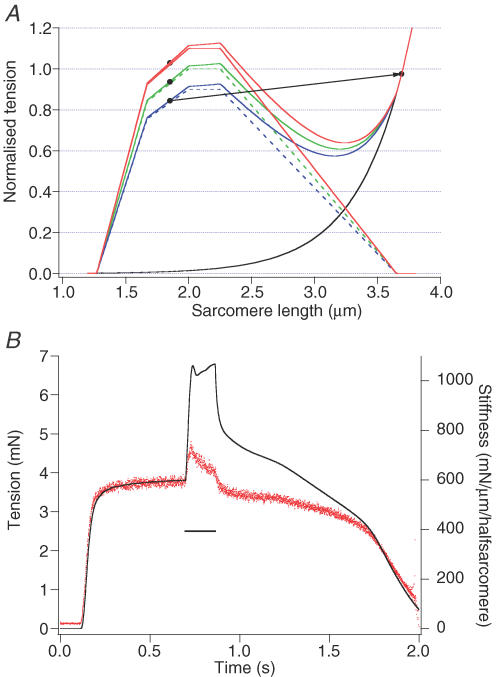
Consider, however, a fibre or myofibril with an initial non-uniform distribution of strengths in half-sarcomeres along its length, arising, for example, from some random variation in the number of thick and thin filaments in the half-sarcomeres. These half-sarcomeres will have length–tension curves that are scaled versions of each other. Consider, for example, the situation where all sarcomeres have a length slightly below optimum. An isometric contraction will produce a tension that is a weighted average of the sarcomere isometric tensions, with stronger sarcomeres shortening and weaker sarcomeres lengthening. If a stretch is applied, the weaker sarcomeres will lengthen rapidly. Initially, on the ascending limb, the rising isometric tension will slow down their movement, but if they reach their plateau length, no further increase is possible and they will rapidly, uncontrollably, extend to lengths beyond filament overlap (pop). The tension after the stretch will then be determined by the remaining active sarcomeres which, on average, will be stronger than the initial population. This will lead to the apparent development of extra tension. Similarly, if the initial length is on the plateau, the weakest sarcomeres will rapidly pop, and the tension after the stretch will be determined by the remaining active sarcomeres, that is, the original population minus the few popped ones. This would be consistent with Herzog's results. A three half-sarcomere example is shown in Fig. 1A.
Figure 1. Tension and stiffness changes during and after an active stretch.
A, half-sarcomeres (red, green and blue) with scaled length–tension curves (Gordon et al. 1966), but with the same initial length. (Dashed lines are active tension and continuous lines are total tension.) The recorded tension will be the average of the isometric capabilities of the three, weighted according to the slope of their force–velocity relation. As a simple approximation, this can be taken as the average of the three, that is, equal to that for the middle marker. After stretch, the weakest sarcomere will be beyond overlap as shown by the right hand marker, and the isometric tension will be the average of the upper two left hand markers, showing a small amount of apparent permanent extra tension. B, stiffness (red trace) and tension (blue trace) for a single fibre of the frog tibialis anterior muscle recorded at 3°C, during the first stretch. Stiffness was measured by the response to a 2 kHz sinusoid of about 1 nm per sarcomere amplitude. There is some rise in stiffness at the onset of stretch. Throughout most of the stretch, tension rises, but stiffness is clearly seen to fall. Immediately after the stretch, stiffness is well below its previous isometric value while tension remains well above the isometric level. Stimulation was stopped at 1.1 s. Subsequent repetitions of the measurement had stiffness reduced progressively more than tension, especially during and after the stretch. (Redrawn from Morgan et al. 1996.)
In addition, differences in the numbers of sarcomeres in series for different myofibrils could be important, with some myofibrils reaching their optimum force at lengths below the whole fibre optimum. This is similar to differences in fibre length in a whole muscle.
Damage
A number of the records shown in the review suggest inconsistencies in the measurement of passive tension at a particular length. This is primarily what leads to the conclusion that passive changes contribute to the ‘permanent extra tension’. The records all appear to be from whole muscles, although in some cases this can only be inferred from the legend. In the absence of experiments to investigate such changes in passive tension, it is difficult to fully explain them. However, knowledge of the damage that can be caused by eccentric contractions would suggest that the increased ‘passive’ tension may be the result of injury contractures in some fibres within the muscle (Whitehead et al. 2003). This would explain the failure to observe similar phenomena in single fibres.
Increased stiffness
The second observation used in the review to challenge the consensus position is a report, from the authors' own laboratory and using whole muscles, that stiffness in the ‘enhanced state’ exceeded that during an isometric contraction. This is clearly contrary to all observations made on single fibres, and may relate to the presence of damage in some fibres.
The best measurements of stiffness during a stretch are those of Morgan et al. (1996). These records, not cited in the review, clearly show stiffness decreasing during most of the first stretch (Fig. 1B), and, after the stretch, falling below the previous isometric value even when using such a relatively brief contraction. This paper also showed stiffness decreasing from one contraction to the next, in accord with the observations of Katz (1939).
Careful examination of the records published by the Sugi laboratory (Tsuchiya & Sugi, 1988) reveals that stiffness in the ‘enhanced’ state actually falls below that measured during a preceding fixed end contraction at the initial length or at the final length. (Sugi concluded that stiffness fell to near its value for a fixed end contraction at the final length, but his stiffness values were still falling at that point.) It is apparent that stiffness varies substantially from one contraction to the next, consistent with the idea of a postulated progressive accumulation of disrupted half-sarcomeres, but which cannot be explained by any theory ascribing the extra tension to extra cross-bridges, or even extra tension per cross-bridge.
Other observations
As has been mentioned already, the explanation of permanent extra tension resulting from non-uniform half-sarcomere lengthening is part of a broader hypothesis that is able to account for a number of additional phenomena, including the progressive changes observed by Katz, the fibre damage that often results from stretching active muscle, and the length dependence of all these phenomena (Morgan, 1994; Morgan et al. 2000). No explanation for these findings is provided in terms of either of the proposed mechanisms put forward in the review.
In particular, the increase in extra tension with increased amplitude of stretch, for amplitudes of stretch beyond the limits of distortion for an attached cross-bridge, follows immediately if final tension is essentially equal to isometric tension at the initial length, but it is much more difficult to see how a stretch could modify the actin–myosin interactions as suggested in the review. Such dependence of actin–myosin interactions on the history of activity is difficult to reconcile with any ‘sliding filament–independent force generator’ theory.
It is also notable that the alternative hypotheses put forward in the review come with no details and no attempt is made to incorporate them into more general models.
Conclusion
The single fibre measurements reported in this review show nothing new, and, in fact, continue to support the current consensus view about such phenomena when considered in terms of the limits of accuracy permitted by the measurements. The whole-muscle experiments suffer from obvious additional problems. The small discrepancies reported for single fibres can be explained in terms of sarcomere strength non-uniformities. The whole collection of observations is readily explained by non-uniformity of half-sarcomere lengthening, which also explains many other, otherwise puzzling observations. None of the alternative explanations offered in this review provide any additional insight into our understanding of these phenomena.
References
- Edman KA, Elzinga G, Noble MI. Enhancement of mechanical performance by stretch during titanic contractions of vertebrate skeletal muscle fibres. J Physiol. 1978;281:139–155. doi: 10.1113/jphysiol.1978.sp012413. [DOI] [PMC free article] [PubMed] [Google Scholar]
- Edman KA, Elzinga G, Noble MI. Residual force enhancement after stretch of contracting frog single muscle fibres. J Gen Physiol. 1982;80:769–784. doi: 10.1085/jgp.80.5.769. [DOI] [PMC free article] [PubMed] [Google Scholar]
- Gordon AM, Huxley AF, Julian FJ. Tension development in highly stretched vertebrate muscle fibres. J Physiol. 1966;184:143–169. doi: 10.1113/jphysiol.1966.sp007908. [DOI] [PMC free article] [PubMed] [Google Scholar]
- Herzog W, Lee EJ, Rassier DE. Residual force enhancement in skeletal muscle. J Physiol. 2006;574:635–642. doi: 10.1113/jphysiol.2006.107748. [DOI] [PMC free article] [PubMed] [Google Scholar]
- Julian FJ, Morgan DL. The effect on tension of non-uniform distribution of length changes applied to frog muscle fibres. J Physiol. 1979;293:379–392. doi: 10.1113/jphysiol.1979.sp012895. [DOI] [PMC free article] [PubMed] [Google Scholar]
- Katz B. The relationship between force and speed in muscular contraction. J Physiol. 1939;96:45–64. doi: 10.1113/jphysiol.1939.sp003756. [DOI] [PMC free article] [PubMed] [Google Scholar]
- Morgan DL. An explanation for residual increased tension in striated muscle after stretch during contraction. Exp Physiol. 1994;79:831–838. doi: 10.1113/expphysiol.1994.sp003811. [DOI] [PubMed] [Google Scholar]
- Morgan DL, Claflin DR, Julian FJ. The effects of repeated active stretches on tension generation and myoplasmic calcium in frog single fibres. J Physiol. 1996;497:665–674. doi: 10.1113/jphysiol.1996.sp021798. [DOI] [PMC free article] [PubMed] [Google Scholar]
- Morgan DL, Gregory JE, Proske U. The influence of fatigue on damage from eccentric contractions in the gastrocnemius muscle of the cat. J Physiol. 2004;561:841–850. doi: 10.1113/jphysiol.2004.069948. [DOI] [PMC free article] [PubMed] [Google Scholar]
- Morgan DL, Whitehead NP, Wise AK, Gregory JE, Proske U. Tension changes in the cat soleus muscle following slow stretch or shortening of the contracting muscle. J Physiol. 2000;522:503–513. doi: 10.1111/j.1469-7793.2000.t01-2-00503.x. [DOI] [PMC free article] [PubMed] [Google Scholar]
- Tsuchiya T, Sugi H. Muscle stiffness changes during enhancement and deficit of isometric force in response to slow length changes. Adv Exp Med Biol. 1988;226:503–511. [PubMed] [Google Scholar]
- Whitehead MP, Morgan DL, Gregory JE, Proske U. Rise in whole muscle passive tension of mammalian muscle after eccentric contractions at different lengths. J App Physiol. 2003;95:1224–1234. doi: 10.1152/japplphysiol.00163.2003. [DOI] [PubMed] [Google Scholar]


