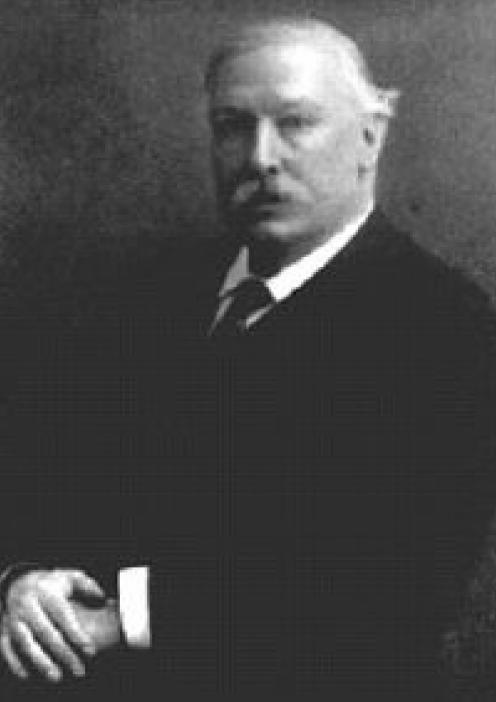
William Allen Sturge (fig 1) first described a syndrome in 1879 in a 6½‐year‐old girl.1
Figure 1 William Allen Sturge (1850–1919).
“She started to have twitching on [the] left side of her body as a six months old baby. The attacks worsened but without loss of consciousness. Later, the twitching started to spread to the other side and she would lose consciousness. She benefited from potassium bromide. Of particular interest was that the child had what was described as [a] “mother's mark” on the right side of the head and face. The skin lesion was accurately demarcated in the midline and involved the upper lip, nose, forehead, scalp and back of the neck, extending a little beyond the midline on the chin and on the upper part of the sternum. It extended as low as the third or fourth dorsal vertebra.
The lips, gums, tongue, roof of mouth, floor of mouth, uvula and pharynx were all similarly affected. The right eye was larger (buphthalmos, congenital glaucoma) and the sclera, choroid and retina were all affected by a vascular malformation. In addition, there was a patch about the size of the palm of the hand over the left eye, frontal and temporal regions. The mark was of a deep purple colour, the colour partially disappearing on firm pressure. The affected parts were distinctly larger than the corresponding parts on the other side”.
Sturge called this mark a “port‐wine” stain. Sturge presented his case to the Clinical Society in London on 18 April 1879. His colleagues doubted his suggestion that the neurological deficit was caused by a lesion on the ipsilateral surface of the brain, but not of the brain parenchyma, because he argued, such pathology would have caused epilepsy from the outset. Sturge's speculation proved correct. But it was not until 1901 that Siegfried Kalischer2 provided pathological proof.
Parkes Weber,3 in 1922, showed the intracranial calcifications. In an elegantly written, self‐deprecating account, he says
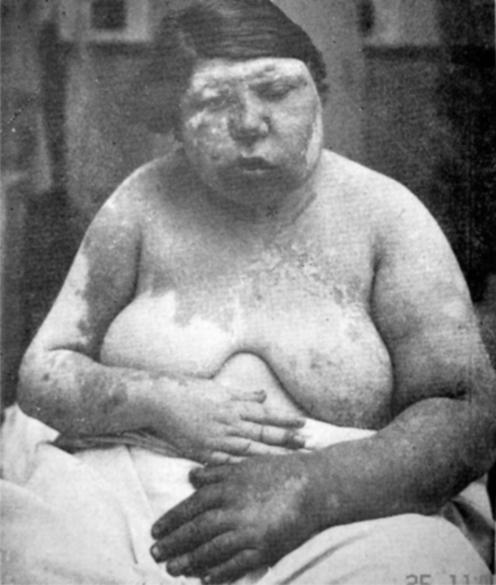
“The patient (fig 2) was indeed a remarkable sight. She was a rather obese young woman with a fleshy face, much of which, especially the left side, was scarlet or purple owing to a very large telangiectatic naevus (fig 2); on the left side she had an ‘ox‐eye' (buphthalmos, congenital glaucoma), and the right half of her body was smaller than the left half and also hemiparetic, pointing to a lesion on the left side of the brain. Because the X‐ray findings by Dr James Metcalfe in my case proved the existence of a more or less calcified and apparently ‘festooned' lesion on the surface of the left cerebral hemisphere, KH Krabbe4 suggested the term ‘Parkes Weber‐Dimitri disease', V Dimitri5 in the Argentine, having described the X‐ray findings in a similar case in 1923. But in 1935 Professor Hilding Bergstrand called the condition the ‘Sturge‐Weber disease, because of the priority of Dr W. Allen Sturge's account in 1879”.6
Figure 2 Parkes Weber's photograph shows a very extensive naevus affecting both sides (the left more than the right) of the face, neck and chest down to T2‐3, both arms and the left hand.
Rudolf Schirmer (1831–96)7 had earlier described a 36‐year‐old man with a left facial naevus and buphthalmos, but he made no allusion to epilepsy or brain disease.
Sturge–Kalischer–Weber–Dimitri syndrome (usually abbreviated to Sturge–Weber syndrome (SWS)), sometimes called the fourth phakomatosis, is characterised by naevus flammeus of the face and angioma of the meninges.8 It is congenital, but unlike other phakomatoses there is no evidence of a hereditary basis. However, the Klippel–Trenaunay–Weber syndrome is sometimes associated.
Fibronectin expression regulates angiogenesis. The reproducible increased fibronectin expression in Sturge–Weber syndrome fibroblasts and brain tissue is consistent with the presence of a hypothetical somatic mutation.9
William Allen Sturge (1850–1919) was born in Bristol, UK, and attended a Quaker school in London. While playing soccer there, he injured his knees. Staying with his uncle, a doctor, after this accident, he became interested in medicine, which he studied at Bristol in 1868. He contracted diphtheria in June 1869 and while convalescing developed rheumatic fever. He returned to his studies, but a second episode in 1872 enforced a prolonged rest. Eventually, he resumed his studies and qualified from University College, London, in 1873.
He became research medical officer and then registrar at the National Hospital for Paralysis and Epilepsy. In 1876, he studied with Charcot (1825–93) at the Salpetrière and with Jean Alfred Fournier (1832–915) at the St Louis Hospital.
Both Charcot and Fournier were impressed with Sturge's intelligence and originality. In Paris he met his wife, (Dr) Emily Bovell they married in September 1877 and set up a thriving practice together in Wimpole Street. He was appointed assistant physician and pathologist to the Royal Free Hospital and physician to the Royal Infirmary for Women and Children. He was an excellent teacher.10 He wrote several neurological papers, receiving the silver medal of the Medical Society of London for a paper on progressive muscular atrophy.
In 1880, his wife became ill with tuberculosis and Sturge moved to Nice, where he lived for 27 years. Emily Bovell died in her early 40s in 1885. Sturge looked after Queen Victoria and her family during their four visits to Cimez. In recognition, she awarded him the MVO (Member Victorian Order). Sturge married Julia Sherriff in 1886.
In his later years, he lived in Icklingham Hall, Suffolk, where he set up a unique museum of flint implementsi more than 100 000, which are now in the British Museum. Sturge was a founder and first president of the Society of Prehistoric Archaeology of East Anglia. His collection of Greek amphora is housed in the Toronto Museum, Canada, as the Sturge Collection.
Sturge died childless in March 1919.
Footnotes
Competing interests: There are no conflicting interests or financial support in this work. This paper has not been submitted to any other journal.
References
- 1.Sturge W A. A case of partial epilepsy, apparently due to a lesion of one of the vasomotor centres of the brain. Trans Clin Soc Lond 187912162 [Google Scholar]
- 2.Kalischer S. Ein Fall von Telangiektasie (Angiom) des Gesichts und der weichen Hirnhäute. Arch Psychiatr Nervenkrankheiten Berl 190134171–180. [Google Scholar]
- 3.Weber F P. Right‐sided hemi‐hypertrophy resulting from right‐sided congenital spastic hemiplegia, with a morbid condition of the left side of the brain, revealed by radiograms. J Neurol Psychopathol (Lond) 19223134–139. [DOI] [PMC free article] [PubMed] [Google Scholar]
- 4.Krabbe K H. Facial and meningeal angiomatosis associated with calcification of the brain cortex. A clinical and an anatomopathologic contribution. Arch Neurol Psychiatry (Chicago) 193432737–755. [Google Scholar]
- 5.Dimitri V. Tumor cerebral congénito (angioma cavernosum). Rev Ass Med Argent 19233663 [Google Scholar]
- 6.Parkes Weber F.Rare diseases and some debatable subjects. The Sturge‐Kalischer Disease. London: Staples, 19469–11.
- 7.Schirmer R. Ein Fall von Telangiektasia. Albrecht von Graefes Arch Ophtalmol 18607119–121. [Google Scholar]
- 8.Thomas‐Sohl K A, Vaslow D F, Maria B L. Sturge‐Weber syndrome: a review. Pediatr Neurol 200430303–310. [DOI] [PubMed] [Google Scholar]
- 9.Comi A M, Hunt P, Vawter M P.et al Increased fibronectin expression in Sturge‐Weber syndrome fibroblasts and brain tissue. Pediatr Res 200353762–769. [DOI] [PubMed] [Google Scholar]
- 10.Barlow T. William Allen Sturge. MVO, MD. BMJ 19191468–469. [Google Scholar]


