Susac's syndrome is a rare disease predominantly affecting young women. The syndrome is characterised by the triad of encephalopathy, branch retinal artery occlusions (BRAO) and hearing loss.1,2,3 An inflammatory or vascular background, or both, for this small vessel disease is controversially discussed. Therapeutic options are rare.
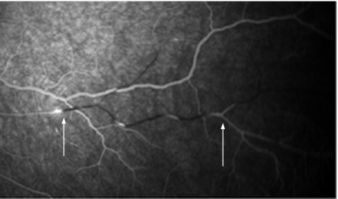
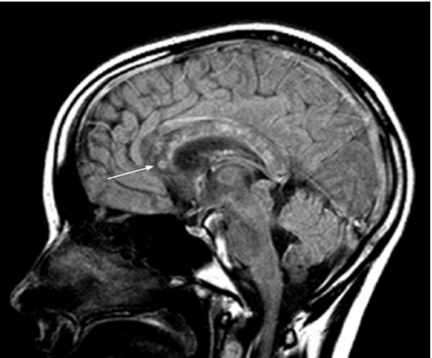
A 17‐year‐old girl without a remarkable medical history presented with several episodes of acute psychosis, nausea, dysarthria, ataxia, visual field defects, asymmetrical hearing loss and prodromal migrainous headache. Fluorescence angiography showed branch retinal artery occlusions (fig 1). Audiometry showed sensorineural pancochlear bilateral hypacusis. Magnetic resonance imaging showed hyperintense snowball‐like lesions of the corpus callosum (fig 2).4 These clinical investigations confirmed the diagnosis of Susac's syndrome. Risk factors such as smoking, reduced protein S activity, oral contraception and prodromal viral infection of the respiratory tract were found. Treatment with high‐dose glucocorticoids during the acute episodes, and a secondary prophylactic treatment with nimodipine and aspirin, was effective in both reducing the severity of acute symptoms and preventing further episodes.5
Figure 1 Hyperfluorescent retinal arterial wall plaques, branch retinal arterial occlusion and retrograde vessel filling. Arrows indicate leakage of retinal arteries.
Figure 2 Sagittal T2‐weighted image (fluid‐attenuated inversion recovery) showing multiple microinfarctions in the corpus callosum. Note the ring‐shaped lesion in the rostral genu, possibly representing a residual snowball lesion (indicated by an arrow).
From the presented case, we conclude that (1) the triad of encephalopathy, branch retinal artery occlusions and sudden hearing loss in young women strongly suggests Susac's syndrome; (2) snowball‐shaped lesions in the corpus callosum appear highly specific for the clinical diagnosis of Susac's syndrome; and (3) the use of high‐dose glucocorticoids in the acute phase of the disease and prophylactic treatment with aspirin and nimodipine seem to be beneficial for these patients, but has to be evaluated prospectively in larger studies.
Footnotes
Competing interests: None declared.
References
- 1.Ringelstein E B, Knecht S. Cerebral small vessel diseases: manifestations in young women. Curr Opin Neurol 20061955–62. [DOI] [PubMed] [Google Scholar]
- 2.Susac J O, Hardman J M, Selhorst J B. Microangiopathy of the brain and retina. Neurology 197929313–316. [DOI] [PubMed] [Google Scholar]
- 3.Susac J O. Susac's syndrome: the triad of microangiopathy of the brain and retina with hearing loss in young women. Neurology 199444591–593. [DOI] [PubMed] [Google Scholar]
- 4.Xu M S, Tan C B, Umapathi T.et al Susac syndrome: serial diffusion‐weighted MR imaging. Magn Reson Imaging 2004221295–1298. [DOI] [PubMed] [Google Scholar]
- 5.Wildemann B, Schulin C, Storch‐Hagenlocher B.et al Susac's syndrome: improvement with combined antiplatelet and calcium antagonist therapy. Stroke 199627149–151. [PubMed] [Google Scholar]


