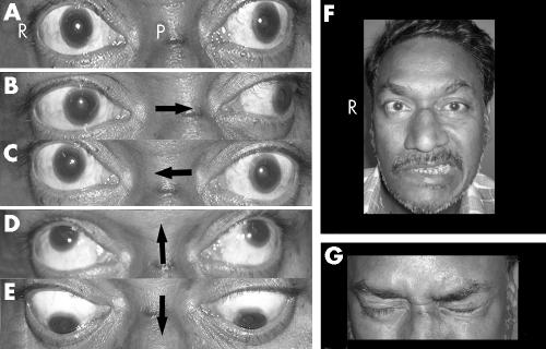
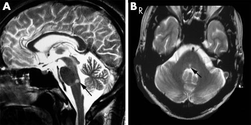
A 52 year old man with hypertension and diabetes mellitus presented with sudden onset of binocular diplopia on looking to the left side, right facial weakness, and epiphora in the right eye. Ocular motor examination revealed combination of right gaze paresis and right internuclear ophthalmoplegia suggestive of horizontal one‐and‐a‐half syndrome (fig 1A–C). Vertical ocular movements from the primary position were normal (fig 1D, E). In addition, he also had right lower motor neurone facial weakness (fig 1F, G). Cranial MRI showed right paramedian tegmental pontine lesion (fig 2A, B). The lesion was hyperintense on diffusion weighted MRI image (b = 1000 s/mm2) and hypointense on apparent diffusion coefficient map, compatible with features of acute infarct. Magnetic resonance angiography of intracranial vasculature was normal. The neurological problem was ascribed to lower pontine tegmental infarct due to occlusion of right paramedian pontine perforators. Electrophysiological studies (direct facial nerve stimulation and blink reflex) performed in the second week of illness, revealed incomplete right facial lesion. During evaluation in the third week, adduction lag in the right eye had slightly improved.
Figure 1 (A–C) Combination of right gaze paresis along with adduction lag in right eye and unimpaired abduction in the left eye (with nystagmus) suggestive of right horizontal one‐and‐half syndrome. Note the normal vertical eye movements from the primary position of gaze (D, E). (P‐ Primary position of gaze, arrows point towards the direction of gaze shifts). (F) Right facial weakness evident on clinical examination. (G) Note the right orbicularis oculi weakness on closure of both eyelids. Consent has been obtained for publication of this figure.
Figure 2 (A) Right paramedian tegmental pontine infarct seen on the T2 weighted saggital magnetic resonance imaging (arrow). (B) The same lesion in transaxial T2 weighted sequence (arrow).
Our patient presented with the unique combination of right sided horizontal one‐and‐a‐half syndrome and lower motor neurone seventh cranial nerve palsy. Such a combination of signs (seven plus one‐and‐a‐half) is known as eight‐and‐a‐half syndrome.1 Involvement of right abducens nucleus, right medial longitudinal fasciculus, and right.
Facial nucleus/fascicles in the lower pontine tegmentum contributed to the observed clinical signs. Thus recognition of this syndrome allows precise localisation of the lesion to lower pontine tegmentum ipsilaterally.
Footnotes
Competing interests: none declared
Consent was obtained for publication of figure 1
References
- 1.Eggenberger E. Eight‐and‐a‐half syndrome: one‐and‐a‐half syndrome plus cranial nerve VII palsy. J Neuroophthalmol 199818114–116. [PubMed] [Google Scholar]


