Focal damage to the fornices is uncommon and may be due to surgical removal of ventricular cysts and tumours.1 We report a case of bilateral fornix infarction with reduced fractional anisotropy values at 3 T after anterior communicating artery aneurysm clipping.
A healthy 33‐year‐old woman was admitted to our hospital with the incidental finding of an anterior communicating artery (ACoA) aneurysm on magnetic resonance angiography. Neurological examination was normal. Digital subtraction angiography visualised a broad based, tapered and 4 mm sized aneurysm of the ACoA and a median callosal artery (fig 1C). The ACoA aneurysm was treated with surgical clipping because of its irregular configuration. After surgery, the patient was drowsy with fluctuating impaired vigilance. She was disoriented in time, space and person, and revealed anterograde amnesia and amnesic aphasia. Her relatives noticed personality changes, psychomotor slowing and decreased spontaneity of speech and behaviour. Apart from transient mild right sided facial paresis, motor function of the limbs, deep tendon reflexes, sensory and coordinative examination and cranial nerves were normal. During the next 5 weeks of neurological rehabilitation, cognitive performance improved considerably. Seven weeks after the operation, she was orientated in all qualities and initial deficits in attentional performance and executive functions recovered. However, neuropsychological testing at this time revealed an average performance on general and selective attention, whereas the performance on divided attention and on the Wisconsin Card Sorting Test was impaired. The spatial–constructional abilities tested by the Rey–Osterrieth Complex Figure test and the mosaic test of Hamburg Wechsler Intelligence Test for Adults were normal. In the Auditory–Verbal Learning Test, she could initially recall the words in five trials with borderline values but over time or with interference of other words she could not. The storage process for long term memory was more impaired than for short term memory. The activities of daily living were not disturbed.
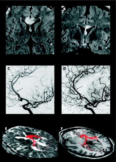
Figure 1 (A–E) Axial (A) and coronar (B) MRI sections demonstrating a hyperintense lesion on fluid attenuated inversion recovery images of the corpus callosum and the fornix. Initial (C) and postoperative (D) digital subtraction angiography with oblique projection revealed diffuse severe vascular narrowing of the median callosal artery. Fibre tracking of the partially infarcted fornix (E) and in a healthy 34‐year‐old woman (F). Several erroneous tracts were traced which were excluded in a second step (see frontal fibres on the right).
The postoperative angiogram demonstrating narrowing of the median callosal artery is shown in fig 1D.
MRI performed 1 day after surgery showed a nearly symmetric signal increase of the body of the fornix and of the adjacent part of the genu and anterior body of the corpus callosum on fluid attenuated inversion recovery images (fig 1A, 1B). Diffusion weighted images also revealed hyperintense signal changes with reduced values on apparent diffusion coefficient (ADC) maps using a region of interest (ROI) analysis with 32 voxels (mean (SD): 0.49 (0.04)×10−3 mm2/s; normal value from a separate control subject 0.76 (0.11)×10−3 mm2/s). Diffusion tensor images (DTI: single shot echoplanar imaging sequence with TR 7200 ms; TE 80 ms; 12 non‐collinear diffusion directions; 128×128 matrix, voxel dimensions 1.6×1.6×1.6 mm3; b = 700 s/mm2) and fibre tracking were performed 13 days later. Fibre tracking was performed using DTI studio (Johns Hopkins University, Baltimore, Maryland, USA). The propagation algorithms were medium lengths of fibres, FA>0.3 for each voxel, and line deviated by an angle >70°. DTI revealed an increase in ADC (1.21 (0.19)×10−3mm2/s; normal value 0.71 (0.16)×10−3mm2/s) and a decrease in FA (0.32 (0.06)) in the region of interest of the lesion in the fornix whereas ADC (0.72 (0.16)×10−3mm2/s) and FA were normal in the body of the fornix outside the lesion (0.62 (0.13); FA value from a separate control subject 0.64 (0.12)). Fibre tracking using a low threshold (FA 0.3) showed that some fibres could be followed even in the infarction of the anterior fornix (fig 1E, 1F).
Discussion
Amnesia is an uncommon, but increasingly recognised syndrome after damage of the anterior fornices. The fornix forms an integral part of the hippocampal–anterior thalamic pathway mediating memory retrieval, so that lesions of the fornix may selectively impair recall processes.1 Infarction of the anterior fornices in conjunction with the rostrum and genu of the corpus callosum has been discussed previously by Moussouttas et al.2 The vascular supply of these structures emanates from the subcallosal or the median callosal artery.3 In our patient, a dominant median callosal artery supplied the fornix. The damage of the basal forebrain, including the septohippocampal complex supplied by the anterior perforation arteries, is a well known complication of rupture or repair of ACoA aneurysms. However, the presented type of focal infarction has not been reported previously. Amnesic syndromes often complicate the clinical outcome after rupture and surgery of ACoA aneurysms. They are commonly explained by damage of the nuclei of the medial septum and of the diagonal band of BROCA, whereas detailed information about the fornices is lacking. The initial disturbances of the motivated behaviour and arousal in our case are explained by the concomitant involvement of the precallosal region.4 Anterograde amnesia and compromised verbal recall in retrieval processes are described both in fornix infarction and after rupture and repair of ACoA aneurysms.2,4,5 The memory impairment of our patient is consistent with this memory profile. Therefore, it may be supposed that fornix infarction after treatment of ACoA aneurysms is a complication that has not been assessed to date.
Changes in ADC values reflect alterations at the cellular level whereas DTI mirrors the functional integrity of axonal structures. Our case is the first in which DTI and diffusion tensor tractography in infarction of the fornix were performed. Thus the significance of the FA changes in this fibre bundle has not been investigated previously. Despite half the reduction of FA in the lesion compared with normal, the clinical outcome as well as fibre tracking indicated that some fibres might be preserved. These findings imply that fibre tracking may predict the outcome after damage to this fibre system. Nonetheless, the tracing of designated fibre bundles has technical pitfalls because of the curved course and the partial volume effect of the adjacent CSF. However, increased signal resulting from the higher field strengths of 3 T in the presented case allows acquisition with smaller voxel sizes, improving substantially the tracking conditions.
Footnotes
Competing interests: None.
References
- 1.Gaffan E A, Gaffan D, Hodges J R. Amnesia following damage to the left fornix and to other sites. A comparative study. Brain 19911141297–1313. [DOI] [PubMed] [Google Scholar]
- 2.Moussouttas M, Giacino J, Papamitsakis N. Amnestic syndrome of the subcallosal artery: a novel infarct syndrome. Cerebrovasc Dis 200519410–414. [DOI] [PubMed] [Google Scholar]
- 3.Ture U, Yasargil M G, Krisht A F. The arteries of the corpus callosum: a microsurgical anatomic study. Neurosurgery 1996391075–1084. [DOI] [PubMed] [Google Scholar]
- 4.Böttger S, Prosiegel M, Steiger H J.et al Neurobehavioural disturbances, rehabilitation outcome, and lesion site in patients after rupture and repair of anterior communicating artery aneurysm. J Neurol Neurosurg Psychiatry 19986593–102. [DOI] [PMC free article] [PubMed] [Google Scholar]
- 5.Moudgil S S, Azzouz M, Al‐Azzaz A.et al Amnesia due to fornix infarction. Stroke 2000311418–1419. [DOI] [PubMed] [Google Scholar]


