



Abstract
Damage to the spinal cord may be caused by a wide range of pathologies and generally results in profound functional disability. Therefore, a reliable diagnostic workup of the spine is very important because even relatively small lesions in this part of the central nervous system can have a profound clinical impact. This is primarily due to the dense arrangement of long fiber tracts extending to and from the extremities within the spinal cord. Because of its inherent sensitivity to soft tissues and its capability of displaying long segments of the vertebral column in one examination, MRI has become the method of choice for the detection and diagnosis of many disorders in the spine. A variety of innovative MRI methods have been developed to improve neuroimaging. Nevertheless, the application of these new methods to the spinal cord is, compared to its cephalad cousin, still not used that frequently. These techniques include the development of better pulse sequences and new MR contrast parameters that offer a wider spectrum of biophysical parameters in deriving a diagnosis. Overall, these new “cutting-edge” technologies have the potential to profoundly impact the ease and confidence of spinal disease interpretation and offer a more efficient diagnostic work-up of patients suffering from spinal disease.
Introduction
In the past twenty years, imaging technology has revolutionized medical care, establishing radiologic evaluation as a vital part of patient management. General practitioners as well as medical and surgical subspecialists now rely heavily on imaging to establish and confirm diagnoses, and plan and monitor treatments. Magnetic resonance imaging (MRI) is at the forefront of ever changing and improving technology and has now become a practical and widely available tool for diagnosis of a range of diseases. In the spine, MRI is the primary imaging modality for detecting disease because no other modality can provide adequate contrast resolution to differentiate the intraspinal soft tissue structures, and reveal spinal cord or canal pathology. In addition, MR has proven to be the most sensitive tool for detecting infiltration of bone marrow [1]. While there have been major strides in the development of MDCT affording rapid imaging and outstanding spatial resolution, application to the spine has been limited thus far due to limited tissue contrast and artifacts from adjacent bones and surgical material.
Standard structural MRI sequences, including T1 and T2 weighted spin echo and fast spin echo, (spoiled) gradient echo, and contrast enhanced images provide the majority of information required for detecting and characterizing spinal pathology and achieving a differential diagnosis. Therefore, improvements to these basic sequences, generating greater tissue contrast, better spatial resolution and decreased motion and susceptibility artifact, likely will provide the most significant gains in MR evaluation of spinal disease. However, as demonstrated in the brain, advanced techniques can provide additional information that increases sensitivity and specificity of diagnosis and provide more detailed physiologic or anatomic information that can help the referring clinician in guiding management. Diffusion weighted imaging (DWI), diffusion tensor imaging (DTI) and tractography, perfusion, MR Spectroscopy (MRS), and functional MRI (fMRI) sequences are now often part of a routine work up in the brain for assessment of strokes, tumors and inflammatory lesions. Although equally promising for the diagnostic work up of spine patients, these techniques, however, are seldom used in spine imaging due to technical challenges that limit image quality, including the highly magnetically inhomogeneous material surrounding the spinal canal, small size of spinal structures, the relative large cranio-caudal extent of the spine, CSF and blood pulsation, respiration, swallowing, and bulk motion. In addition, a substantial amount of spine patients who would require radiologic workup have to be turned away or receive inadequate imaging results because of metal artifacts adjacent to the diagnostically relevant regions.
Advances in spinal imaging depend on improvements in both MRI hardware and software. In the last decade, spine MRI has benefited the most from the introduction of phased array coil technology [2] and increased field strength, both of which increase baseline signal-to-noise ratio (SNR) of a study which is a major factor for successful spine studies that are notoriously SNR deprived. With the recent advent of parallel imaging [3-4], multi-element RF coils have been improved and have enhanced the SNR of high resolution MRI over a large CC-extent. With the availability of combined head and spine arrays, the entire spine can be imaged (by either stepwise or continuously moving the patient table) without repositioning the patient and changing the coil (Fig. 1). This is of great relevance because often the disease of a patient (e.g. MS or Neurofibromatosis) requires a total workup of the entire CNS and repositioning the patient is often tedious and associated with additional patient discomfort. In addition to the application of new contrast mechanisms and functional studies, structural imaging sequences have matured further, providing better SNR and spatial resolution within dramatically shortened imaging times. Similar to MDCT, there is a new trend towards volumetric acquisition of spine MRI data on the horizon and will afford multiplanar or curved-planar reformations (Fig. 2).
Figure 1.
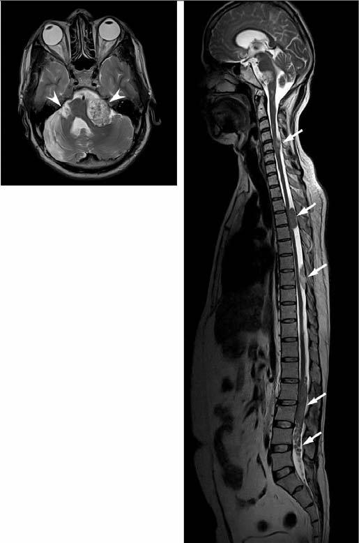
Sagittal T2W FSE of the entire spine and axial T2W FSE through the skull base in a patient with neurofibromatosis type 2. Bilateral masses in cerebello-pontine angles (arrowheads) as well as throughout the spine (arrows) are clearly visible. New multicoil technology affords seemless imaging of the entire CNS without repositioning the patient. (images courtesy Drs. Krueger and Mohr, Siemens Medical Systems, Erlangen, Germany).
Figure 2.
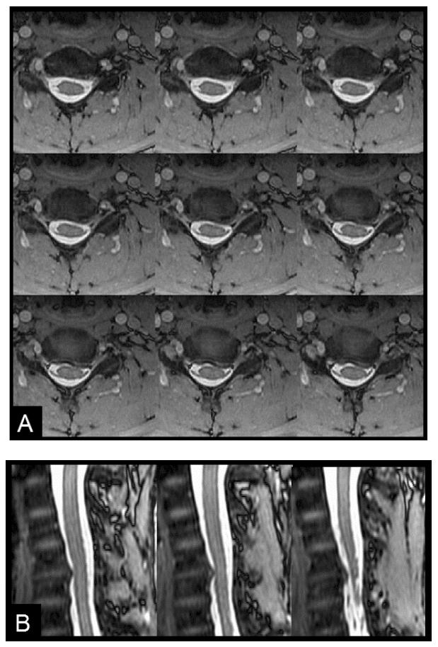
Axial views from a modified 3D balanced SSFP (COSMIC) scan of the cervical spine (A) and sagittal reformats (B). The interface between CSF and cord is well identified with superb visualization of nerve roots. Gray and white matter conspicuity is improved over T2W FSE and GRE sequences.
Although “Cutting-Edge” technology often implies that the technology being developed is not readily available to every user or is still under investigation, most of the sequences discussed are in fact currently or soon available from most MRI vendors and could be implemented in typical radiology practices. The techniques discussed below can be supported at field strength of 1.5T, but their utility at 3T will make them more popular in clinical settings, so they could be incorporated into routine protocols. We describe these advanced sequences in the context of common clinical scenarios, addressing how such sequences may help to answer clinical questions with greater accuracy and precision.
Degenerative Disease and chronic instability
One of the most common indications for spine imaging is evaluation of back pain and radicular symptoms caused by degenerative disc disease and facet arthropathy [5]. The established role of MRI in this clinical setting is to identify causes of nerve root compression (including disc protrusions, osteophytes and synovial cysts) and to assess severity of spinal stenosis, as well as to exclude other conditions such as infection and neoplasm that may not be clinically suspected [6].
High sensitivity of MR imaging studies is important as additional invasive testing such as nerve root and facet blocks or discography may be required in cases where standard MRI sequences fail to reveal a specific source of pain. Improved SNR with imaging at 3T (Fig. 3) and decreased CSF pulsation artifact as well as improved spatial and contrast resolution using new pulse sequences described below are likely to improve sensitivity for identifying subtle abnormalities. Such advances will provide the greatest advantage in the cervical spine where small disc spaces and relative paucity of epidural fat make delineation of disc protrusions more challenging than in the thoracic and lumbar spine.
Figure 3.
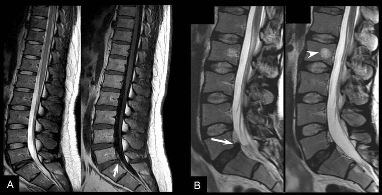
3T Imaging. A. Sagittal T2w (left) and T1w (right) images demonstrate relatively normal spine with mild degenerative changes of the L5-S1 disk (arrow). B. Sag T2w image of the lumbar spine demonstrates hemangioma at L2 (arrowhead) and a mildly protruded disc at L5-S1 (arrow) on T2w scans. In both patients the better resolution afforded by the higher SNR at 3T allows for better delineation of the conus, anatomy of the vertebral bodies and disks.
In addition to identifying a possible source of back pain and radicular symptoms, referring clinicians also look to MRI findings to guide surgical intervention. Detailed information regarding spinal canal stenosis, neural foraminal narrowing, and compression of nerve roots and the spinal cord can tip the balance in favor of surgical rather than conservative treatment. Identification of an extruded or sequestered disc fragment is vital information for the surgeon prior to intervention. MRI findings must be interpreted together with clinical information as symptomatic lesions cannot always be differentiated from asymptomatic lesions purely on the basis of imaging [7-8]. However, accurate delineation of the contents of the spinal canal and neural foramina may improve the diagnostic yield of MRI. Lesions that are associated with nerve root compression are more likely to be symptomatic and therefore improved visualization of individual nerve roots and surrounding CSF as well as more accurate assessment of caliber of the spinal canal and neural foramina are likely to improve correlation of MR findings with treatment outcomes and help guide appropriate therapy [9].
For practical purposes a short standard protocol, including sagital T1 and T2 weighted sequences as well as axial GRE or T2 is desirable for screening the large number of patients referred for back pain. Improving the speed of these acquisitions with parallel imaging [3-4] is helpful not only to improve patient throughput but also to minimize discomfort for patients who suffer back pain and may have difficulty lying in a fixed position for long periods of time. In short, parallel imaging uses multiple small coils that are part of a phased array coil, each of which having a different signal reception characteristic, to provide complementary image encoding through coil sensitivity in addition to regular gradient encoding [10].
Axial GRE is typically used to assess degenerative disease in the cervical spine. Unlike with spin echo (SE) and fast spin echo (FSE) sequences (also known as turbo spin echo or TSE), disk material (which is hyperintense on GRE) and osteophytes (which are hypointense) can usually be differentiated with GRE regardless of flip angle [11]. In addition, small size of the cervical disc spaces requires use of contiguous slices which is less feasible with FSE or SE. However, GRE often suffers from limited GM/WM contrast and contrast to better visualize nerve roots and foraminal stenoses. Recently, a GRE method with multiple bipolar gradient echo formations has been introduced that combines the signal from the individual echoes. Here, early echoes provide increased SNR, whilst later echoes boost contrast (Fig. 4). This sequence type is known as Multiple Echo Recombined Gradient Echo (MERGE) or Multi Echo Data Image Combination (MEDIC) and can be performed either as 2D or 3D sequence. For the cervical spine typical scan parameters are as follows: TR/TE=650ms/27ms, 4mm section thickness, FOV=16cm, 512 matrix, 2× parallel imaging acceleration). 3D imaging confers an extra advantage in the cervical spine, enabling sagital oblique reformats to be generated that can demonstrate the obliquely oriented cervical neural foramina en face.
Figure 4.
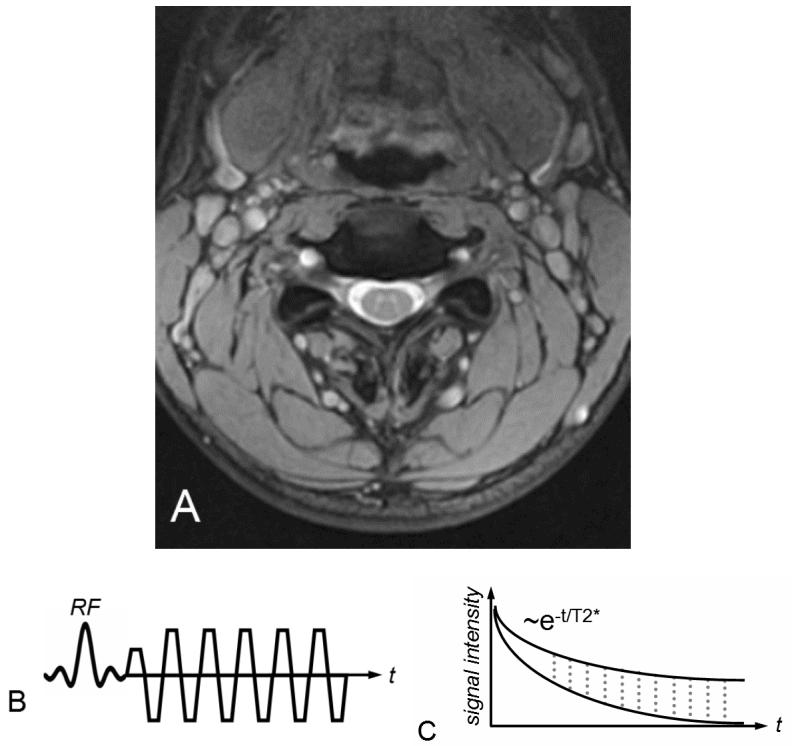
Axial MERGE image of the cervical spine demonstrates excellent gray/white contrast in the spinal cord as well as good contrast between CSF and the cord (A). The good SNR and contrast helps to demonstrate nerve roots extremely well. MERGE acquires multiple echoes during an oscillatory gradient echo readout (B), which become increasingly T2* weighted (C). The echoes are combined in a way that the early echoes provide increased SNR whilst the later improve contrast.
Balanced steady-state free precession (SSFP) sequences have also an inherently high contrast between tissue and fluid. Moreover, compared to unbalanced (i.e. the net gradient area within one TR is not zero) SSFP sequences, bSSFP provides high baseline SNR. Thus, it would provide an efficient alternative sequence to better detect herniations, sequestrations or nerve root compression. Conversely, GM/WM contrast of conventional bSSFP is relatively poor. Modifications to the sequence, specifically ramping up and down the flip angles to increase contrast and SNR, have been suggested under the name Coherent Oscillatory State acquisition for the Manipulation of Image Contrast (COSMIC) (Fig. 2).
SE and FSE sequences provide good anatomic detail in spine-imaging and are favored for evaluation of spinal canal diameter as well as for detection of spinal cord abnormalities, with less susceptibility artifact from bone and improved contrast between grey and white matter structures within the cord compared with GRE. Given abundance of epidural fat within the lumbar spine and relatively large disk spaces, FSE sequences are favored over GRE in the lumbar spine to assess for focal disc protrusions and nerve root compression. However, CSF adjacent to the cord and together with cord motion often causes ghosting artifacts in conventional Cartesian imaging, and is particularly problematic in FSE T2 weighted images. Such artifacts create a substantial challenge for the radiologist in identification of small structures within the CSF space, such as nerve roots. Radial sampling, in particular when combined with PROPELLER-type [12] acquisitions is less sensitive to these types of distortions and improves the diagnostic quality of such sequences (Fig. 5). This method has been recently combined with fast-recovery (FR) FSE sequences [13]. The benefit of FR-FSE over conventional FSE is the dramatically reduced TR, at similar or even improved T2 contrast between tissue and fluids [14]. With FR-FSE, assuming that the transverse signal of tissue (short T2) has decayed away at the end of the FSE train, a negative 90deg (echo reset) pulse orients spins with long-T2 (e.g. fluid) from the transverse plane back along the longitudinal direction leading to a much faster recovery of long T2 components to the equilibrium signal and thus better contrast between long and short T2 species. FR-FSE has demonstrated great utility also for 3D acquisitions as the TR can be substantially reduced and thus the imaging time [14].
Figure 5.
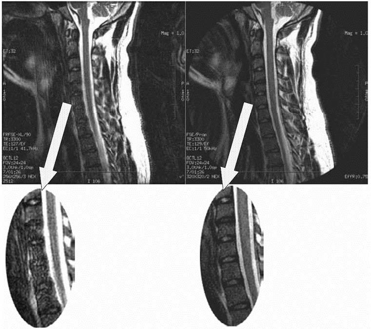
Sagittal T2w FSE with conventional Fourier encoding (left) and driven equilibrium T2w FSE (FR-FSE) with PROPELLER readout (right). Conventional Fourier encoding is sensitive to pulsation and motion. Such artifacts demonstrate multiple ghosts along the phase encode direction (left, see insert). Since PROPELLER excessively oversamples the center of k-space with each PROPELLER blade, the pulsatile and motion distortions are essentially averaged out (right, see insert). (images courtesy Dr. A. Gaddipati, GE Healthcare).
Additional sequences may be helpful in the face of persistent unexplained symptoms or for more complex or specific questions about anatomy or influence of patient position on alignment and stenoses. Contrast enhanced T1-weighted images with fat saturation can reveal facet joint pathology, spondylolysis, spinal degenerative/inflammatory changes and changes within the paraspinal muscles that are not always evident on conventional imaging [15]. Some studies also suggest that MR performed with axial load may add sensitivity and specificity to evaluation of spinal stenosis and nerve root compression [16-17]. Dynamic imaging with patients positioned to reproduce symptoms, similarly, may improve our ability to identify significant lesions. Very often abnormalities are only apparent during weight bearing in upright position or by flexion or extension of the spine. While weight bearing can be studied in certain interventional magnets that have enough aperture to allow the patients to sit in an upright position, flexion and extension or lateral flexion can be also accomplished in conventional magnets by using specific positional devices that allow different degrees of flexion/extension (Fig. 6). Certainly, great care has to be exercised when applying these maneuvers and the selection of those patients who might qualify for these kind of tests should be made only together with the referring orthopedic surgeon or neurologist.
Figure 6.

Positional device (A-D) to A/P (B) or L/R (C) flex and rotate (D) a patient's head and keep it in the desired position for scanning. By performing scans in different head positions, such as anterior flexion (E) or posterior flexion (F) bulging disks (arrows) can be detected that may appear as normal in a neutral position of the neck. Such positional devices allow for an optimal assessment of spinal canal stenosis and neuroforamenal narrowing. This improves correlation between imaging findings and clinical symptoms as patients may experience pain only in certain positions. (images courtesy Drs. Krueger and Mohr, Siemens Medical Systems, Erlangen, Germany).
MRI may provide important prognostic information regarding potential of recovery following decompressive surgery in patients with long standing myelopathic symptoms. High signal within the compressed cord on conventional T2 weighted sequences is non-specific, representing a combination of myelopathic changes and surrounding edema. DWI may provide greater specificity than conventional sequences regarding which changes in the cord are irreversible. DWI is an MR technique that is sensitive to the random motion of water molecules in tissues over microscopic distances and can generate a map of the average apparent diffusion coefficient (ADC). Reduced ADC is seen in acute cerebral stroke and aids early detection of infarcts, improving accuracy over conventional MR imaging. DWI has also become an invaluable technique for assessing other intracranial diseases, including applications in infection, tumors and cysts. In one of our studies [18], we found that spondylotic myelopathy presented with reduced ADC values, whereas the surrounding cord demonstrated elevated diffusivity. The former is presumably either due to cord compression or due to vascular compromise, while the latter is due to surrounding edema.
Post-Operative evaluation
The challenge presented to radiologists by the post-operative spine is two-fold: 1. Metallic surgical implants may produce artifacts that obscure anatomic detail; and 2. post-surgical reactive inflammatory changes can be difficult to distinguish from residual or recurrent disease or post-surgical complications such as abscess or seroma.
Metal in the area being imaged is problematic for both CT and MR. The concern for MR is significant susceptibility distortions caused by the metal adjacent to the tissue. While the metal itself cannot be imaged, these susceptibility changes in the proximity of the metal (e.g. pedicle screws, metal fixation rods, metal cages or endplates of artificial discs, etc.) can lead to geometric distortions and signal loss/pile-up. Signal loss and geometric distortions can be reduced by smaller voxel sizes/thinner slices and excessive RF refocusing. Recently, new variants of 3D FSE sequences (FSE-XETA, T2-SPACE, VISTA) have been introduced that differ from conventional FSE sequences by their excessively long FSE readout. Here, a readout train comprises up to 200 echoes obtained at a minimum echo spacing and allows image formation very rapidly, altogether diminishing artifacts (Fig. 7). A specific hallmark of these sequences is the flip angle modulation during the FSE readout. Here the focus is to carry along magnetization as long as possible to avoid blurring and provide optimal signal at the effective TE (i.e. when the center k-space lines are acquired) (Fig. 8). Using that regime the specific absorption rate can be also substantially diminished. The volume acquisition follows a new trend that has been carried over from MDCT. By acquiring high resolution 3D volumes and subsequently generating multiplanar reformats or even curved planar reformats imaging could be made much more efficient. However, it needs to be shown whether or not these reformats are of sufficient quality and detail to replace additional conventional cuts. By additional modification to the pulse sequence different contrast and better gray/white differentiation can be achieved (Fig. 9).
Figure 7.
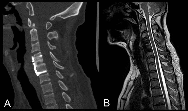
A. Sagittal CT reformation of the cervical spine in a patient with C4-5 anterior cervical disk fusion. B. The new volumetric FSE sequences (FSE-XETA, VISTA, SPACE) afford much smaller voxel sizes and thus less intravoxel dephasing, improving MR imaging in the presence of metal which is typically problematic because of the field perturbations created by the metal. That in combination with short echo spacing and excessive RF refocusing dramatically reduces distortions in the spine even in the presence of surgical hardware. (images courtesy Dr. Ripart, Siemens Medical Systems, Erlangen, Germany, and Dr. Ricolfi, CHU Dijon, France).
Figure 8.
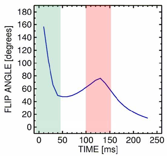
Flip angle sweep during FSE readout. With high flip angles the signal in FSE readouts is primarily determined by the primary echoes, whilst with lower flip angles the signal becomes increasingly dominated by higher order echoes and stimulated echoes. In order to maximize signal for long echo train lengths the flip angle of the refocusing pulses are continuously ramped down almost 50deg and slightly raised when the center of k-space is acquired to optimize contrast and SNR.
Figure 9.
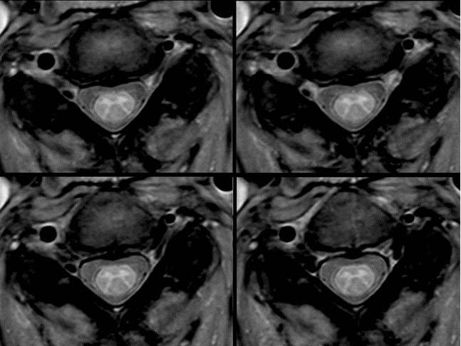
Axial view of a volumetric proton-density weighted FSE scan (VISTA) provides excellent gray/white matter contrast (images courtesy Dr. Hoogenraad, Philips Medical Systems, Eindhoven, Netherlands).
Gadolinium enhanced imaging is vital following surgery. Enhancement identifies areas of inflammatory post-surgical change as well as recurrence of disease. Following discectomy, recurrent disc protrusions are identified due to their lack of contrast enhancement in comparison to uniformly enhancing scar tissue. After tumor resection, non neoplastic enhancement can develop quickly due to post surgical inflammation and neovascularity, and has been described even within the first 24hours after surgery in the brain [19]. Early imaging following tumor resection maximizes the radiologist's ability to distinguish residual enhancing tumor from post surgical changes and helps to establish a post-operative baseline for the patient. In on-going tumour surveillance it is important to have high quality fat saturated enhanced images to be able to identify areas of subtle new nodular enhancement representing tumour on the background of post-surgical scarring. Homogeneous fat saturation is critical when obtaining contrast enhanced T1-weighted images to avoid obscuring enhancement with signal from epidural fat and fatty vertebral body marrow.
Due to the harsh magnetic environment in and around the spine or due to the presence of surgical material, frequency-selective fat suppression techniques or spectrally-selective excitation pulses are often suboptimal (Fig. 10). Therefore, STIR techniques are frequently used despite their obvious SNR penalty and the potential for altered contrast in the presence of contrast material. Recently, a variant of Dixon imaging has been introduced that has been proven to be very robust (Fig. 11) and provides increased SNR [20] due the combination of measurements. Especially for scans with NEX>1 it appears to make sense to acquire data at slightly different echo times and combine the data with the aforementioned iterative Dixon technique. One challenge for the Dixon method is too rapid field fluctations so that the underlying phase maps needed for fat water separations fail.
Figure 10.
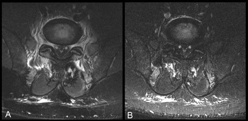
Axial T2w FSE images with spectral fat saturation (A) and short-tau inversion recovery (STIR) (B) in a patient with intrapedicular screws for posterior fusion (image below the screws). The geometric distortions from the titanium screws are clearly apparent on spectral fat saturation as increased signal in perivertebral soft tissues as well as in neural foramina. The field perturbations induced by the screws impair chemical fat saturation and lead to difficulties separating between fat and edema or fluid collections, which is significantly reduced on STIR.
Figure 11.
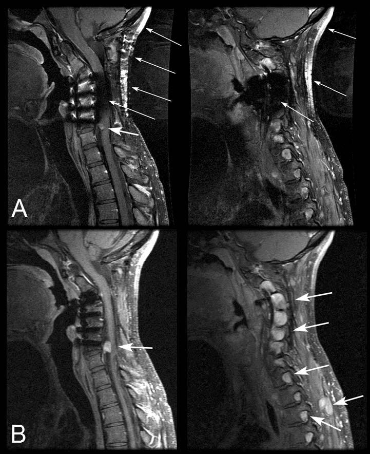
Midline sagittal and parasagittal postcontrast T1W fat-saturated SE images (A) and corresponding sagittal and parasagittal postcontrast T1W IDEAL-FSE water images (B) in a patient with neurofibromatosis type 1 and spinal hardware. Numerous enhancing lesions are seen within the neural foramina (large arrows), abutting the spinal cord, and in the paraspinal soft tissues. The lesion adjacent to the spinal cord is better visualized in the IDEAL image (B) compared to the fat-saturated image (A), where failed fat saturation from severe B0 inhomogeneities from metallic hardware degrades signal in the spinal canal near this mass. Large areas of failed fat saturation (small arrows) show uniform suppression of fat in the IDEAL water images. (images courtesy Dr. Reeder, University of Wisconsin).
Diffusion weighted imaging may also be helpful in early post operative follow up. After surgery, patients may develop non-specific fluid collections in the paraspinal soft tissues. Often these represent seromas that will gradually resolve over time with conservative management. Such benign collections will not demonstrate restricted diffusion, whereas frank pus within an abscess typically will show high signal on DWI with reduced ADC [21]. Diffusion weighted imaging may also be helpful to distinguish ischemic injury to the paraspinal muscles from reactive enhancement due to retraction during surgery [22].
Trauma
In the acute setting, CT is the primary imaging modality used to assess traumatic spine injury. CT of the entire spine with multiplanar reformats can be performed rapidly with high spatial resolution and is much less sensitive to patient motion than MR. CT is more sensitive than MRI for detection of cortical disruption due to fractures and can show subtle malalignment due to subluxation of facets or vertebral bodies. MRI, however, can demonstrate ligamentous and cord injury, displaced disc fragments and intraspinal hematomas not visible on CT, and is not infrequently requested in a patient with persistent neurologic deficit despite normal CT or prior to treatment in a patient with abnormal CT findings.
A comprehensive MRI examination for spine trauma is quite demanding, requiring sequences that demonstrate anatomic detail to delineate ligamentous structures, disc spaces and the spinal cord as well as sequences that highlight edema indicating areas of acute injury. T2 weighted images with homogeneous fat saturation are key in imaging the trauma patient, as these will demonstrate high signal extending through disc spaces and ligaments due to injury and high signal from edema within vertebral bodies due to microtrabecular fractures without being obscured by high signal from fat. High resolution T1 and T2 weighted images are critical to define any focal disruption of the anterior and posterior longitudinal ligaments or of the interspinous ligaments, and to identify traumatic sequestered disc fragments that may cause cord or nerve root compression or can become dislodged and cause neurologic injury if precautions are not taken during surgery. Focal areas of cord contusion and cord swelling are best demonstrated with axial T2 weighted images, but T2*GRE sequences are more sensitive to detect hemorrhagic shear injury. Vascular injury may be suspected in some cases, and addition of MRA and T1 weighted fat saturated sequences may be needed to rule out arterial dissection.
Techniques that reduce scanning time and reduce motion artifacts are critical to obtaining a complete and diagnostic MR examination. We described PROPELLER imaging with the FR FSE sequence as a means to reduce image acquisition time and decrease motion artifacts from CSF pulsation [13]. This is also a useful method to decrease artifacts from gross patient movement.
Accelerated data acquisition is a powerful method of decreasing study times and consequently reducing motion. Currently, parallel imaging offers at least 2 to 4 fold acceleration relative to regular gradient encoding. Typically, the scan acceleration without significant residual reconstruction artifacts is limited by the number of coils, their arrangement and their size relative to the field of view. Here, parallel imaging capitalizes on the spatially inhomogeneous coil sensitivity profiles of individual coils that adds additional image encoding to the regular gradient encoding. For 3D sequences acceleration applied to both phase encode directions (e.g. R=2×2) works better than if all acceleration is applied to a single direction (e.g. R=4). Parallel imaging methods, such as SENSE [3], GRAPPA [4] or any of its variants, can be applied to reduce scan time, but also to diminish blurring in FSE and EPI scans and to reduce geometric distortions in EPI, e.g. DWI [10]. Parallel imaging has had certainly a major role in the development and improvement of spine array coils that afford imaging from the brain to the lumbar spine.
MRI can be challenging in trauma patients not only due to poor patient cooperation and patient motion, but also due to critical injuries requiring immobilization, presence of fresh blood requiring universal precautions and necessity of monitoring equipment which make repositioning extremely difficult. In trauma, more than any other clinical application, it is therefore vital to be able to complete a comprehensive MR exam as quickly as possible without repositioning the patient.
Often trauma patients have distracting orthopedic injuries or are unresponsive and require imaging of the entire neural axis to rule out neurologic injury that is not apparent clinically. As mentioned earlier, the use of combined head and neck coils in combination with moving table technology (including either stepwise or continuous table movement [23]), can facilitate imaging of the brain and spine without patient repositioning or changing the coil. This not only reduces the danger to the patient of being moved with multiple unstable injuries, but also limits the time the patient must be maintained in the poorly accessible environment of the magnet as well as protecting the MR technologist from occupational injuries sustained while moving patients.
In addition to acute diagnosis, MRI is also useful in ongoing assessment and predicting prognosis of patients with traumatic cord injury. Traumatic injury may result in cellular swelling and degeneration, the disruption of myelin membranes, or even more severe damage causing functional deficits. Increased functional loss is also related to “secondary injury” [24], resulting in increased lesional size, swelling, and ultimately, the additional degeneration of axonal fiber tracts. The exact stage of traumatic injury is often difficult to characterize by conventional MRI and cannot detect possible therapeutic responses to neuroprotective drugs. Here, Wallerian degeneration above and below the site of injury is known to be indicative of axonal loss, but occurs only with advanced progression of tissue damage and is not differentiable from edema. It has been suggested that DWI might be better able to define the type and extent of spinal cord injury than conventional MRI, because different pathophysiologies may affect diffusion properties differently. In experimental animal models of spinal cord injury, a decrease of longitudinal ADC and an increase of transverse ADC were observed [25]. A spinal trauma can be complicated further if syringomyelia develops. In animal models, changes can be seen on ADC maps soon after 1 week, while conventional MRI is first positive only 4 weeks after the injury [26].
Infection and inflammation
A common role for urgent MR imaging of the spine is to rule out spinal infection, including epidural abscess and spondylitis/diskitis. Imaging of the entire spinal axis is recommended to assess for multiple sites of involvement [27] and the use of moving table technology is valuable in this setting. Contrast-enhanced T1w images and T2w short-tau inversion recovery (STIR) are helpful in identifying areas of active disease, but can be non-specific. Enhancement with contrast material and T2-hyperintensity due to degenerative or inflammatory change may be mistaken for infection, leading to inappropriate treatment or need for invasive procedures such as bone or soft tissue biopsy. As well, in the post surgical patient, enhancement of scar tissue might be difficult to distinguish from enhancement due to infectious disease. High signal on DWI with reduced ADC has been demonstrated in spinal epidural abscesses and may be helpful to confirm the diagnosis of infection in the presence of an abscess [28]. Sequences such as 3D COSMIC or MERGE may be helpful to assess involved structures as these provides superb intervertebral disc visualization. Presence of disk involvement may help to further narrow a differential diagnosis of abnormalities, and may favor infection over inflammatory or neoplastic etiologies in the correct clinical setting.
Inflammatory etiologies mostly involve the intramedullary space. Several inflammatory conditions affect the spine, with multiple sclerosis likely being the most common of all in adults. While multiple sclerosis affects the brain in the majority of cases, there is a group of patients who present with only spinal lesions at the time of diagnosis. It is in these cases where imaging of the spine is particularly important if MS is clinically suspected.
For MS as well as for other inflammatory conditions such as acute disseminated encephalomyelitis, and non specific transverse myelitis, signal abnormalities visualized by conventional MRI are non specific and cannot be attributed to a particular etiology. Therefore, the primary role of MR is to help detect a lesion, characterize its morphology, and determine its extent. Several studies have compared different pulse sequences and their ability to detect intramedullary lesions. Some studies conclude that fast STIR sequence is more sensitive than T2 FSE and MT [29], however, others find similar sensitivity between STIR and FSE [30]. Magnetization transfer technique is used by some institutions as it may provide additional value in disorders affecting myelin integrity. Magnetic transfer (MT) imaging is based on the differences between “bound” water protons associated with macromolecules (proteins and cell membranes) and free or “bulk” water protons and their respective pool exchange [31]. Either an off-resonant or on-resonant MT RF pulse can saturate the bound water protons. Depending on the tissue's susceptibility to magnetization transfer this will lead to more or less signal reduction. Hence, the addition of an MT pre-pulse to a sequence (typically T1-weighted sequences) can enhance the contrast between healthy and abnormal tissue. If the same sequence is repeated with and without MT pulses, the MT effect in tissue can be mapped as an MT ratio (MTR). The MTR has to be carefully considered because it can be confounded by various parameters, such as the type of MT pulse, continuous vs. pulsed MT saturation, saturation efficacy, etc. Nevertheless, MTR can be seen as the logical next step towards a more quantitative MT imaging without taking extra pain and going through true quantitative MT experiments [32].
It should be noted, however, that besides lesion detection other morphological parameters were shown to correlate with patient's prognosis and disability, such as focal versus diffuse lesions, where diffuse abnormality correlated with a progressive clinical course and greater disability [33], and degree of spinal cord atrophy [34].
New advanced techniques described in this article may allow for better detection and characterization of signal abnormality within the cord by improving grey/white matter differentiation (VISTA and MERGE), reduction in CSF pulsation artifact (PROPELLER), and increased conspicuity between CSF and peripheral matter of the cord (FR-FSE). These techniques combined with high field MRI provide for an excellent evaluation of inflammatory conditions. 3T MRI essentially doubles the baseline SNR and is especially important when imaging small structures such as the cord. Additionally, comprehensive imaging of the brain and total spine without repositioning shortens the exam time, and makes it more comfortable for patients who repeatedly undergo extensive MRI work ups for evaluation of their disease.
Ischemic/Vascular
Compared to ischemic events in the brain, ischemic cord injuries are relatively uncommon. Embolic or thrombotic events can be triggered by typical risk factors for stroke, but also by traumatic or interventional events, including spine surgery, vertebroplasty, or stenting. The most advanced technique in early diagnosis of ischemic tissues is diffusion weighted imaging (DWI), which has been shown to be highly sensitive for detection of hyperacute infarcts in the brain Recently, similar findings were also reported for the spinal cord [35-36]. Although the number of patients included was very small in each of these studies, there is convincing evidence that cord ischemia demonstrate a very similar characteristic on DWI as in the brain. The exact time course following the onset of cord ischemia is not yet known, which should be considered when trying to determine the age of a lesion. Thus far, quantitative diffusion measurements in healthy volunteers confirmed the assumption that diffusion coefficients in the spinal cord are comparable to those of the brain and demonstrate diffusion anisotropy [37]. Despite that DWI is well established for imaging the brain, its use in the spine is somewhat limited. This is due mostly to the small size of the cord, CSF pulsation, and susceptibility artifacts induced by the magnetically inhomogeneous environment adjacent to the cord. Analogous to the brain, anisotropic diffusion is characterized most accurately by diffusion tensor imaging (DTI) [37], but DTI is challenged even more by the small cord size and motion.
Unlike the brain to date tPA treatment of cord ischemia is less common and to our knowledge no study except a case study [38] currently exists documenting the efficacy or pharmacokinetics of iA or iV tPA for clot lysis in the spinal cord. An early diagnosis for early treatment initiation is therefore much less of an issue for spinal cord ischemia than in the neurocranium. Here, one is concerned rather about ruling out other causes for stroke-like symptoms that would require alternative therapies, especially when conventional MRI is equivocal. Moreover, with conventional MR sequences, it may take days to observe intramedullary signal changes following spinal cord ischemia. Even then, it is often hard to discriminate such changes from those caused by other etiologies such as myelitis.
Aside from the lower incidence rates for cord ischemia, the small number of patients included in DWI studies of the cord also reflects existing difficulties in applying DWI to the spinal cord in the routine clinical setting. The DWI technique most frequently available is diffusion-weighted single-shot EPI, which is notoriously difficult to apply to the spinal cord. Similar to conventional MRI, the small size of the spinal cord, the limited spatial resolution of EPI, and the adjacent CSF space sometimes make it difficult to quantify diffusion and to distinguish between gray and white matter. Improved imaging techniques, such as navigated interleaved EPI [39] or parallel imaging enhanced EPI [10] provide much better resolution and less artifacts than conventional EPI and make use of DWI in spinal cord more relevant.
Most vascular anomalies of the spine are dural arteriovenous fistulas and arteriovenous malformations. Initial radiological evaluation depends on presenting symptoms which are most commonly pain or neurological deficit. Depending on patients' demographics and location of symptoms, cross-sectional imaging initially may be done to evaluate for degenerative changes such as disk disease and nerve root compression. Usually, MRI protocols for these purposes are done without the use of contrast and would certainly not include MRA. In these cases where there are no significant degenerative changes to explain patient's symptoms a careful evaluation of spinal canal structures is appropriate for possible vascular anomaly. If no abnormality is seen in the intramedullary space and no obvious flow voids are present, careful attention should be paid to evaluating the extramedullary/intradural space for numerous tiny hypointensities that may represent flow voids in the presence of a dAVF (Fig. 12). These are best appreciated on T2 weighted images. FSE T2 weighted sequences may have significant artifacts from CSF pulsation obscuring these flow voids, however, with the use of PROPELLER FR-FSE [13] technique, the sensitivity of making this finding could be dramatically improved. Increased SNR on 3T should be utilized to improve spatial resolution which could further help to define the subarachnoid space in search of small flow voids.
Figure 12.
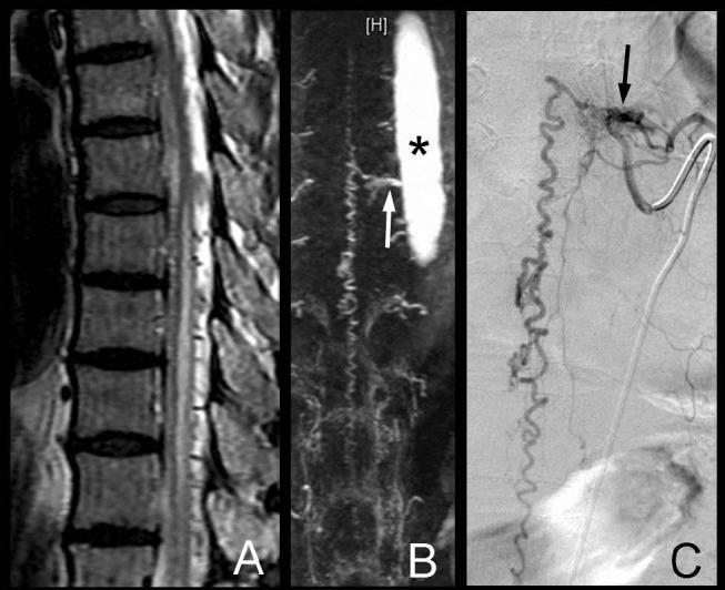
70 year-old male who presented with progressive myelopathy. A. Sag T2 FR FSE images of the thoracic spine demonstrate edema within the thoracic cord. Multiple intradural serpiginous flow voids are seen consistent with enlarged vascular channels. B. Contrast enhanced MRA with contrast bolus timed for maximal enhancement of the aortic arch was performed to evaluate for suspected dural AV fistula. MIP reformats show the feeding artery arising from the left T8 intercostal artery (arrow) (Aorta is indicated by a star). C. Digital subtraction spinal angiography confirmed the MRA findings (arrow) and embolization of the dural AV Fistula was subsequently performed.
Once the diagnosis of a vascular malformation is made or suspected on the basis of the initial study, further characterization should be made by angiography. Conventional angiogram is still performed at some point in the work up as it delineates vascular anatomy with the highest spatial resolution. Additionally, it provides temporal resolution making it possible to interrogate arterial supply, capillary phase and nidus, and venous drainage. However, it is an invasive procedure and requires the presence of interventional radiologists, nurses, technologists, and sometimes anesthesiologists.
MRA techniques have been dramatically improved to allow for better SNR which allows the use of a larger matrix to improve spatial resolution of small vessels within the spinal canal. Here, specifically contrast enhanced MRA's (Fig. 12) and 3T might provide the conspicuity to reliably characterize vascular malformations. Specifically, 3T offers increased baseline SNR and prolonged tissue T1 (relative to blood), which in turn boosts vascular contrast. Together with contrast agents with increased relaxivity and with better RF coils, this extra gain in contrast-to-noise might allow to appreciate even very small vessels. Another non-invasive angiographic technique is CTA, however, it does not provide distinction between arterial and venous structures that could be obtained from a time-resolved MRA, and is confounded in the spine by presence of bone which is not easy to eliminate during 3D post-processing of CTA images. Time-resolved MRAs offer to define the site of an arterio-venous fistula and resolve feeding arteries from dilated draining veins. Compared to conventional MRAs there are, however, advantages to CTA that include, increased spatial resolution and lack of artifacts that exist with performing an MRI. Cutting-edge MRA methods can easily compete with CTA particularly when parallel imaging is added. The high baseline SNR of MRAs is ideal for parallel imaging and allows to significantly speed up the acquisition during the bolus passage allowing better spatial resolution and arterio-venous separation. Here, the introduction of recent contrast agent with shorter T1 relaxivity can provide an ever better vessel delineation for the same amount of contrast agent injected.
Tumors
MR imaging of spinal tumours is required not only for initial diagnosis but also for guiding therapy and monitoring response to treatment. Classically, radiologists focus on localizing spinal lesions to extradural, intradural extramedullary and intramedullary compartments in order to generate an appropriate differential diagnosis. This is a surprisingly important step and highlights the importance of generating high quality T1 and T2-weighted images that are not degraded by patient motion or CSF pulsation and that have high spatial resolution. Extradural neoplastic lesions are far more likely to be due to secondary or metastatic disease, whereas primary lesions are more common than metastases in the intradural and intramedullary compartments. Primary lesions also differ between compartments, with lesions mainly arising from bone, muscle, fat, marrow or notochord remnants occurring in the epidural compartment and lesions arising from nerve roots, meninges, neuronal or glial elements within the intradural and intramedullary spaces. Within respective compartments, exact location of lesions can be helpful as well. For example, identification of a lesion that surrounds or abuts a nerve root supports the diagnosis of a nerve sheath tumor. Diagnosis can be further refined if this lesion can be shown to envelop the adjacent nerve root, as occurs with neurofibromas but not schwannomas.
Sometimes lesions transgress compartments or the exact location of a lesion is difficult to resolve on MR images. In such cases, one relies on identifying some of the specific features that point to a specific location. Extradural lesions cause focal displacement of the thecal sac and its contents away from the mass. Extrinsic compression of the thecal sac occurs and there is decrease in the CSF space between the lesion and the cord. Dura draped over the mass and an epidural fat-cap are helpful signs that one looks for on MRI to determine that a lesion is extradural. Intradural extramedullary lesions arise inside the dura but outside of the cord and cauda equina. These lesions tend to displace the spinal cord and enlarge the ipsilateral subarachnoid space with a sharp interface between the surface of the mass and the CSF space, creating a CSF-cap. Intramedullary lesions tend to expand the spinal cord but may grow exophytically. Balanced steady-state free precession (SSFP) sequences that have high contrast between tissue and fluid, including modifications to the sequence under the name Coherent Oscillatory State acquisition for the Manipulation of Image Contrast (COSMIC), the new volumetric FSE methods (FSE-XETA, SPACE, VISTA), or merely the better spatial resolution afforded by 3T as previously described, may be particularly helpful to accurately characterize lesion location in tricky cases.
Additional conventional imaging features may help to favor one diagnosis over another, but rarely are definitive. For example, chordomas classically show bright signal on T2-weighted images with a low signal rim and septations whereas lymphoma is characterized by relatively low T2 signal due to high cellularity [40, 41] However, such features are not definitive. Post-gadolinium images improve detection of intradural extramedullary disease and help to characterize and delineate intramedullary lesions, distinguishing enhancing tumour from associated non-enhancing cysts and from the non-enhancing spinal cord [42-43]. In the post operative setting, enhancement may be due either to reactive changes or residual or recurrent tumor. In tumors that involve the vertebrae or epidural space, effective uniform fat saturation is required as high signal from fat may otherwise obscure enhancing lesions.
Generally, the goals of new sequences in imaging spinal tumors include: 1. to improve lesion detection and delineation; 2. to differentiate different tumor histologies and grades; 3. to separate residual/recurrent tumor from post-surgical and post-treatment changes with greater accuracy; and 4. to determine the proximity of lesions to key spinal cord tracts for surgical planning and prognostic information.
Newer pulse sequences that have been investigated for use in spinal imaging have had mixed results. These include Fluid-attenuation inversion recovery (FLAIR), T2w STIR , MRS, DWI/DTI, and fMRI. FLAIR is a heavily T2-weighted sequence that nulls signal from CSF and thus is expected to increase conspicuity of hyperintense lesions in close proximity to CSF. Despite its utility in brain imaging, FLAIR has been less successful in the spine and has shown to be less sensitive than standard T2-weighted sequences in assessment of cord lesions [44-47] and is not recommended for tumour imaging. T2w STIR (a fat suppressed T2-weighted sequence) is commonly used to assess for vertebral body involvement in the setting of trauma or infection, due to increased conspicuity of edema in the vertebral marrow, and may similarly be helpful to identify vertebral body involvement with neoplastic disease especially in poorly enhancing lesions. Despite several studies that have demonstrated increased sensitivity of STIR for cord disease in multiple sclerosis relative to other T2-weighted sequences [47- 48], application to intramedullary tumours may be limited by poor signal to noise and greater sensitivity to motion than standard T2-weighted sequences [45]. Fat demonstrates hyperintensity on FSE, which is due to J-coupling. In certain cases, switching to true spin echoes is a possible alternative.
Perhaps the greatest advantage of newer T2 weighted sequences in tumor imaging will be conferred by the reduced artifacts from patient motion and CSF pulsation that will give a much more accurate visualization of lesions present within the cord and within the CSF space. This can be critical when following patients for subtle changes in T2 signal that may indicate tumour recurrence or when assessing intradural disease. In addition, as mentioned above, SSFP sequences including COSMIC with high contrast between CSF and soft tissue may provide a myelographic type sequence that is effective for lesion localization and assessment of the intradural space.
In brain imaging, MR spectroscopy is used frequently to interrogate tumors. MRS is a technique which can obtain biochemical information about tissues being studied by creating a spectrum of metabolites within a region of interest. Elevation of choline can be helpful to indicate increased cellular turnover and is therefore a marker of neoplastic disease rather than inflammatory disease or post-treatment related changes that can appear similar on T2w sequences. However, like for all other methods the small size and the harsh magnetic environment of the spine pose a big challenge, but future advances may make this technique both possible and useful in spinal cord imaging.
DWI, which can detect the altered cellular matrix of neoplastic tissues, may add to the staging of tumors and could help to differentiate different types of mass lesions. Promising results have been shown for the brain, where researchers reported DWI's ability to differentiate between cerebral tumor types; similar results might be anticipated for the spine. For example, in one patient suffering from an astrocytoma in the cervical cord, we found that the lesion had a significantly elevated ADC [18]. However, in high grade, heterogeneous tumors, such as glioblastoma multiforme or high grade astrocytomas, ADC values can vary over a large range and a general differentiation based on ADC can be difficult. Tumors with high cellularities, like lymphomas, usually demonstrate with massively decreased ADC. Also abscesses and epidermoids present with hyperintensities on DWI due to restricted diffusion (Fig. 13), which ultimately makes the diagnosis.
Figure 13.
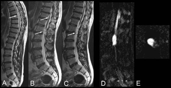
Sagittal T2w FSE images of the spine (A-C) demonstrate two non specific extramedullary/intradural masses requiring a differential diagnosis of several neoplastic etiologies. Sagittal and axial diffusion weighted images (D and E) show these masses to have significantly reduced diffusion thereby making a diagnosis of epidermoids. (images courtesy Dr. M. M. Thurnher, Medical University Vienna, Vienna, Austria).
Another approach is to use the orientational information obtained from DTI to perform fibertracking to determine whether a tumor invades or displaces fiber tracts. The latter might have consequences for the planning of surgical intervention and is currently the focus of several brain studies. Although fiber tracking is possible in the spine in the research setting, its accuracy has not yet been validated and it is uncertain whether it will translate well to the clinical setting.
Functional MRI has been employed in brain tumour imaging in order to map out eloquent areas of cortex so as to predict and minimize morbidity of tumor resection by possibly limiting resection or altering the surgical approach. Such information is likely to be helpful in spine tumor imaging. However, at the current time spinal fMRI still remains a research tool for reasons that are discussed below.
Functional MRI (FMRI) in the Spinal Cord
The application of fMRI [49] to the spinal cord appears to be a logical extension to its cephalad cousin, but in comparison has received relatively little attention thus far. In addition to the usual challenges of obtaining high quality fMRI data, the relatively low number of publications appears to be a consequence of the considerable challenge of acquiring MRIs of the spinal cord. However, the urgent need for an fMRI method adapted for demonstrating function in the spinal cord arises from the fact that there is no other non-invasive, global method available that can measure cord function. Since the cord is contained within the vertebral column, it is relatively inaccessible without opening the spinal canal and risking injury to the cord by inserting electrodes or needles and inflicting pain that could confound the study. The only means of assessing the function of the cord relies on the patient being able to feel a stimulus or having the proper reflexes. However, this assumes that the sensory receptors, peripheral nerves, and relevant areas of the brain, are all functioning normally. Even with normal function of these areas, very little information can be garnered about the cord's function distal to the location of an injury and relevant physical and physiological information that may be needed for proper assessment of a patient's condition or the effectiveness of treatment is masked. Two possible options to improve diagnosis are fMRI and evoked potentials. With evoked potentials (MEP, SEP, etc.) one can allocate a lesion in the spinal cord and distinguish it from a peripheral lesion. However, it doesn't provide imaging information of activation in concert with structural imaging. Most challenges of spinal cord fMRI arise from differences in the magnetic environment between the bone, cartilage, and tissues [49]. The net effect is subtle magnetic field variations within these materials and field gradients at their boundaries, which can cause distortion and loss of signal. Respiration, cardiac and CSF motion are another confounder that causes the field distortions to fluctuate rhythmically. After correcting for all these difficulties Drs. Mackey and Glover at our laboratory were for example able to demonstrate activation in the cervical/thoracic cord after presenting a nociceptive pain stimulus at the upper arm (Fig. 14).
Figure 14.
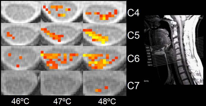
Functional MRI (fMRI) in the cervical spinal cord during performance of nociceptive pain stimulation on the right upper extremity using a Peltier element to generate local heating. Clearly, activation increases with increased temperature of the Peltier element. To isolate BOLD effect extra caution has to be exercised to minimize the influence from cord pulsation and respiratory artifacts. (images courtesy Drs. Mackey and Glover, Stanford University, Stanford, CA).
Non-traumatic Vertebral Body Compression Fractures
Vertebral compression fractures in the absence of trauma are a common clinical problem in the elderly population. Although clinical history is helpful, up to one third of fractures in patients with known primary malignancy are benign, and approximately one quarter of fractures in apparently osteopenic patients are due to metastases [49]. Diagnosis of an underlying lesion is important as it influences clinical staging, treatment planning and prognosis for the patient. In the chronic setting, the differentiation between pathologic fracture due to underlying malignancy and benign osteoporotic fracture is fairly simple and can be made with a high level of certainty [50-51]. Acute compression fractures, however, may share many of the imaging findings of metastatic lesions and differentiation is more challenging [52-53] Morphological signs, such as complete replacement of vertebral marrow, involvement of the posterior elements, and epidural or paraspinal masses, can be used to improve the diagnostic accuracy in predicting metastatic disease but may be equivocal. Results from recent studies have raised hope that DWI might be able to differentiate benign from malignant acute vertebral fractures. It has been reasoned that proton diffusivity is elevated in osteoporotic fractures because of bone marrow edema. Conversely, metastatic lesions might change diffusivity only moderately or even decrease it. It was postulated that a high cellularity of metastatic lesions, especially of actively growing tumors, would reduce proton diffusivity. Initial studies on DWI of the osseous spine to separate benign compression fractures from metastatic lesions were performed with a rather “exotic” diffusion-weighted SSFP sequence that is notoriously sensitive to confounders (e.g. relaxation times, B1 field, etc.) and that has an impressive discrimination capacity. However, subsequent studies using the more established Stejskal-Tanner based approach reported less enthusiastic and more mixed results. To date, the diagnostic utility of DWI to differentiate acute compression fractures is still controversial. MR perfusion curves have received some interest, and a pattern of rapid wash-in and wash-out of contrast may be predictive of metastatic compression fractures rather than benign compression fracture [54]. In-phase and out-of-phase Gradient echo imaging, which has been used for a long time to assess adrenal lesions in body imaging, is perhaps the most promising new technique suggested for the separation of metastatic spread from acute osteoporotic fractures. The use of in-phase and out-of-phase imaging to differentiate benign and malignant lesions is based on the assumption that malignant lesions completely replace vertebral body fat whereas in benign lesions fat is still present. Recently, Erly et al. showed that a signal intensity ratio for in- and out-of-phase images of >0.8 was able to predict metastatic disease whereas a ratio of <0.8 could predict benign compression fractures [55].
HIGH FIELD MR OF THE SPINE (3T)
As mentioned previously, high field imaging of the spine is appealing as 3T MRI essentially doubles the baseline SNR which can help when imaging small structures such as the cord or using sequences that require rapid-acquisition such as MRA. Issues that have to be addressed when migrating to high field MRI are increased SAR and stronger sensitivity to susceptibility distortions. While currently T2w-FSE scanning achieves outstanding imaging quality, there are still some unsolved issues with reduced T1 contrast at higher field. Although the spectrum of T1 values widens with increased field strengths many radiologist complain about the shallow T1 contrast at 3T. A simple remedy is to change the flip angle from 90 to improve contrast, but by doing that the SNR benefit is partially lost [56]. At higher field strength the T1 relaxation times of semisolid tissue increases, thus requiring longer TRs to fully relax. CSF on the other hand doesn't change much. This has to be considered as well, especially when setting up FLAIR sequences.
Summary
Size and extent of the spinal cord pose a substantial challenge to the process of MR image formation in this area. Although similar contrast parameters as in the brain can be used for the diagnostic work-up of the, new sequences, tailored to spine imaging provide better results than adapting conventional pulse sequences. In addition, more emphasis has to be made on SNR and pulsation. During the last few years major strides have been made in the development of new structural imaging sequences. The utility of more advanced methods, such as perfusion, diffusion, functional, or spectroscopic imaging still needs to be shown. For many of these methods further development in hard and software is needed before such an assessment can be made and a method is rejected prematurely.
Table 1.
Acronyms of new pulse sequences†
| GE | Philips | Siemens | |
|---|---|---|---|
| Multi-echo gradient echo acquisition | MERGE | N/A | MEDIC |
| Volumetric FSE | FSE-XETA | VISTA | SPACE |
other vendors might have similar sequences but these were not known to the authors at the time this manuscript was compiled.
Footnotes
Publisher's Disclaimer: This is a PDF file of an unedited manuscript that has been accepted for publication. As a service to our customers we are providing this early version of the manuscript. The manuscript will undergo copyediting, typesetting, and review of the resulting proof before it is published in its final citable form. Please note that during the production process errors may be discovered which could affect the content, and all legal disclaimers that apply to the journal pertain.
REFERENCES
- 1.Ghanem N, Uhl M, Brink I, Schafer O, Kelly T, Moser E, Langer M. Diagnostic value of MRI in comparison to scintigraphy, PET, MS-CT and PET/CT for the detection of metastases to bone. European Journal of Radiology. 2005;55:41–55. doi: 10.1016/j.ejrad.2005.01.016. [DOI] [PubMed] [Google Scholar]
- 2.Roemer PB, Edelstein WA, Hayes CE, Souza SP, Mueller OM. The NMR phased array. Magn Reson Med. 1990 Nov;16(2):192–225. doi: 10.1002/mrm.1910160203. [DOI] [PubMed] [Google Scholar]
- 3.Pruessmann KP, Weiger M, Scheidegger MB, Boesiger P. SENSE: sensitivity encoding for fast MRI. Magn Reson Med. 1999 Nov;42(5):952–62. [PubMed] [Google Scholar]
- 4.Griswold MA, Jakob PM, Heidemann RM, Nittka M, Jellus V, Wang J, Kiefer B, Haase A. Generalized autocalibrating partially parallel acquisitions (GRAPPA) Magn Reson Med. 2002 Jun;47(6):1202–10. doi: 10.1002/mrm.10171. [DOI] [PubMed] [Google Scholar]
- 5.Hollingworth W, Todd CJ, Bell MI, Arafat Q, Girling S, Karia KR, Dixon AK. The Diagnosis and Therapeutic Impact of MRI: an Observational Multi-centre Study. Clinical Radiology. 2000;55:825–831. doi: 10.1053/crad.2000.0546. [DOI] [PubMed] [Google Scholar]
- 6.Saal JS. General principles of diagnostic testing as related to painful lumbar spine disorders: A critical appraisal of current diagnostic techniques. Spine. 2002;27:2538–2545. doi: 10.1097/00007632-200211150-00027. [DOI] [PubMed] [Google Scholar]
- 7.Jensen MC, Brant-Zawadzki MN, OBuchowski N, Modic MT, Malkasian D, Ross JS. Magnetic resonance imaging of the lumbar spine in people without back pain. N Engl J Med. 1994;331:69–73. doi: 10.1056/NEJM199407143310201. [DOI] [PubMed] [Google Scholar]
- 8.Beattie PF, Meyers SP, Stratford P, Millard RW, Hollenberg GM. Association between patient report of symptoms and anatomic impairment visible on lumbar magnetic resonance imaging. Spine. 2000;25:819–28. doi: 10.1097/00007632-200004010-00010. [DOI] [PubMed] [Google Scholar]
- 9.Pfirrmann CW, Dora C, Schmid M, Zanetti M, Hodler J, Boos N. MR Image-based grading of lumbar nerve root compromise due to disk herniation: reliability study with surgical correlation. Radiology. 2004;230:583–8. doi: 10.1148/radiol.2302021289. [DOI] [PubMed] [Google Scholar]
- 10.Bammer R, Schoenberg SO. Current concepts and advances in clinical parallel magnetic resonance imaging. Top Magn Reson Imaging. 2004 Jun;15(3):129–58. doi: 10.1097/01.rmr.0000139666.23921.27. [DOI] [PubMed] [Google Scholar]
- 11.Yousem D, Atlas SW, Goldberg HI, Grossman RI. Degenerative narrowing of the cervical spine neural foramina: evaluation with high resolution 3D FT, gradient-echo MR imaging. Am J Neuroradiol. 1991;12:229–236. [PMC free article] [PubMed] [Google Scholar]
- 12.Forbes KP, Pipe JG, Bird CR, Heiserman JE. PROPELLER MRI: clinical testing of a novel technique for quantification and compensation of head motion. J Magn Reson Imaging. 2001 Sep;14(3):215–22. doi: 10.1002/jmri.1176. [DOI] [PubMed] [Google Scholar]
- 13.Gaddipati A, Kumar A, Peters R, Francis R, Kanal E. Proc. Intl. Soc. Mag. Reson. Med. Vol. 14. Seattle, WA: 2006. Motion Robust T2-weighted imaging of C-spine with Propeller with Fast Recovery Modification; p. 3140. [Google Scholar]
- 14.Melhem ER, Itoh R, Folkers PJ. Cervical spine: three-dimensional fast spin-echo MR imaging--improved recovery of longitudinal magnetization with driven equilibrium pulse. Radiology. 2001 Jan;218(1):283–8. doi: 10.1148/radiology.218.1.r01ja38283. [DOI] [PubMed] [Google Scholar]
- 15.D'Aprile P, Tarantino A, Jinkins JR, Brindicci D. The value of fat saturation sequences and contrast medium administration in MRI of degenerative disease of the posterior/perispinal elements of the lumbosacral spine. Eur Radiol. 2007 Feb;17(2):523–31. doi: 10.1007/s00330-006-0324-0. Epub 2006 May 30. [DOI] [PubMed] [Google Scholar]
- 16.Willen J, Danielson B, Gaulitz A. Dynamic effects on the lumbar spinal canal: axially loaded CT myelography and MRI in patients with sciatica and/or neurogenic claudication. Spine. 1997;22:2968–76. doi: 10.1097/00007632-199712150-00021. [DOI] [PubMed] [Google Scholar]
- 17.Danielson B, Willen J. Axially loaded magnetic resonance image of the lumbar spine in asymptomatic individuals. Spine. 2001;26:2601–6. doi: 10.1097/00007632-200112010-00015. [DOI] [PubMed] [Google Scholar]
- 18.Bammer R, Fazekas F, Augustin M, Simbrunner J, Strasser-Fuchs S, Seifert T, Stollberger R, Hartung HP. Diffusion-weighted MR imaging of the spinal cord. AJNR Am J Neuroradiol. 2000;21(3):587–91. [PMC free article] [PubMed] [Google Scholar]
- 19.Henegar MM, Moran CJ, Silbergeld DL. Early postoperative magnetic resonance imaging following nonneoplastic cortical resection. J Neurosurg. 1996;84:174–9. doi: 10.3171/jns.1996.84.2.0174. [DOI] [PubMed] [Google Scholar]
- 20.Reeder SB, Yu H, Johnson JW, Shimakawa A, Brittain JH, Pelc NJ, Beaulieu CF, Gold GE. T1- and T2-weighted fast spin-echo imaging of the brachial plexus and cervical spine with IDEAL water-fat separation. J Magn Reson Imaging. 2006 Oct;24(4):825–32. doi: 10.1002/jmri.20721. [DOI] [PubMed] [Google Scholar]
- 21.Guo AC, Provencale JM, Cruz LCH, Jr, Petrella JR. Cerebral Abscesses: investigation using apparent diffusion coefficient maps. Neuroradiology. 2001;43:370–4. doi: 10.1007/s002340000519. [DOI] [PubMed] [Google Scholar]
- 22.Stevens KJ, Spenciner DB, Griffiths KL, Kim KD, Zwienenberg-Lee M, Alamin T, Bammer R. Comparison of minimally invasive and conventional open posterolateral lumbar fusion using magnetic resonance imaging and retraction pressure studies. J Spinal Disord Tech. 2006 Apr;19(2):77–86. doi: 10.1097/01.bsd.0000193820.42522.d9. [DOI] [PubMed] [Google Scholar]
- 23.Kruger DG, Riederer SJ, Grimm RC, Rossman PJ. Continuously moving table data acquisition method for long FOV contrast-enhanced MRA and whole-body MRI. Magn Reson Med. 2002 Feb;47(2):224–31. doi: 10.1002/mrm.10061. [DOI] [PubMed] [Google Scholar]
- 24.Liu XZ, Xu XM, Hu R, et al. Neuronal and glial apoptosis after traumatic spinal cord injury. J Neurosci. 1997;17:5395–5406. doi: 10.1523/JNEUROSCI.17-14-05395.1997. [DOI] [PMC free article] [PubMed] [Google Scholar]
- 25.Ford JC, Hackney DB, Alsop DC, et al. MRI characterization of diffusion coefficients in a rat spinal cord injury model. Magn Reson Med. 1994;31:488–494. doi: 10.1002/mrm.1910310504. [DOI] [PubMed] [Google Scholar]
- 26.Schwartz ED, Yezierski RP, Pattany PM, Quencer RM, Weaver RG. Diffusion-weighted MR imaging in a rat model of syringomyelia after excitotoxic spinal cord injury. AJNR Am J Neuroradiol. 1999;20:1422–1428. [PMC free article] [PubMed] [Google Scholar]
- 27.Solomou E, Maragkos M, Kotsarini C, Konstantinou D, Maraziotis T. Multiple spinal epidural abscesses extending to the whole spinal canal. Magn Reson Imaging. 2004;22:747–50. doi: 10.1016/j.mri.2004.01.072. [DOI] [PubMed] [Google Scholar]
- 28.Eastwood JD, Vollmer RT, Provenzale JM. Diffusion-weighted imaging in a patient with vertebral and epidural abscesses. AJNR Am J Neuroradiol. 2002;23:496–8. [PMC free article] [PubMed] [Google Scholar]
- 29.Rocca MA, Mastronardo G, Horsfield MA, Pereira C, Iannucci G, Colombo B, Moiola L, Comi G, Filippi M. Comparison of Three MR Sequences for the detection of the cervical cord lesions in patients with multiple sclerosis. AJNR. 1999;20:1710–1716. [PMC free article] [PubMed] [Google Scholar]
- 30.Thorpe JW, Kidd D, Moseley IF, Thompson AJ, MacManus DG, Compston DA, McDonald WI, Miller DH. Spinal MRI in patients with suspected multiple sclerosis and negative brain MRI. Brain. 1996;119:709–714. doi: 10.1093/brain/119.3.709. [DOI] [PubMed] [Google Scholar]
- 31.Balaban RS, Ceckler LS. Magnetization transfer contrast in magnetic resonance imaging. Magn Reson Q. 1992;8:116–137. [PubMed] [Google Scholar]
- 32.Ramani A, Dalton C, Miller DH, Tofts PS, Barker GJ. Precise estimation of fundamental in-vivo MT parameters in human brain in clinically feasible times. Magn Reson Imaging. 2002;20:721–731. doi: 10.1016/s0730-725x(02)00598-2. [DOI] [PubMed] [Google Scholar]
- 33.Lycklamaa Nijehold GJ, Barkhof F, Scheltens P, Castelijns JA, Ader H, van Waesberqhe JH, Polman C, Jonqen SJ, Valk J. MR of the spinal cord in multiple Sclerosis: relation to clinical subtype and disability. AJNR. 1997;18:1041–1048. [PMC free article] [PubMed] [Google Scholar]
- 34.Losseff NA, Webb SL, O'Riordan JI, Page R, Wang L, Barker GJ, Tofts PS, McDonald WI, Miller DH, Thompson AJ. Spinal cord atrophy and disability in multiple sclerosis. A new reproducible and sensitive MRI method with potential to monitor disease progression. Brain. 1996;119:701–708. doi: 10.1093/brain/119.3.701. [DOI] [PubMed] [Google Scholar]
- 35.Ford JC, Hackney DB, Alsop DC, et al. MRI characterization of diffusion coefficients in a rat spinal cord injury model. Magn Reson Med. 1994;31:488–494. doi: 10.1002/mrm.1910310504. [DOI] [PubMed] [Google Scholar]
- 36.Schwartz ED, Yezierski RP, Pattany PM, Quencer RM, Weaver RG. Diffusion-weighted MR imaging in a rat model of syringomyelia after excitotoxic spinal cord injury. AJNR Am J Neuroradiol. 1999;20:1422–1428. [PMC free article] [PubMed] [Google Scholar]
- 37.Ries M, Jones RA, Dousset V, Moonen CTW. Diffusion tensor MRI of the spinal cord. Magn Reson Med. 2000;44(6):884–92. doi: 10.1002/1522-2594(200012)44:6<884::aid-mrm9>3.0.co;2-q. [DOI] [PubMed] [Google Scholar]
- 38.Restrepo L, Guttin JF. Acute spinal cord ischemia during aortography treated with intravenous thrombolytic therapy. Tex Heart Inst J. 2006;33(1):74–7. [PMC free article] [PubMed] [Google Scholar]
- 39.Butts K, de Crespigny A, Pauly JM, Moseley M. Diffusion-weighted interleaved echo-planar imaging with a pair of orthogonal navigator echoes. Magn Reson Med. 1996;35(5):763–70. doi: 10.1002/mrm.1910350518. [DOI] [PubMed] [Google Scholar]
- 40.Wippold FJ, Koeller KK, Smirniotopoulos JG. Clinical and imaging features of the cervical chordoma. AJR Am J Roentgenol. 1999;172:1423–6. doi: 10.2214/ajr.172.5.10227531. [DOI] [PubMed] [Google Scholar]
- 41.Mulligan ME, McRae GA, Murphey MD. Imaging features of primary lymphoma of bone. AJR AM J Roentgenol. 1999;173:1691–7. doi: 10.2214/ajr.173.6.10584821. [DOI] [PubMed] [Google Scholar]
- 42.Sze G. Gadolinium-DTPA in the evaluation of intradural extramedullary spinal disease. AJR Am J Roentgenol. 1988;150:911–21. doi: 10.2214/ajr.150.4.911. [DOI] [PubMed] [Google Scholar]
- 43.Sze G, Krol G, Zimmermen RD, Deck MD. Intramedullary disease of the spine: diagnosis using gadolinium-DTPA-enhanced MR imaging. AJR Am J Roentgenol. 1988;151:1193–1204. doi: 10.2214/ajr.151.6.1193. [DOI] [PubMed] [Google Scholar]
- 44.Ross JS. Newer sequences for spinal MR imaging: smorgasbord or succotash of acronyms? AJNR Am J Neuroradiol. 1999;20:361–73. [PMC free article] [PubMed] [Google Scholar]
- 45.Lowe GM. Magnetic resonance imaging of intramedullary spinal cord tumors. Journal of Neuro-Oncology. 2000;47:195–210. doi: 10.1023/a:1006462321234. [DOI] [PubMed] [Google Scholar]
- 46.Keiper MD, Grossman RI, Brunson JC, Schnall MD. The low sensitivity of fluid-attenuation inversion-recovery MR in the detection of multiple sclerosis of the spinal cord. AJNR Am J Neuroradiol. 1997;18:1035–9. [PMC free article] [PubMed] [Google Scholar]
- 47.Hitmair K, Mallek R, Prayer D, Schindler EG, Kolleger H. Spinal cord lesions in patients with multiple sclerosis: comparison of pulse sequences. AJNR Am J Neuroradiol. 1996;17:1555–65. [PMC free article] [PubMed] [Google Scholar]
- 48.Rocca MA, MAstronardo G, Horsfield MA, Pereira C, Iannucci G, Colombo B, Moiola L, Comi G, Filippi M. Comparison of three MR sequences for detection of cervical cord lesions in patients with multiple sclerosis. AJNR Am J Neuroradiol. 1999;20:1710–6. [PMC free article] [PubMed] [Google Scholar]
- 49.Ross JS, Brant-Zawadski M, Moore KR, Crim J, Chen MZ, Katzman GL. Amirsys Inc; Salt Lake City: 2004. Diagnostic Imaging: Spine. [Google Scholar]
- 50.Baker LL, Goodman SB, Perkash I, Lane B, Enzmann DR. Benign versus pathologic compression fractures of vertebral bodies: assessment with conventional spin-echo, chemical-shift and STIR MR Imaging. Radiology. 1990;174:495–502. doi: 10.1148/radiology.174.2.2296658. [DOI] [PubMed] [Google Scholar]
- 51.An HS, Andreshak TG, Nguyen C, Williams A, Daniels D. Can we distinguish between benign vs malignant compression fractures of the spine by magnetic resonance imaging? Spine. 1995;20:1776–82. doi: 10.1097/00007632-199508150-00005. [DOI] [PubMed] [Google Scholar]
- 52.Rupp RE, Ebraheim NA, Coombs RJ. Magnetic resonance imaging differentiation of compression spine fractures or vertebral lesions caused by osteoporosis or tumour. Spine. 1995;23:2499–2503. doi: 10.1097/00007632-199512000-00007. discussion 2504. [DOI] [PubMed] [Google Scholar]
- 53.Yuh WT, Zachar CK, Barloon TJ, Sato Y, Sickels WJ, Hawes DR. Vertebral compression fractures: distinction between benign and malignant causes with MR imaging. Radiology. 1989;172:215–8. doi: 10.1148/radiology.172.1.2740506. [DOI] [PubMed] [Google Scholar]
- 54.Chen WT, Shih TT, Chen RC, Lo HY, Chou CT, Lee JM, Tu HY. Blood perfusion of vertebral lesions evaluated with gadolinium-enhanced dynamic MRI: in comparison with compression fracture and metastasis. J Magn Reson Imaging. 2002;15:308–14. doi: 10.1002/jmri.10063. [DOI] [PubMed] [Google Scholar]
- 55.Erly WK, Oh ES, Outwater EK. The utility of in-phase/opposed-phase imaging in differentiating malignancy from acute benign compression fractures of the spine. AJNR Am J Neuroradiol. 2006;27:1183–88. [PMC free article] [PubMed] [Google Scholar]
- 56.Schmitz BL, Gron G, Brausewetter F, Hoffmann MH, Aschoff AJ. Enhancing gray-to-white matter contrast in 3T T1 spin-echo brain scans by optimizing flip angle. AJNR Am J Neuroradiol. 2005 Sep;26(8):2000–4. [PMC free article] [PubMed] [Google Scholar]