

More than 80 years ago, Cooley and Lee described skeletal changes associated with haemolytic anaemias.1 Hair‐on‐end appearance of the skull is a characteristic feature of chronic haemolysis usually seen in patients with thalassaemia and sickle cell anaemia. It results from accentuated vertical trabeculae between the inner and outer tables of the skull because of excessive bone marrow hyperplasia. An incidence of 8.3% of hair‐on‐end appearance in thalassaemic patients has been reported.2 Though, rare, these appearances have been described in iron deficiency anaemia and cyanotic congenital heart disease.3,4
This young child had been diagnosed at age 9 months with thalassaemia major (parents and siblings were normal). At presentation, in respiratory distress, he was undergoing monthly blood transfusions. Skull x ray showed characteristic hair‐on‐end appearance (figs 1 and 2). With the availability of effective treatments for thalassaemia in developed countries, this complication is rarely seen.

Figure 1 Hair‐on‐end appearance: lateral view.
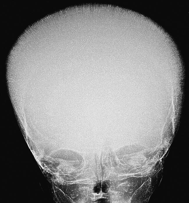
Figure 2 Hair‐on‐end appearance: frontal view.
Footnotes
Competing interests: none
References
- 1.Cooley T B, Lee P. A series of cases of anemia with splenomegaly and peculiar bone changes. Trans Am Pediatr Soc 19253729–30. [Google Scholar]
- 2.Wisetsin S. Cepalography in thalassemic patients. J Dent Assoc Thai 199040260–268. [PubMed] [Google Scholar]
- 3.Britton H A, Canby J P, Kohler C M. Iron deficiency anemia producing evidence of marrow hyperplasia in the calvarium. Pediatrics 196025621–628. [PubMed] [Google Scholar]
- 4.Walor D M, Berdon W E, Westra S J. ‘Hair‐on‐end' skull changes resembling thalassemia caused by marrow expansion in uncorrected complex cyanotic heart disease. Pediatr Radiol 200535698–701. [DOI] [PubMed] [Google Scholar]