A 3‐year‐old girl presented with a 12 hour history of left sided weakness. The parents reported that she had experienced episodic partial amnesia in the preceding weeks, being unable to recognise her favourite cartoon characters and family friends.
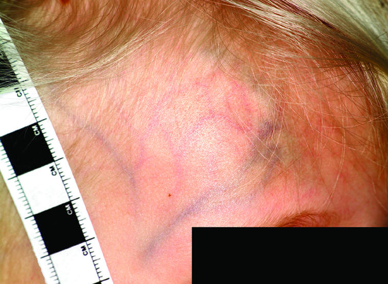
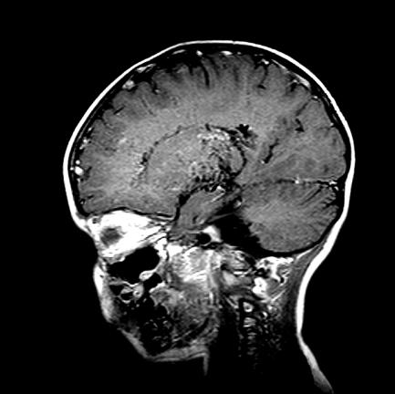
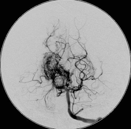
The examination revealed reduced power in the left upper and lower limb (4/5) and unsteady gait, without other neurological abnormalities. Prominent dilated, pulsating veins in the right parietal region and a audible bruit in the same area were noted on examination (fig 1). A cranial MRI (fig 2) scan showed a large, dysplastic arteriovenous malformation with moyamoya‐like appearance extending throughout most of the right hemisphere and crossing the midline. An angiogram (fig 3) confirmed shunting between this malformation and the extracranial circulation.

Figure 1 Clinical photograph showing prominent scalp veins. Consent was obtained for publication of this figure.
Figure 2 Initial MRI scan showing an extensive vascular malformation in the right hemisphere.
Figure 3 Angiogram showing the dysplastic character of the arteriovenous malformation. Further sequences showed shunting between intra‐ and extracerebral circulation.
The patient recovered fully from this episode but continues to experience transient ischaemic attacks with varying symptoms including amnesia, mood swings, motor deficits, and visual impairment. The treatment options are limited by her young age and the extent of the malformation. Since interventional endovascular closure is deemed to be technically unfeasible, alternatives including microsurgery and radiosurgery are currently being considered.
Arteriovenous malformations are congenital disorders with an estimated prevalence of 0.68 per 100 000.1 Data from postmortem examinations suggest that as few as 12% become symptomatic during life.2 Although the majority of cases present before 40 years of life, symptomatic manifestation this young is extremely rare. Intracranial haemorrhage is the most common presenting feature, followed by seizures, recurrent headaches, and progressive neurological deficit.3 Heart failure, macrocephaly, and prominent scalp veins are less commonly seen in this group of patients.
Footnotes
Competing interests: None declared.
References
- 1.Stapf C, Mast H, Sciacca R R.et al The New York Islands AVM Study: design, study progress, and initial results. Stroke 200334e29–e33. [DOI] [PubMed] [Google Scholar]
- 2.McCormick W F. Classification, pathology, and natural history of angiomas of the central nervous system. Wkly Update Neurol Neurosurg 1978142–7. [Google Scholar]
- 3.Nataf F, Schlienger M, Lefkopoulos D.et al Radiosurgery of cerebral arteriovenous malformations in children: a series of 57 cases. Int J Radiat Oncol Biol Phys 200357184–195. [DOI] [PubMed] [Google Scholar]


