

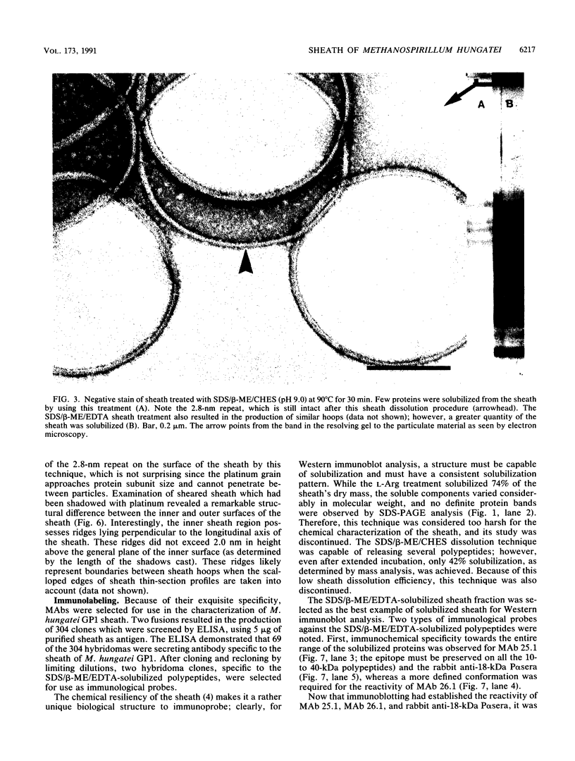
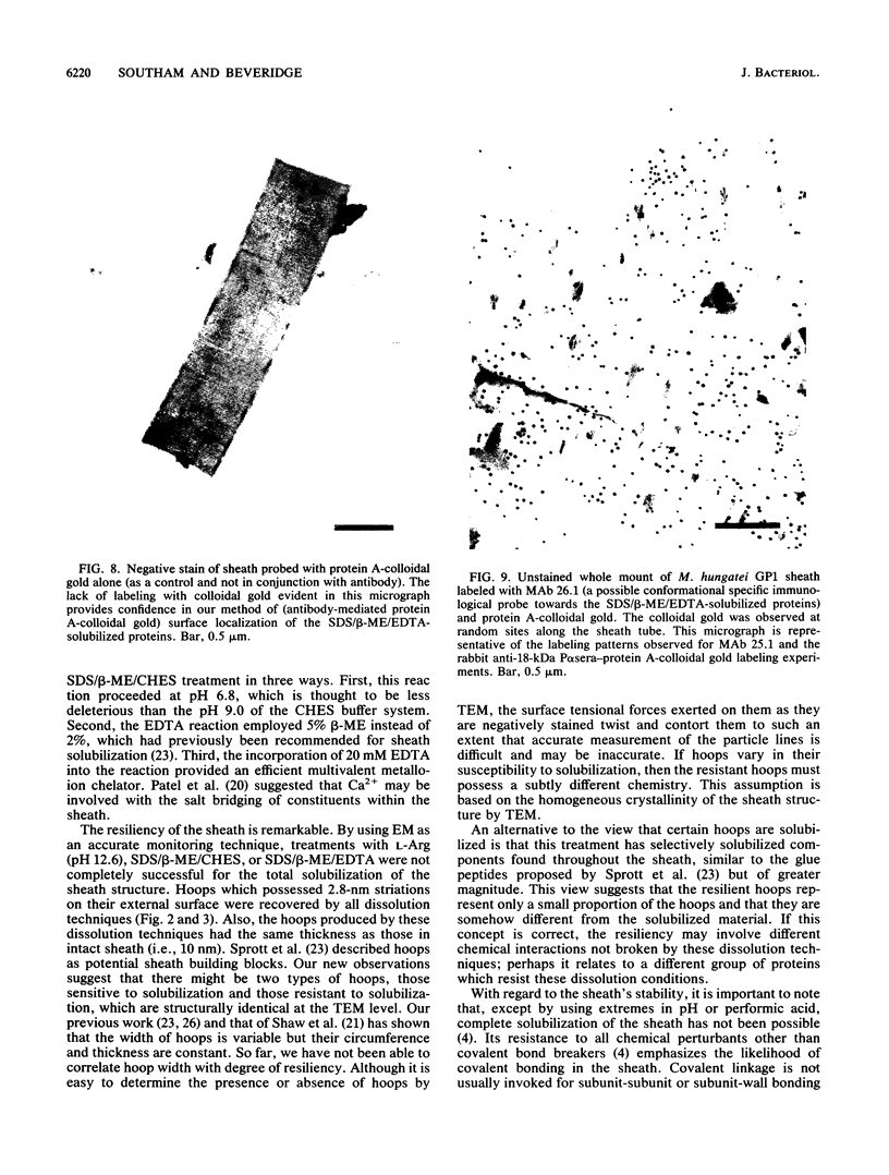
Abstract
The sheath of Methanospirillum hungatei GP1 was degraded by three dissolution techniques, which produced a range of soluble products. By using 0.05 M L-arginine buffer (pH 12.6) at 90 degrees C for 10 min, 74% (dry weight) of the sheath was dissolved; however, the solubilized polypeptides were extensively degraded. Treatment with 2% beta-mercaptoethanol and 2% sodium dodecyl sulfate at 90 degrees C in 0.05 M 2(N-cyclohexylamino)ethanesulfonic acid (CHES) buffer (pH 9.0) solubilized 42% (dry weight) of the sheath as a group of polypeptides of 30 to 40 kDa. At 100 degrees C for 2 h, 5% beta-mercaptoethanol, 2% sodium dodecyl sulfate (SDS), and 20 mM EDTA released 74% of the sheath's mass as a group of polypeptides of 10 to 40 kDa. All solubilized products were examined by SDS-polyacrylamide gel electrophoresis, and a range of high- and low-molecular-weight polypeptides was identified. None were glycoproteins. Hoops, which comprise the sheath's structure, were seen by electron microscopy after all of the attempted dissolutions. Monoclonal antibodies were produced against the 10- to 40-kDa range of solubilized products and against the approximately 40-kDa polypeptides, and polyclonal antiserum was produced against an 18-kDa polypeptide. These immunological markers were used in Western immunoblotting and protein A-colloidal gold-antibody probing by electron microscopy to identify the structural location of the various polypeptides. Native sheath, which possesses 2.8-nm particles on its outer surface (M. Stewart, T.J. Beveridge, and G.D. Sprott, J. Mol. Biol. 183:509-515, 1985; P.J. Shaw, G.J. Hills, J.A. Henwood, J.E. Harris, and D.B. Archer, J. Bacteriol. 161:750-757, 1985), presented a gentle wave-form surface in platinum-shadowed specimens. In contrast, the inner face of the sheath was highlighted by ridges lying perpendicular to the longitudinal axis of the sheath and likely corresponded to hoop boundaries. Both the polyclonal and monoclonal antibodies were specific for different faces; polyclonal antibodies labeled the inner face, whereas monoclonal antibodies labeled the outer face. Accordingly, the apparent asymmetry of structure between the two faces of the sheath can be correlated by our immunochemical probing with a distinct asymmetry in the distribution of exposed polypeptides between the faces. The possible implications of this asymmetry for growth and maturation of the sheath are explained.
Full text
PDF


Images in this article


Selected References
These references are in PubMed. This may not be the complete list of references from this article.
- Bendayan M. Double immunocytochemical labeling applying the protein A-gold technique. J Histochem Cytochem. 1982 Jan;30(1):81–85. doi: 10.1177/30.1.6172469. [DOI] [PubMed] [Google Scholar]
- Beveridge T. J., Southam G., Jericho M. H., Blackford B. L. High-resolution topography of the S-layer sheath of the archaebacterium Methanospirillum hungatei provided by scanning tunneling microscopy. J Bacteriol. 1990 Nov;172(11):6589–6595. doi: 10.1128/jb.172.11.6589-6595.1990. [DOI] [PMC free article] [PubMed] [Google Scholar]
- Beveridge T. J., Stewart M., Doyle R. J., Sprott G. D. Unusual stability of the Methanospirillum hungatei sheath. J Bacteriol. 1985 May;162(2):728–737. doi: 10.1128/jb.162.2.728-737.1985. [DOI] [PMC free article] [PubMed] [Google Scholar]
- Burnette W. N. "Western blotting": electrophoretic transfer of proteins from sodium dodecyl sulfate--polyacrylamide gels to unmodified nitrocellulose and radiographic detection with antibody and radioiodinated protein A. Anal Biochem. 1981 Apr;112(2):195–203. doi: 10.1016/0003-2697(81)90281-5. [DOI] [PubMed] [Google Scholar]
- Fairbanks G., Steck T. L., Wallach D. F. Electrophoretic analysis of the major polypeptides of the human erythrocyte membrane. Biochemistry. 1971 Jun 22;10(13):2606–2617. doi: 10.1021/bi00789a030. [DOI] [PubMed] [Google Scholar]
- Laemmli U. K. Cleavage of structural proteins during the assembly of the head of bacteriophage T4. Nature. 1970 Aug 15;227(5259):680–685. doi: 10.1038/227680a0. [DOI] [PubMed] [Google Scholar]
- Lam M. Y., McGroarty E. J., Kropinski A. M., MacDonald L. A., Pedersen S. S., Høiby N., Lam J. S. Occurrence of a common lipopolysaccharide antigen in standard and clinical strains of Pseudomonas aeruginosa. J Clin Microbiol. 1989 May;27(5):962–967. doi: 10.1128/jcm.27.5.962-967.1989. [DOI] [PMC free article] [PubMed] [Google Scholar]
- MacDonald R. I. Membrane fusion due to dehydration by polyethylene glycol, dextran, or sucrose. Biochemistry. 1985 Jul 16;24(15):4058–4066. doi: 10.1021/bi00336a039. [DOI] [PubMed] [Google Scholar]
- Markwell M. A., Haas S. M., Bieber L. L., Tolbert N. E. A modification of the Lowry procedure to simplify protein determination in membrane and lipoprotein samples. Anal Biochem. 1978 Jun 15;87(1):206–210. doi: 10.1016/0003-2697(78)90586-9. [DOI] [PubMed] [Google Scholar]
- Mutharia L. M., Hancock R. E. Surface localization of Pseudomonas aeruginosa outer membrane porin protein F by using monoclonal antibodies. Infect Immun. 1983 Dec;42(3):1027–1033. doi: 10.1128/iai.42.3.1027-1033.1983. [DOI] [PMC free article] [PubMed] [Google Scholar]
- Patel G. B., Roth L. A., van den Berg L., Clark D. S. Characterization of a strain of Methanospirillum hungatti. Can J Microbiol. 1976 Sep;22(9):1404–1410. doi: 10.1139/m76-208. [DOI] [PubMed] [Google Scholar]
- Shaw P. J., Hills G. J., Henwood J. A., Harris J. E., Archer D. B. Three-dimensional architecture of the cell sheath and septa of Methanospirillum hungatei. J Bacteriol. 1985 Feb;161(2):750–757. doi: 10.1128/jb.161.2.750-757.1985. [DOI] [PMC free article] [PubMed] [Google Scholar]
- Sleytr U. B., Messner P. Crystalline surface layers in procaryotes. J Bacteriol. 1988 Jul;170(7):2891–2897. doi: 10.1128/jb.170.7.2891-2897.1988. [DOI] [PMC free article] [PubMed] [Google Scholar]
- Sprott G. D., Colvin J. R., McKellar R. C. Spheroplasts of Methanospirillum hungatii formed upon treatment with dithiothreitol. Can J Microbiol. 1979 Jun;25(6):730–738. doi: 10.1139/m79-106. [DOI] [PubMed] [Google Scholar]
- Sprott G. D., McKellar R. C. Composition and properties of the cell wall of Methanospirillum hungatii. Can J Microbiol. 1980 Feb;26(2):115–120. doi: 10.1139/m80-017. [DOI] [PubMed] [Google Scholar]
- Stewart M., Beveridge T. J., Sprott G. D. Crystalline order to high resolution in the sheath of Methanospirillum hungatei: a cross-beta structure. J Mol Biol. 1985 Jun 5;183(3):509–515. doi: 10.1016/0022-2836(85)90019-1. [DOI] [PubMed] [Google Scholar]
- Towbin H., Staehelin T., Gordon J. Electrophoretic transfer of proteins from polyacrylamide gels to nitrocellulose sheets: procedure and some applications. Proc Natl Acad Sci U S A. 1979 Sep;76(9):4350–4354. doi: 10.1073/pnas.76.9.4350. [DOI] [PMC free article] [PubMed] [Google Scholar]