

Abstract
By explanting tissues isolated microsurgically from implanting strain 129 mouse blastocysts individually on STO feeder cells we have established that embryonic stem (ES) cells originate from the epiblast (primitive ectoderm). Isolated early epiblasts yielded ES cell lines at a substantially higher frequency than intact blastocysts regardless of whether they were explanted whole or as strictly single-cell suspensions. When explanted from delayed-implanting 129 blastocysts, epiblasts gave lines consistently in 100% of cases. If primary embryonic fibroblasts rather than STO cells were used as feeders, germline-competent ES cell lines were obtained readily from epiblasts of delayed-implanting blastocysts of several hitherto refractory strains, particularly when recombinant leukemia inhibitory factor was included in the medium during the initial period of culture. Because lines were obtained from the nonpermissive CBA/Ca strain at a rate of up to 56%, this approach to the derivation of germline-competent ES cell lines may not only prove generic for the mouse but also worth pursuing in other species of mammal.
Considering how widely they are now used in transgenesis, surprisingly little is known about the origin and biology of mouse embryonic stem (ES) cells (1). Thus, while these cells are generally held to originate from the early epiblast or primitive ectoderm, this has not been demonstrated unequivocally because the starting material for their derivation has invariably been either dissociated morulae (2), intact blastocysts (3), or entire inner-cell masses (ICMs) (4). Even ICMs that have been exposed to immunosurgery (5) cannot be assumed to be composed of epiblast tissue that is entirely free from contaminating primitive endoderm (6). At present, the case that ES cells are of epiblastic origin depends on comparison of their properties with those of the different tissues of periimplantation conceptuses (1, 7). Although particular weight is assigned to their similarity to epiblast in developmental potential, this cannot be regarded as definitive because yolk sac teratomas, which can yield as rich a variety of differentiated tissues as ectopically grafted ES cells (8), evidently originate from primitive, endoderm-derived cells (9).
One reason for establishing the origin of ES cells is to determine whether the efficiency of obtaining them can be improved by explanting their precursor cells alone rather than, as hitherto, together with other tissues of the blastocyst. So far, even in the 129 strain, which is held to be most permissive in yielding ES cell lines, a success rate of 30% is regarded as high (10). ES cell lines of proven ability to colonize the germ-line have been obtained at very low frequency in only a few mouse strains other than 129 and, as yet, in no other species of mammal (1). These strain and species limitations severely restrict the scope of transgenesis via ES cells for elucidating gene function and for obtaining appropriate animal models of human genetic diseases.
Very recently, blastocysts carrying a transgene consisting of the Neo coding region linked to regulatory sequences of the Oct3/4 gene have been used in an attempt to overcome the barrier to ES cell production in a purportedly nonpermissive stock of mice (11). The rationale of the approach is based on the assumption that because expression of Oct3/4 is restricted to undifferentiated cells of the early conceptus, selection against cells in which the gene is down-regulated may aid the establishment of ES lines by enriching for their precursor cells. However, while the background of the mouse stock that was used was predominantly CBA, a reputedly nonpermissive strain, it also included a significant minor component from the permissive C57BL/6 strain. Given the low percentage of blastocysts from which lines were obtained and the persisting ignorance about the genetic basis of permissivity, the claim of success in a nonpermissive strain is not compelling. A further problem with this approach is that expression of Neo in all the resulting ES cell lines precludes the use of this very valuable selection system in subsequent transfection experiments.
Here we describe a simpler and more direct approach to the problem of devising a generic technique for deriving ES cell lines in the mouse and hence, possibly, in other mammals. This was to establish unequivocally the identity of the ES progenitors so that they could be allowed to interact directly with feeders after isolation from all other cells of the periimplantation conceptus. As well as enabling ES cell lines to be established from 100% of conceptuses of the 129 strain, this approach also gave high success rates in several hitherto nonpermissive strains, including pure CBA/Ca.
MATERIALS AND METHODS
Mice.
Natural matings between mice of the 129/Ola, C57BL/6, and CBA/Ca inbred strains and of the PO or MF1 random-bred strains were used to provide blastocysts. Some PO females were mated with males of a heterogenous stock that were homozygous for the ROSA26-B geo transgene (12). Midgestation fetuses that were used as a source of primary embryonic fibroblasts (PEFs) were from matings between PO or CBA/Ca mice. PO females mated to vasectomized males of the same strain were used as pseudopregnant recipients for blastocyst transfer. Mice were kept either on a 12-hr light/dark cycle in which the dark period was from 1900 hr to 0700 hr, or on a 14-hr light/10-hr dark cycle in which the dark period was from 1300 hr to 2300 hr. Implantation was delayed by ovariectomizing females in the afternoon of the 3rd day of pregnancy and injecting them subcutaneously with 1 mg of Depo-Provera (Upjohn) immediately thereafter. Delayed-implanting blastocysts were recovered 1 week after ovariectomy. Nondelayed blastocysts were recovered from intact females on either the 4th or 5th day after mating.
Recovery and Microdissection of Blastocysts.
Mouse tubal fluid (MTF)–Hepes medium (13) was used for the recovery, storage at room temperature, and micromanipulation of blastocysts. Fifth-day-postcoitum (p.c.) implanting and 9th-day-p.c. delayed-implanting blastocysts were microdissected into their component tissues using a pair of solid, fine-tipped, siliconized (Repelcote, BDH, Poole, UK) glass needles, essentially as described elsewhere (6, 14, 15). Briefly, the mural trophectoderm was torn open with the needles and the blastocysts then were incubated for 18–24 min in a mixture of 0.5% (wt/vol) trypsin (Difco; 1/250) and 2.5% (wt/vol) pancreatin (Difco). After a rinse in MTF–Hepes, each blastocyst was opened up with the outer surface of the trophectoderm pinned against the undersurface of the coverslip of the Puliv (Leitz) manipulation chamber with one needle, while the other needle was used first to gently scrape the primitive endoderm from the epiblast and then scrape the epiblast from the polar trophectoderm. Tissue fractions were then explanted individually into four-well plates (Nunc) that contained fresh ES medium (10) and had been seeded the previous afternoon with γ-irradiated STO cells or PEFs. For dissociation before explantation, isolated epiblasts were incubated for 6–8 min at 37°C in 0.25% pronase (grade B, Calbiochem) in PBS before being rinsed and then incubated for 30–40 min in calcium-free OC medium containing EGTA at 0.2 mg/ml.
Isolation, Culture, Sexing, and Karyotyping of ES Cell Lines.
The culture medium was DMEM-supplemented with 15% fetal calf serum, and with 2-mercaptoethanol, antibiotics, nonessential amino acids, and nucleosides, as detailed elsewhere (10). It was also supplemented with recombinant murine leukemia inhibitory factor (LIF) (ESGRO, GIBCO/BRL) at 103 units per ml for culturing some epiblasts from delayed-implanting blastocysts of strains other than 129.
Intact 4th-day blastocysts and individual epiblasts or other tissues microdissected from 5th-day implanting blastocysts were plated on STO feeder cells, and ES lines were isolated and expanded as described elsewhere (10). Dissociated epiblasts were seeded on STO feeders and cultured for 7 days when any ES-like colonies were dissociated individually into fresh wells, and then expanded as for intact blastocysts and epiblasts. Epiblasts from delayed-implanting blastocysts were plated onto either STO or PEF feeder cells and cultured for 6 days before the colonies were dissociated into fresh wells. After a further 4 days of culture, when ES colonies were visible, each well was trypsinized and its entire contents replated in a 35-mm dish of fresh feeders. Lines were then expanded as before (10).
The sex of cultures growing on STO feeder cells was determined by using PCR to amplify a Y chromosome-specific sequence within the Zfy locus (16). Karyotyping of lines was done as described elsewhere (17).
Production of ES Chimeras.
Between 10 and 15 ES cells were injected per blastocyst either as described by Bradley (18), but with a pipette whose tip had been bevelled by machine (Narishige, Tokyo) at an angle of 45°, or as described by Gardner (19), but with omission of the third needle.
To test for germline transmission, overt chimeras were mated to mice of a genotype that enabled donor coat color to be distinguished from that of the host.
RESULTS
ES Potential of Tissues from Implanting Blastocysts.
Initially, 5th-day-p.c. implanting 129 blastocysts were microdissected and their constituent tissues then explanted individually onto STO feeder cells without the contents of the wells being known to the person undertaking their subsequent culture. As a control for the baseline rate of ES cell production, a series of intact 4th-day-p.c. 129 blastocysts was also explanted individually in each experiment. Several points are evident from the results, which are summarized in Table 1. First, the proportion of intact 4th-day-p.c. blastocysts giving ES lines was about one-quarter and varied remarkably little between experiments, arguing that satisfactory culture conditions were achieved routinely. Second, the only tissue from implanting blastocysts to yield ES lines was the epiblast and, although the frequency with which it did so varied considerably between experiments, the mean success rate was more than twice that for intact 4th-day blastocysts. Strictly single-cell suspensions of epiblasts also gave a higher yield of ES lines than intact 4th-day-p.c. blastocysts (Table 1). Because of lysis during dissociation and the removal of residual clumps and pairs, the number of cells explanted per well was lower for dissociated than for whole epiblasts. When allowance is made for this, the ability of the epiblast to generate ES cells does not seem to be compromised by its dissociation. Interestingly, 2 separate primary colonies were obtained from one dissociated epiblast and 3 from each of a further three, all 11 colonies being established as separate ES lines. Because no more than 25 single cells were seeded per feeder well of ca. 1 cm2 and epiblast cells show little tendency to re-adhere immediately following dissociation, each primary colony almost certainly originated clonally from a different progenitor cell.
Table 1.
ES lines isolated from tissues of implanting strain 129 blastocysts
| Starting material | Total no. of explants | No. giving ES lines (%)* | No. giving male lines† | No. giving female lines |
|---|---|---|---|---|
| Epiblast from 5th-day blastocyst | 48 | 25 (52) | 9 | 13 |
| Endoderm from 5th-day blastocyst | 41 | 0 | — | — |
| Trophectoderm from 5th-day blastocyst | 9 | 0 | — | — |
| Whole ICM from 5th-day blastocyst | 18 | 0 | — | — |
| Fourth-day intact blastocyst (baseline controls) | 31 | 7 (23) | 1 | 5 |
| Single-cell suspension of 5th-day epiblast | 47 | 16 (34)‡ | 9 | 6 |
| Fourth-day intact blastocyst (baseline controls) | 22 | 5 (23) | 3 | 2 |
| ICM from 5th-day blastocyst with outer endoderm layer removed prior to first dissociation | 9 | 0 | — | — |
| Intact ICM from 5th-day blastocyst | 10 | 0 | — | — |
| Fourth-day intact blastocyst (baseline controls) | 12 | 3 (25) | ND | ND |
ND, not determined.
Lines were identified morphologically. Details of chimera formation and germline transmission are presented in Table 3.
Altogether, five lines were lost before they could be sexed.
One gave two and another three each gave three clonal lines.
Third, not only did the trophectoderm and primitive endoderm fail to yield any ES lines, but so did explanted whole ICMs, suggesting that the presence of primitive endoderm impaired the ability of epiblast to produce such cells. Primary colonies derived from whole ICMs did not yield ES cells even when the outer endoderm layer was removed before their first, partial dissociation (Table 1). Therefore, the effect of the endoderm did not seem to be attributable simply to its forming a tight epithelial rind, thus making dissociation impossible to achieve without destroying nascent ES cells.
Fourth, while trophoblastic giant cells were observed routinely in early passage ES lines derived from intact 4th-day-p.c. blastocysts, they were not seen at any stage in lines derived from isolated epiblasts.
Finally, a substantial proportion of the ES lines obtained from isolated epiblast were male, alleviating concern that flushing blastocysts from the uterus during implantation, when it is seldom possible to recover entire litters, might select against the possibly faster developing males (20).
Tissues from Delayed-Implanting 129 Blastocysts.
A general finding using whole blastocysts as starting material for ES cell production is that the success rate is greater if they are recovered from females in which implantation has been delayed (10). To investigate whether delay affects the epiblast directly rather than indirectly by, for example, reducing the apparent inhibitory influence of the primitive endoderm, a further series of epiblast explantations was undertaken using delayed-implantating blastocysts of the 129 strain. ES lines were obtained from all delayed epiblasts explanted in three separate experiments, two in which STO cells and one in which PEFs were used as feeders (Table 2). A striking feature of cultures with STO feeder cells was the number of undifferentiated colonies obtained following the first partial dissociation of the epiblasts, which ranged from 30 to >300. This contrasted very markedly with the 5–30 colonies that were obtained from nondelayed epiblasts cultured on STO feeders or delayed epiblasts cultured on PEFs.
Table 2.
ES lines from epiblast of delayed-implanting blastocysts
| Type of feeders | Strain | No. of epiblasts | No. giving ES lines (%) |
|---|---|---|---|
| Explanted onto STO feeders | 129 | 17 | 17 (100) |
| PO | 7 | 0 | |
| [PO × ROSA26]F1 | 4 | 0 | |
| C57BL/6 | 3 | 1 (33) | |
| Explanted onto primary embryonic fibroblast feeders | 129 | 6 | 6 (100) |
| PO | 9 | 5 (56) | |
| [PO × ROSA26]F1 | 4 | 0 | |
| CBA/Ca | 61 | 13 (21) | |
| Explanted onto primary embryonic fibroblast feeders + LIF | [PO × ROSA26]F1 | 8 | 4 (50) |
| CBA/Ca | 18 | 10 (56) |
Explantation of Epiblasts from Blastocysts of Other Genotypes.
No ES lines were obtained from epiblasts from implanting blastocysts in either the PO random-bred or CBA/Ca inbred strains, or in the C57BL/6 inbred strain. The latter was unexpected because the C57BL/6 strain is generally regarded as second to 129 in permissiveness. In a limited series of explantations of epiblasts from delayed-implanting blastocysts onto STO feeders, C57BL/6 was the only one of three genotypes, other than 129, to give a positive result (Table 2). However, when epiblasts from delayed-implanting blastocysts were seeded onto PEF feeders, ES cell lines were obtained from both PO and CBA/Ca blastocysts, although not those of [PO × ROSA26]F1 genotype (Table 2). Use of recombinant LIF in conjunction with PEF feeders not only enabled derivation of ES lines from epiblasts of delayed-implanting [PO x ROSA26]F1 blastocysts but also more than doubled the success rate with the corresponding CBA/Ca material (Table 2).
Validation of ES Lines.
The only practicable way of scoring large numbers of cultures for the production of ES cell lines is according to whether or not they show sustained growth with the retention of a characteristic undifferentiated colony morphology (10). However, experience in other mammals in particular has revealed the inadequacy of these properties as indicators of genuine, germline-competent ES cells (1). Hence, subsets of the 129, PO, and CBA/Ca ES lines, as well as the single C57BL/6 line, were assayed for chimera formation and germline transmission by blastocyst injection. From the details presented in Table 3 and Fig. 1, it is evident that satisfactory rates of chimerism and germ-line transmission were obtained with all three genotypes of ES cells. These data also underscore the profound effect of choice of the genotype of the host blastocyst on both parameters (1), since PO was clearly inferior to both C57BL/6 and MF1 (Table 3).
Table 3.
Efficiency of chimera formation and germline colonization by ES cell lines
| Source | Strain | Sex | No. of lines giving chimerism/ no. tested | Range in % of chimeras among liveborn young | No. of lines giving germline colonization/no. tested | Range in % of chimeras that transmitted | Strain of host blastocysts |
|---|---|---|---|---|---|---|---|
| Fourth-day intact blastocyst | 129 | Male | 2/2 | 38–44 | 2/2 | 25–75 | PO |
| 129 | Female | 2/2 | 10–36 | 1/1 | 100† | PO | |
| Single-cell suspension of 5th-day epiblast | 129 | Male | 4/4 | 9–24 | 1/4 | 50† | PO |
| 2/2 | 71–100 | 2/2 | 75–100 | C57BL/6 | |||
| Epiblast from delayed-implanting blastocyst | 129 | ND | 5/7 | 40–75 | 3/4 | 33–100 | MF1 |
| Epiblast from delayed-implanting blastocyst | PO | ND | 1/3 | 13 | 1/1 | 100 | C57BL/6 |
| Epiblast from delayed-implanting blastocyst | C57BL/6 | Male | 1/1 | 46 | ?/1* | MF1 | |
| Epiblast from delayed-implanting blastocyst | CBA/Ca | Male | 2/4 | 21–33 | 2/2 | 25–50 | C57BL/6 |
| 5/5 | 15–100 | 0/2 | MF1 | ||||
| 3/3 | 31–70 | 3/3 | 20–50 | PO |
Test breeding in progress.
These were female chimeras; all other chimeras tested were male.
Figure 1.
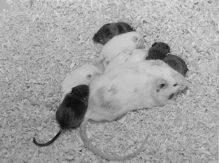
Chimeric mouse, produced by injecting ES cells of strain CBA/Ca into a PO blastocyst. When mated to a PO female, half the resulting offspring were of CBA origin, as judged by coat color.
DISCUSSION
This study shows that the primitive ectoderm or epiblast is the sole source of ES progenitor cells in the late blastocyst. The possibility remains, however, that cells at an earlier stage in the ontogeny of the epiblast lineage [i.e., those of the ICM of the 4th-day blastocyst (3, 4) or ICM precursors inside the morula (2)] can form ES cells directly rather than via differentiation into epiblast cells. The present findings also show that the derivation of ES cells is greatly facilitated in the permissive 129 strain by culturing the cells’ progenitors in isolation from other tissues of the blastocyst. In particular, primitive endoderm seems to exert an inhibitory effect that is not due simply to its forming a tough “rind,” which severely reduces the yield of viable cells from the epiblast during the first dissociation. A more likely hypothesis is that primitive endoderm promotes the differentiation of early epiblast cells to a stage when they can no longer form ES cells. Very close proximity is established between early postimplantation epiblast and primitive endoderm in vivo through discontinuities in their shared basal lamina (21), which could provide a basis for their interaction. Furthermore, there is compelling evidence that the visceral endoderm is essential for normal growth and differentiation of the epiblast in the intact conceptus (22).
That complete dissociation had no discernible effect on the efficiency with which epiblast gave rise to ES lines argues against a requirement for homotypic cell interactions in their production. These experiments also show that more than one cell per epiblast is able to form ES cells. Because the standard practice in establishing ES lines is to pool all undifferentiated colonies obtained from a single blastocyst (10, 23), at least some of the lines in general use will be polyclonal in origin. Whether this contributes to the heterogeneity of ES lines, which, according to current views, is held to arise entirely in culture (1), is clearly open to investigation now that we have shown that multiple clonal ES cell lines can be obtained from individual dissociated epiblasts.
Trophoblast giant cells, which are commonly observed in early-passage ES cell lines derived from whole blastocysts, were absent from lines established from isolated epiblasts. This supports the contention that it is contaminating trophectoderm stem cells with a limited proliferative potential, rather than ES cells, that account for their presence (24).
The finding that delayed implantation is beneficial even when isolated epiblasts rather than whole blastocysts are used as starting material argues against its effect being entirely an indirect one through, for example, negating an inhibitory effect of endoderm. Whether arrest of cell cycling or differentiation (25), an increase in cell number (24), or some other facet of delay accounts for this remains to be established.
Use of epiblasts from delayed-implanting blastocysts in conjunction with PEFs rather than STO feeder cells has enabled us to obtain ES cell lines reproducibly from several strains of mice, including pure CBA/Ca, that have proved refractory to the conventional approach, particularly when LIF was included in the culture medium from explantation to beyond the second dissociation. Since lines sampled from all these disparate strains colonized the germline as well as somatic tissues following blastocyst injection, the present approach may be not only of general utility for the mouse but also applicable to other mammals.
Finally, the finding that ES cell lines can be obtained efficiently from single, dissociated epiblast cells should aid the investigation of the changes that such an uncoupling of proliferation from differentiation entails. A thorough understanding of these changes may be essential for the development of an effective general strategy for the efficient derivation of ES cell lines both in mammals and in other vertebrates.
Acknowledgments
We thank Ann Yates for help in preparing the manuscript, Andy Forkner and his colleagues for their expert care of our mice, and the Royal Society, E.P. Abraham Research Fund, and the Imperial Cancer Research Fund for support.
ABBREVIATIONS
- ES cells
embryonic stem cells
- LIF
leukemia inhibitory factor
- ICM
inner cell mass
- PEF
primary embryonic fibroblast
References
- 1.Gardner, R. L. & Brook, F. A. (1997) Int. J. Dev. Biol., in press. [PubMed]
- 2.Eistetter H R. Dev Growth Differ. 1989;31:275–282. doi: 10.1111/j.1440-169X.1989.00275.x. [DOI] [PubMed] [Google Scholar]
- 3.Evans M J, Kaufman M H. Nature (London) 1981;292:154–156. doi: 10.1038/292154a0. [DOI] [PubMed] [Google Scholar]
- 4.Martin G R. Proc Natl Acad Sci USA. 1981;78:7634–7638. doi: 10.1073/pnas.78.12.7634. [DOI] [PMC free article] [PubMed] [Google Scholar]
- 5.Handyside A H, O’Neill G T, Jones M, Hooper M L. Roux’s Arch Dev Biol. 1989;198:48–55. doi: 10.1007/BF00376370. [DOI] [PubMed] [Google Scholar]
- 6.Gardner R L. J Embryol Exp Morph. 1985;88:303–326. [PubMed] [Google Scholar]
- 7.Hooper M L. Embryonal Stem Cells: Introducing Planned Changes into the Animal Germline. Switzerland: Harwood; 1992. [Google Scholar]
- 8.Sobis H, Vandeputte M. Int J Cancer. 1974;13:444–453. doi: 10.1002/ijc.2910130403. [DOI] [PubMed] [Google Scholar]
- 9.Sobis H, Verstuyf A, Vandeputte M. Development. 1991;111:75–78. doi: 10.1242/dev.111.1.75. [DOI] [PubMed] [Google Scholar]
- 10.Robertson E J. In: Teratocarcinomas and Embryonic Stem Cells: A Practical Approach. Robertson E J, editor. Oxford: IRL; 1987. pp. 71–112. [Google Scholar]
- 11.McWhir J, Schnieke A E, Ansell R, Wallace H, Colman A, Scott A R, Kind A J. Nat Genet. 1996;14:223–226. doi: 10.1038/ng1096-223. [DOI] [PubMed] [Google Scholar]
- 12.Friedrich G, Soriano P. Genes Dev. 1991;5:1513–1523. doi: 10.1101/gad.5.9.1513. [DOI] [PubMed] [Google Scholar]
- 13.Gardner D K, Sakkas D. Hum Reprod. 1993;8:288–295. doi: 10.1093/oxfordjournals.humrep.a138039. [DOI] [PubMed] [Google Scholar]
- 14.Gardner R L, Rossant J. J Embryol Exp Morph. 1979;52:141–152. [PubMed] [Google Scholar]
- 15.Gardner R L, Barton S C, Surani M A H. Genet Res Camb. 1990;56:209–222. doi: 10.1017/s001667230003531x. [DOI] [PubMed] [Google Scholar]
- 16.Nagamine C M, Chan K, Kozak C A, Lau Y. Science. 1989;243:80–83. doi: 10.1126/science.2563174. [DOI] [PubMed] [Google Scholar]
- 17.Hogan B, Constantini F, Lacy E. Manipulating the Mouse Embryo: A Laboratory Manual. Plainview, NY: Cold Spring Harbor Lab. Press; 1986. pp. 221–225. [Google Scholar]
- 18.Bradley A. In: Teratocarcinomas and Embryonic Stem Cells: A Practical Approach. Robertson E J, editor. Oxford: IRL; 1987. pp. 113–151. [Google Scholar]
- 19.Gardner R L. In: Methods in Mammalian Reproduction. Daniel J C Jr, editor. New York: Academic; 1978. pp. 137–165. [Google Scholar]
- 20.Tsunoda Y, Tokunaga T, Sugie T. Gamete Res. 1985;12:301–304. [Google Scholar]
- 21.Takeuchi I K, Takeuchi Y K. Dev Growth Differ. 1981;23:157–164. doi: 10.1111/j.1440-169X.1981.00157.x. [DOI] [PubMed] [Google Scholar]
- 22.Chen W S, Manova K, Weinstein D C, Duncan S A, Plump A S, Prezioso V R, Bachvarova R F, Darnell J E., Jr Genes Dev. 1994;8:2466–2477. doi: 10.1101/gad.8.20.2466. [DOI] [PubMed] [Google Scholar]
- 23.Abbondanzo S J, Gadi I, Stewart C L. Methods Enzymol. 1993;225:803–823. doi: 10.1016/0076-6879(93)25052-4. [DOI] [PubMed] [Google Scholar]
- 24.Evans M J, Kaufman M H. Cancer Surv. 1983;2:185–207. [Google Scholar]
- 25.Gardner R L, Davies T J. J Exp Zool. 1993;265:54–60. doi: 10.1002/jez.1402650108. [DOI] [PubMed] [Google Scholar]