Abstract
Objective
Rats exposed to high airborne mass concentrations of low‐solubility low‐toxicity particles (LSLTP) have been reported to develop lung disease such as fibrosis and lung cancer. These particles are regulated on a mass basis in occupational settings, but mass might not be the appropriate metric as animal studies have shown that nanoparticles (ultrafine particles) produce a stronger adverse effect than fine particles when delivered on an equal mass basis.
Methods
This study investigated whether the surface area is a better descriptor than mass of LSLTP of their ability to stimulate pro‐inflammatory responses in vitro. In a human alveolar epithelial type II‐like cell line, A549, we measured interleukin (IL)‐8 mRNA, IL8 protein release and glutathione (GSH) depletion as markers of pro‐inflammatory effects and oxidative stress after treatment with a range of LSLTP (fine and nanoparticles) and DQ12 quartz, a particle with a highly reactive surface.
Results
In all the assays, nanoparticle preparations of titanium dioxide (TiO2‐np) and of carbon black (CB‐np) produced much stronger pro‐inflammatory responses than the same mass dose of fine TiO2 and CB. The results of the GSH assay confirmed that oxidative stress was involved in the response to all the particles, and two ultra‐fine metal dusts (cobalt and nickel) produced GSH depletion similar to TiO2‐np, for similar surface‐area dose. As expected, DQ12 quartz was more inflammatory than the low toxicity dusts, on both a mass and surface‐area basis.
Conclusion
Dose–response relationships observed in the in vitro assays appeared to be directly comparable with dose–response relationships in vivo when the doses were similarly standardised. Both sets of data suggested a threshold in dose measured as surface area of particles relative to the surface area of the exposed cells, at around 1–10 cm2/cm2.
These findings are consistent with the hypothesis that surface area is a more appropriate dose metric than mass for the pro‐inflammatory effects of LSLTP in vitro and in vivo, and consequently that the high surface area of nanoparticles is a key factor in their inflammogenicity.
Keywords: low‐solubility low‐toxicity particles, nanoparticles, surface area, inflammation, IL8
Low‐solubility low‐toxicity particles (LSLTP), regulated in the USA as “particulate not otherwise classified” and in the UK as “nuisance dusts”, include particles of non‐reactive material with neither fibres nor free silica. Occupational exposure limits for the (8‐hour time‐weighted average) respirable dust concentrations of such particles are set at 5 mg/m3 in the USA1 and 4 mg/m3 in the UK.2 In the last decade or so, it has become clear from rat studies that chronic exposure to high airborne mass concentration of LSLTP such as carbon black (CB) or titanium dioxide (TiO2) can lead to development of the features characteristic of “rat lung overload”, namely failed clearance with build‐up of particles, leading to persistent inflammation, epithelial cell proliferation, fibrosis and cancer.3,4
The breakdown in normal alveolar macrophage (AM)‐mediated particle clearance seen in overload was initially thought to be a consequence of volumetric overload of the AMs and the resulting loss of AM mobility.5 As volumetric particle lung burden increased, the clearance was considered to become progressively more impaired. Thus particle‐laden AM, activated to release harmful oxidants and proteases as well as mitogens for epithelial cells, aggregate in the alveolar region, allowing interaction of their secretions and unphagocytosed particles with the epithelium, so enhancing injury, inflammation and tissue remodelling, leading to the typical pathology.5 However, recent studies suggest that volumetric overload is not a complete explanation for impairment of clearance. Exposure of rats to nanoparticles, with a large surface area per unit mass, showed features of overload at a lower lung burden (in terms of mass and volume of particles) compared with larger size particles with same mineral and chemical properties.6
Tran et al7 compared the inflammatory response to two very different LSLTP types with different particle size. At equal lung mass burden, the smaller particles produced more inflammation but the two LSLTP types were equally inflammogenic when the lung burden was expressed as particle‐surface area.7 For a fixed mass of particles, their surface area increases as particle size becomes smaller, thus a dose‐dependence on surface area may explain the well‐documented greater toxicity of nanoparticles compared with the same mass of fine particles of the same material.8 The finding that surface area rather than mass or volume appears to be a better metric of dose for predicting overload inflammation may imply a need to reconsider the regulation of workplace exposure, as current occupational exposure limits for airborne dusts are defined in terms of mass per m3 of air.
The mechanism underlying surface area‐driven LSLTP‐induced inflammation is believed to be the generation of reactive oxygen species (ROS), leading to oxidative stress in lung cells. Nanoparticles have been shown to induce oxidative damage both in vivo9 and in vitro.10 Nanoparticle surface‐derived free radical activity has been shown in cells10 and in a cell‐free system.11
Quartz is an insoluble mineral particle that has a very high surface reactivity, generating free radicals,12,13 and consequently it produces inflammation at much lower mass and surface area dose than do LSLTP particles.14
Particles can induce oxidative stress,15 seen as depletion of GSH, which in turn activates transcription factors such as nuclear factor (NF)‐κB and activator protein‐1 (AP‐1) that control the transcription of pro‐inflammatory genes.16 Recent in vivo studies from our laboratory confirmed that for a range of LSLTP, particle surface area was the driving factor in the initiation of the inflammatory response in vivo.14 Quartz showed enhanced inflammation relative to surface‐area dose, consistent with its surface reactivity.
The nature of the stress response elicited in epithelial cells that line the airspaces of the lungs may be key to understanding the influence of particle characteristics, and it has been studied extensively.17,18 IL8 is an oxidative stress‐responsive pro‐inflammatory chemokine, released from epithelial cells following particle‐induced oxidative stress leading to neutrophil influx and inflammation, which has been extensively investigated in particle studies.19,20,21,22
We selected assays and time points to elucidate the key sequence of events following contact between particles and cells. We measured oxidative stress in the form of GSH depletion at 4 h, IL8 mRNA levels at 6 h and IL8 protein release at 12 and 24 h in A549 human alveolar type II epithelial cells in vitro. The 12 and 24 hour time points were chosen to allow translation, secretion and accumulation of IL8 protein without allowing time for the excessive accumulation in the culture of cell products that might produce effects on the cells that are secondary to treatment effects.
We hypothesised that type II alveolar epithelial cells might show pro‐inflammatory responsiveness to LSLTP surface‐area dose in vitro, as has been seen in vivo. We used A549 lung epithelial cells and determined the above responses in relation to mass and surface‐area dose of the challenge dusts. First, we determined the cell viability after treatment with particles to establish doses suitable for the other assays. We then compared the response obtained with the LSLTP to that obtained with quartz, as a particle with a reactive surface. The aim of the work was to examine the relative importance of particle size, surface area and surface reactivity in producing pro‐inflammatory responses in epithelial cells.
Materials and methods
Particles and their characteristics
The dusts were obtained from either manufacturers or laboratories that had used the material in previous studies: fine CB (Huber 990; H Haeffner, Chepstow, UK); fine TiO2 (Tioxide Europe, London, UK); CB nanoparticles (CB‐np) (Printex 90) and TiO2 nanoparticles (TiO2‐np) (both Degussa, Cheshire, UK; barium sulphate (BaSO4) (Institute of Occupational Medicine);7 DQ12 α quartz particles (characteristics described by Robock23).
For one of the assays, cobalt (Co) nanoparticles, and nickel (Ni) nanoparticles were also included.
The specific surface area characteristics of the particles were measured by a nitrogen gas adsorption method,24 giving the values shown in table 1.
Table 1 Specific surface area of the particles used in the study.
| Particle type | Abbreviation | Surface area (m2/g) |
|---|---|---|
| Carbon black nanoparticle | CB‐np | 254. |
| Titanium dioxide nanoparticle | TiO2‐np | 50.6 |
| Nickel nanoparticles† | Ni | 36.9 |
| Cobalt nanoparticles† | Co | 36.2 |
| Carbon black | CB | 7.92 |
| Titanium dioxide | TiO2 | 6.64 |
| Barium sulphate | BaSO4 | 3.68 |
| Quartz | DQ12 | 10.1 |
†These particles (Ni and Co) were included in the glutathione assays only.
Cell culture
Cells from a human alveolar type II‐like epithelial cell line, A549, obtained from the European Collection of Animal Cell Cultures (Porton Down, UK) were maintained in continuous culture at 37°C with 5% CO2 in Dulbecco's modified Eagle's medium (DMEM) (Sigma‐Aldrich, Poole, Dorset, UK) containing 10% heat‐inactivated fetal calf serum (FCS), 2 mmol/l L‐glutamine (Life Technologies, Paisley, UK) and 100 IU/ml penicillin and 100 µg/ml streptomycin (Life Technologies).For the assays, cells were plated in 96‐well or 6‐well plates at, respectively, densities of 9×10⁴ cells/ml or 0.2×10⁶ cells/ml. The cells were grown to 80% confluency in 10% FCS for 24 h, washed with Ca2+/Mg2+‐free phosphate‐buffered saline and starved of serum for 24 h before treatment.25
A549 cell treatment
A549 cells were treated for 4, 6, 12 or 24 hours, with fine and ultrafine LSLTP (ie all particles except quartz) at final concentrations of 15, 31, 62, 125 and 250 μg/ml and with DQ12 quartz at final concentrations of 48, 96, 192 and 384 μg/ml. Cells were suspended in DMEM without FCS for 24 hours (prior to treatment) to starve them of serum and arrest their growth. The particle suspensions were sonicated for 5 min prior to adding to the wells. TNFα was used as a positive control at a concentration of 10 ng/ml.
Lactate dehydrogenase assay
Cell viability after particle treatment was assessed in triplicate in 96 well plates by measurement of lactate dehydrogenase (LDH) activity released from cytosol of damaged cells into the medium (LDH Cytotoxicity Detection Kit; Roche Molecular Biochemicals, Basel, Switzerland). Data were expressed as a percentage of LDH released by cells totally lysed by detergent treatment.
Measurement of interleukin‐8 mRNA by reverse transcription PCR
Following 6 h or 24 h treatment with particles, human IL8 mRNA was detected using semi‐quantitative reverse transcription PCR. Total RNA was extracted from treated cells (TRIzol reagent; Life Technologies) using the recommended protocol. The obtained RNA was suspended in 50 µl of diethyl pyrocarbonate‐treated H2O and quantified. RNA (2 µg) was reverse transcribed using M‐MLV reverse transcriptase (Promega UK, Southampton, UK) according to the manufacturer's instructions. cDNA sequences, corresponding to IL8 mRNA and to the human housekeeping gene glyceraldehyde‐3‐phosphate (GAPDH), were amplified by PCR. IL8 and GAPDH primers were chosen according to Jiminez et al26 (MWG‐Biotech AG; Milton Keynes, UK) and PCR products were separated on a 1.5% agarose gel containing 1 µg/ml ethidium bromide. The expected sizes for IL8 and GAPDH were 300 and 600 bp, respectively. Bands were scanned using an ultraviolet transilluminator (Ultra Violet Products, Cambridge, UK) and quantified by densitometry. The quantity of IL8 mRNA was obtained as a ratio of the intensity of IL8 band over the corresponding GAPDH band, and expressed as a fold increase over the control.
Measurement of IL8 protein by ELISA
The ELISA assay measures IL8 protein that has been released in the culture medium. As with the LDH assay, all of the LSLTP were analysed on the same day in triplicate. Following incubation with the particles for 6 or 24 h, cell supernatants were recovered and centrifuged at 13 000 rpm for 10 min and the particle‐free cell supernatants were stored at −80°C until analysis. ELISA (ELISA Kit; National Institute for Biological Standards and Control, Hertfordshire, UK) was performed in accordance with the manufacturer's instructions and results expressed as pg/ml.
Assessment of total glutathione
Cells were cultured in 6‐well plates, treated for 4 h with particles and suspended in 1 ml of cold 0.6% sulphosalicyclic acid, sonicated and vortexed several times. They were then centrifuged at 5000 rpm for 5 min at 4°C. The supernatant was immediately used to measure total intracellular glutathione by the glutathione reductase–dinitrothiocyanobenzene recycling assay adapted for microtitre plates.27,28 The rate of 2‐nitro‐5‐thibenzoic acid formation (change in absorbance at 412 nm per minute) in each sample was calculated and expressed in nmol/mg of proteins contained in the sample.
Statistical analysis
Data were summarised as tables and graphs of means with standard errors, and analysed using analysis of variance to assess the significance of trends.
Results
Cytotoxicity in A549 cells
At the different doses tested (15, 31, 62, 125 and 250 µg/ml), the fine particles (CB, TiO2 and BaSO4) and nanoparticles of TiO2 (TiO2‐np) did not produce any substantial cytotoxicity (<20%) (data not shown). In the case of CB‐np there was slight cytotoxicity evident at low concentration (15, 31 and 62 µg/ml) but not at higher concentrations. However, we believe the apparent lack of effect at high concentrations to be caused by interference of CB‐np with the assay, as separate tests suggested adsorption of LDH onto the surface of CB‐np (data not shown). DQ12 quartz produced release of LDH that increased linearly with dose, reaching substantial cytotoxicity at 192 and 384 µg/ml with 27% and 54% respectively. The details of the results are available in the study report25 (http://www.hse.gov.uk/research/rrhtm/rr154.htm). The cytotoxicity results identified or confirmed dose ranges suitable for the main assays.
IL8 gene expression
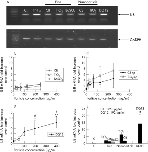
Figure 1A shows an agarose gel of IL8 mRNA expression in A459 cells after 6 h at a dose of 250 µg/ml for the LSLTP particles and 192 µg/ml for DQ12 quartz. The intensity of the IL8 mRNA band is clearly much greater for the nanoparticles and the quartz. The IL8 band intensity expressed as a fold increase over the control (GAPDH) band intensity is plotted against the particle concentration in the assay (figure 1B,C).
Figure 1 IL8 mRNA expression in A549 cells treated for 6 h or 24 h with fine particles, nanoparticles and DQ12 quartz. (A) Agarose gel showing IL8 mRNA intensity upon 6 h treatment with 10 ng/ml of TNFα, 250 µg/ml of fine particles (CB, TiO2, BaSO4) or nanoparticles (CB‐np and TiO2‐np) and 192 µg/ml of DQ12 quartz. (B) fine particles; (C) nanoparticles; and (D) DQ12 quartz. Graphs of dose–response in cells treated for 6 hours, showing mean and standard error from four experiments. #Cytotoxicity at these doses. Significant difference from control: *p<0.05 and **p<0.01. (E) Histogram for cells treated for 24 hours at 250 μg/ml for fine particles and nanoparticles, and at 192 μg/ml for DQ12.
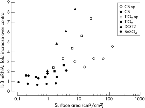
After the 6 h treatment of A549 cells with particles, the dose–response experiments with fine particles showed only slight increases in IL8 mRNA at some of the higher doses (figure 1B). For the nanoparticles, a significant trend of increase in IL8 mRNA with dose (figure 1C) was found by analysis of variance. There was also a general trend towards increase with dose (expressed as surface area) over all particles, as described later. DQ12 quartz also induced a dose‐dependent increase in IL8 mRNA (figure 1D).
Following 24 h treatment, nanoparticles showed a sustained increase in IL8 gene expression whilst quartz alone induced almost doubling of IL8 gene expression by 24 hours compared with 6 hours (figure 1E).
IL8 protein release
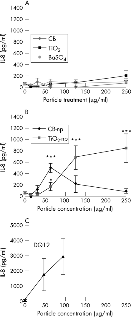
After 24 h treatment (figureure 2), fine particles (figure 2A) showed an indication of an effect only at the highest concentration. The nanoparticles (figure 2B) and quartz (figure 2C) produced increases in IL8 protein that mirrored the increases in IL8 mRNA observed after 6 h. DQ12 quartz induced a higher release of IL8 protein compared with ultrafine particles. In the case of CB‐np, the effect increased with dose initially but then decreased at the highest concentrations (125 and 250 µg/ml) although these concentrations are non‐toxic (with the caveat that adsorption of LDH may have masked some toxicity).
Figure 2 IL8 protein release from A549 cells, after 24 h treatment with (A) fine particles or (B) nanoparticles at 15, 31, 63, 125 and 250 µg/ml, or (C) DQ12 quartz at 48, 96, 192 and 384 µg/ml. Means and standard errors of 4 experiments. Significant difference from control: *p<0.05, **p<0.01, ***p<0.001.
Total glutathione levels
To assess the involvement of oxidative stress in particle‐mediated inflammation, we measured the amount of total glutathione (both oxidised (GSSG) and reduced GSH, with the GSSG converted to GSH) after 4 h treatment with the different particles. These assays included the two extra nanoparticles, the metals Ni and Co.
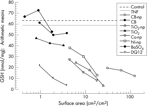
There was a significant depletion of GSH with the fine particles: at 250 μg/ml for BaSO4, 125 μg/ml and 250 μg/ml for CB and at all concentrations for TiO2. The nanoparticles, (CB, TIO2, Ni and Co) and DQ12 quartz produced significant decreases in GSH at each of the three particle concentrations tested. With all particle results taken together, there was a general trend towards depletion being dependent on the surface area dose (figure 3). The dose was expressed as surface area (of particles) per unit surface area of the (cell‐covered) test well.
Figure 3 Effect of particle surface area on GSH depletion in A549 cells exposed for 4 h to LSLTP and DQ12 quartz. The GSH data are expressed as geometric means.
In figure 3, a clear and significant relationship between GSH levels and surface area can be seen. As surface area dose increases above 1 cm2/cm2, the level of GSH decreases below the level measured in controls, and all the LSLTP collapsed almost onto a common trend. DQ12 quartz follows a separate trend; for the same surface area dose as LSLTP, the DQ12 produces more GSH depletion, consistent with extra reactivity at the surface of quartz.
Dependence on surface area dose for IL8 mRNA and protein
We examined the dependence of the response (IL8 protein release, IL8 mRNA) on particle surface area by replotting these data against the dose converted from mass to surface area. Again, the dose was expressed as surface area (of particles) per unit surface area of the (cell covered) test well.
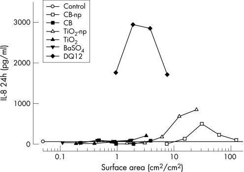
The results for IL8 protein release (in figure 4) show that, for LSLTP, between a dose of 1 and 10 cm2/cm2, the amount of IL8 released starts to increase with dose (except for CB‐np at the two highest doses, which as discussed before is considered to be due to interference at the highest doses). DQ12 starts to rise at a lower dose than the LSLTP and shows higher response than any LSLTP, but shows an unexplained non‐linear dose dependence.
Figure 4 Effect of surface area on IL8 protein release following 24 h exposure of A549 cells to LSLTP and DQ12 quartz. The dose is expressed as surface area (of particles) per unit area of (cell covered) test cell surface.
For IL8 mRNA (figure 5), there is no single relationship between surface area and IL8 mRNA expression, although a surface‐area dose threshold is suggested. Above this dose, three different lines are observed: the steepest is DQ12, followed by TiO2‐np, and finally a flatter line relating to CB‐np.
Figure 5 Effect of surface‐area dose on IL8 mRNA expression after exposure of A549 cells to LSLTP and DQ12 quartz for 6 h.
Pooling the results to distinguish quartz from other particles
The results for IL8 protein release were pooled by normalising the response as IL8 protein per unit of dose, with dose expressed (as for figures 4 and 5) as surface area of particles per unit area of (cell‐coated) well surface. This gives IL8 protein/cm2/cm2 relative to unit dose (1 cm2/cm2). The IL8 protein was measured as a concentration (pg/ml) in the medium in the well, but by multiplication by the volume of medium in the cell (0.2 ml), that concentration converts to a quantity (pg) of protein.
When the IL8 protein production was divided by the particle surface‐area dose to give an IL8 production per unit surface‐area dose, all of the different LSLTP fell into one cluster with a value <10 pg/cm2/cm2. In contrast, quartz gave a value around 240 pg/cm2/cm2, suggesting that a given area of quartz surface is >20 times more active in stimulating IL8 release than the same area of LSLTP surface.
This demonstrates that LSLTP surfaces of very different types have a very similar ability to stimulate epithelial cell pro‐inflammatory response. It also emphasises the extra part that surface reactivity/chemistry plays in the ability of quartz to elicit inflammation.
Discussion
As there is considerable potential for particle exposure in workplaces, there is interest and debate on the basis for regulating LSLTP. Experimental data in rats have shown that exposures at average concentrations around the occupational exposure limits are associated with “overload”‐related adverse effects that are attributed to the particle physical characteristics rather than chemical structure. Studies on nanoparticles (particles <100 nm diameter and sometimes called ultrafine particles) have provided an important part of the evidence leading to the hypothesis that particle surface‐area dose is important in determining pathology29 and inflammation.6
From previous studies in vivo7,14 we hypothesised that (1) surface area of the LSLTP would be the metric that describes their ability to cause epithelial cells to synthesise IL8 protein and (2) a quartz sample would be much more active per unit surface area because of its greater surface reactivity. This study investigated the role of particle surface area and surface reactivity in initiating pro‐inflammatory effects on epithelial cells in vitro, to elucidate their relative importance in the ability of LSLTP to stimulate inflammation.
IL8 is a potent chemoattractant and activator of neutrophils, the transcription of which is NF‐κB‐dependent.30,31 We investigated IL8 gene expression and the subsequent release of IL8 protein by A549 epithelial cells in vitro, as indicators of potential to initiate inflammation. IL8 mRNA expression occurred for cells treated with LSLTP for 6 h, returning to basal levels by 24 h. Only high surface‐area dose switched on substantial IL8 gene expression.
DQ12 quartz upregulated IL8 mRNA at both 6 h and 24 h with low surface‐area dose. In line with these findings, several investigators have reported persistent upregulation of NF‐κB‐controlled genes in response to pathogenic dust particles such as asbestos and quartz.32,33,34
Production of IL8 protein showed contrasts between dusts. After 6 h of treatment, there were significant increases in production with quartz but not with LSLTP. At 24 h of treatment, nanoparticles elicited a modest dose‐dependent increase, most strongly for TiO2‐np. Also at 24 h treatment, quartz had a still more pronounced effect. For the CB‐np at the high (but non‐toxic) doses, an apparent decrease in IL8 protein levels appeared to be a contradiction to the increase in IL8 mRNA observed at those doses. This decrease with CB‐np is a problem that we have previously encountered, and is possibly explained by artefactual adsorption of the cytokine protein onto their large surface.11,35,36 Previous experiments in our laboratory have confirmed significant protein (albumin) adsorption onto the surface of CB‐np.11 Further investigations would be needed to confirm this hypothesis with IL8 protein adsorption.
With TiO2, the IL8 gene expression and protein release showed dose dependence on particle surface area for fine and nanoparticles. Clearly, surface‐area dependence implies that relative to mass dose, the nanoparticles had a greater effect than the fine particles.
Recent studies have shown a role for ROS in promoting the cellular effects of nanoparticles10,37 and that CB‐np has a greater free radical activity than the same mass of fine particles,10 so we measured GSH depletion as a marker of particle‐induced oxidative stress. After 4 h exposure, GSH was depleted in a dose‐ dependent manner relative to dose expressed as surface area. For given mass doses, the nanoparticles produced correspondingly more depletion than fine particles. These findings are consistent with the hypothesis that production of free radicals and the subsequent oxidative stress exerted on epithelial cells is the mechanism by which nanoparticles elicit inflammatory gene expression. A549 cells are known to have a higher level of GSH than primary epithelial cells,38 and they have been in long‐term culture, so can be expected to show variation in phenotype. However, we have used this cell line extensively to show oxidative stressing and pro‐inflammatory effect of a range of harmful particles. These have included welding fumes, where the potency ranking of different welding fume samples for oxidative stress and pro‐inflammatory effects with A549 cells in vitro were replicated in an in vivo model of lung inflammation.39,40
In order to determine whether the surface area is a unifying factor driving the inflammatory response in vitro, we replotted the IL8 protein release, IL8 mRNA and GSH response against particle dose expressed as surface area. For LSLTP there was evidence of a surface area‐related pro‐inflammatory effect with a threshold, but the various particles produced trends with slightly different slopes for IL8 mRNA. For IL8 mRNA, TiO2‐np showed greater potency than other LSLTP including CB‐np, whereas in previous animal studies we have found CB‐np to be more biologically active.41 The greater transcription seen with TiO2 is reflected in the IL8 protein; there were greater levels with TiO2 suggesting that the low IL8 mRNA found with CB might not only be a consequence of adsorption artefact as previously proposed. All of the LSLTP fall on a common general trend for GSH depletion plotted against surface‐area dose. Taken together, these results suggest that the processes dictating gene expression are more complex than the simple depletion of GSH, otherwise, the GSH results would imply that all of the LSLTP should produce approximately the same amount of gene expression.
Plotting the data relative to surface area clearly showed that very low surface area doses of quartz are highly pro‐inflammatory. Additionally, prolonged IL8 expression (at the 24 h time point) was seen only with quartz and this may be a factor in its well‐documented severe and chronic inflammatory effects.12
These in vitro data support the contention that the effects observed with nanoparticles relate to the large surface‐area dose they deliver to the epithelial cells and also that surface area is the dose metric that relates best to the inflammatory response for a range of LSLTP. This finding accords with our previous in vivo studies, in which surface area of LSLTP was found to be the factor driving the inflammatory response in rats.12
In the present study, we also showed that the surface area of the particles is the important factor responsible for oxidative stress‐mediated depletion of GSH in A549 cells. This finding is in keeping with previous studies with nanoparticles, in cell free systems37,42 and in cells in vitro.10,43
The greater ability of quartz to cause inflammation is related to its surface oxidative activity,12 although there is uncertainty over the nature of the surface moieties that are responsible for the increased biological activity in some quartz samples. We predicted that the inflammogenic potential of quartz samples of similar surface reactivity but variable size would also be related to their total surface area,25 and this has been confirmed recently.44
Previous studies have shown both in vitro and in vivo the role of surface area in the harmful effects of particles of various types, including CB,45 manganese dioxide46 and TiO2.47 However, we contend that the present study is the first to describe a quantitatively similar pro‐inflammatory effect of surface area across a range of three different types of LSLTP. We believe that this in vitro approach could form the basis of a screening test that could be used with particles of unknown toxicity, to discriminate LSLTP from those with greater toxicity due to an element of surface reactivity e.g. quartz. An in vitro predictive assay such as this represents a cost‐effective and ethical approach to testing that may reduce testing in animals. There is a need for such a testing system to screen the new generation of nanoparticles that are being produced, in order to identify potential problem particle types; the in vitro model developed in our studies has the potential to fulfil such a need. Such a model would benefit from evidence that the in vitro dosimetry scaled with in vivo dosimetry data on LTLS particles. We have been working on such an analysis and, although this is beyond the scope of the present manuscript, it will form the basis of a separate future publication.
Extension of this research should include a larger array of types of LSLTP to determine their general conformity with the ”surface area” paradigm. Furthermore, additional and more refined endpoints of in vitro pro‐inflammatory effects, such as activation of transcription factors, should be used.
These findings on the role of the surface area of LSLTP in driving the inflammatory response are of special importance with regard to regulation of workplace exposure, which is currently based on airborne mass concentrations. As inflammatory potential is driven by surface area rather than mass, then a given airborne mass concentration of a material in the form of fine particles could be much less inflammogenic than the same airborne mass concentration of same material in the form of ultrafine particles.
In summary, the data suggested a threshold in dose, measured as surface area of LSLTP relative to the surface area of the exposed cells, at around 1–10 cm2/cm2. These findings are consistent with the hypothesis that surface area is a more appropriate dose metric than mass for the pro‐inflammatory effects of LSLTP in vitro and in vivo, and consequently that the high surface area of nanoparticles is a key factor in their inflammogenicity. For particles with a highly reactive surface like quartz, lower surface area doses are required to reach a threshold to stimulate pro‐inflammatory responses.
Acknowledgements
We would like to thank R. Duffin, E. Drost, P.S. Gilmour, H. Jardine, L.A. Jimenez, J. McNeilly and J. Wickenden for their help and support throughout this study and the UK Health and Safety Executive for financial support.
Abbreviations
AM - alveolar macrophage
AP‐1 - activator protein‐1
BaSO4 - barium sulphate
CB - carbon black
Co - cobalt
DMEM - Dulbecco's modified Eagle's medium
IL - interleukin
FCS - fetal calf serum
GAPDH - glyceraldehyde‐3‐phosphate
GSH - glutathione
LDH - lactate dehydrogenase
LSLTP - low‐solubility low‐toxicity particles
NF - nuclear factor
Ni - nickel
np - nanoparticle
ROS - reactive oxygen species
TiO2 - titanium dioxide
Footnotes
Competing interests: none declared
References
- 1.OSHA Chemical Sampling Information Particulates not otherwise regulated (Respirable Fraction). 1993
- 2.HSE Occupational exposure limits 1997 (EH 40/97). Sudbury, UK: HSE Books, 1997
- 3.ILSI Workshop The relevance of the rat lung response to particle overload for human risk assessment. Inhal Toxicol 2000121–17 [DOI] [PubMed] [Google Scholar]
- 4.Mauderly J L, McCunney R J. eds. Particle overload in the rat lung: implications for human risk assessment. Philadelphia, PA: Taylor & Francis, 1996298
- 5.Morrow P E. Possible mechanisms to explain dust overloading of the lungs. Fundam Appl Toxicol 198810369–384. [DOI] [PubMed] [Google Scholar]
- 6.Oberdorster G. Significance of particle parameters in the evaluation of exposure‐dose‐response relationships of inhaled particles. Inhal Toxicol (Suppl 8)73–89. [PubMed]
- 7.Tran C L, Buchanan D, Cullen R T.et al Inhalation of poorly soluble particles. II. Influence Of particle surface area on inflammation and clearance 1. Inhal Toxicol 2000121113–1126. [DOI] [PubMed] [Google Scholar]
- 8.Donaldson K, Stone V, Clouter A.et al Ultrafine particles. Occup Environ Med 200158211–216. [DOI] [PMC free article] [PubMed] [Google Scholar]
- 9.Vallyathan V, Castranova V, Pack D.et al Freshly fractured quartz inhalation leads to enhanced lung injury and inflammation – potential role of free‐radicals. Am J Respir Crit Care Med 19951521003–1009. [DOI] [PubMed] [Google Scholar]
- 10.Stone V, Shaw J, Brown D M.et al The role of oxidative stress in the prolonged inhibitory effect of ultrafine carbon black on epithelial cell function. 1998649–659. [DOI] [PubMed]
- 11.Brown D M, Stone V, Findlay P.et al Increased inflammation and intracellular calcium caused by ultrafine carbon black is independent of transition metals or other soluble components . Occup Environ Med 200057685–691. [DOI] [PMC free article] [PubMed] [Google Scholar]
- 12.Duffin R, Gilmour P S, Schins R P.et al Aluminium lactate treatment of dq12 quartz inhibits its ability to cause inflammation, chemokine expression, and nuclear factor‐kappab activation. Toxicol Appl Pharmacol 200117610–17. [DOI] [PubMed] [Google Scholar]
- 13.Castranova V. Generation of oxygen radicals and mechanisms of injury prevention. Environ Health Perspect 199410265–68. [DOI] [PMC free article] [PubMed] [Google Scholar]
- 14.Duffin R, Clouter A, Brown D.et al The importance of surface area and specific reactivity in the acute pulmonary inflammatory response to particles. Ann Occup Hyg 200246(Suppl)242–245. [Google Scholar]
- 15.Donaldson K, Brown D, Clouter A.et al The pulmonary toxicology of ultrafine particles. J Aerosol Med 200215213–220. [DOI] [PubMed] [Google Scholar]
- 16.Rahman I, MacNee W. Role of transcription factors in inflammatory lung diseases. Thorax 199853601–612. [DOI] [PMC free article] [PubMed] [Google Scholar]
- 17.Driscoll K E, Carter J M, Hassenbein D G.et al Cytokines and particle‐induced inflammatory cell recruitment. Environmental Health Perspectives 19971051159–1164. [DOI] [PMC free article] [PubMed] [Google Scholar]
- 18.Li X Y, Gilmour P S, Donaldson K.et al In vivo and in vitro proinflammatory effects of particulate air pollution (PM10). Environ Health Perspect 1997105(Suppl)1279–1283. [DOI] [PMC free article] [PubMed] [Google Scholar]
- 19.Gilmour P S, Rahman I, Donaldson K.et al Histone acetylation regulates epithelial IL8 release mediated by oxidative stress from environmental particles. Am J Physiol Lung Cell Mol Physiol 2003284L533–L540. [DOI] [PubMed] [Google Scholar]
- 20.Salvi S S, Nordenhall C, Blomberg A.et al Acute exposure to diesel exhaust increases IL8 and GRO‐alpha production in healthy human airways. Am J Respir Crit Care Med 2000161550–557. [DOI] [PubMed] [Google Scholar]
- 21.Hetland R B, Refsnes M, Myran T.et al Mineral and/or metal content as critical determinants of particle‐induced release of IL‐6 and IL8 from A549 cells. J Toxicol Environ Health A 20006047–65. [DOI] [PubMed] [Google Scholar]
- 22.Rosenthal G I, Germolec D R, Blazka M E.et al Asbestos stimulates IL8 production from human lung epithelial‐cells. J Immunol 19941533237–3244. [PubMed] [Google Scholar]
- 23.Robock K. Standard quartz DQ12 <5 um for experimental pneumoconiosis research projects in the Federal Republic of Germany. Ann Occup Hyg 19731663–66. [DOI] [PubMed] [Google Scholar]
- 24.Brunauer S, Emmett P H, Teller E. Adsorption of gases in multi‐molecular layers. 1938309–319.
- 25.Faux S P, Tran C L, Miller B G.et alIn vitro determinants of particulate toxicity: the dose metric for poorly soluble dusts. Research Report RR154. Sudbury, UK: HSE Books, 2003
- 26.Jimenez L A, Drost E M, Gilmour P S.et al PM(10)‐exposed macrophages stimulate a proinflammatory response in lung epithelial cells via TNF‐alpha. Am J Physiol Lung Cell Mol Physiol 2002282L237–L248. [DOI] [PubMed] [Google Scholar]
- 27.Tietze F. Enzymic method for quantitative determination of nanogram quantities of total and oxidised glutathione: application to mammalian blood and other tissues. 196927502–522. [DOI] [PubMed] [Google Scholar]
- 28.Vandeputte C, Guizon I, Genestie‐Denis I.et al A microtiter plate assay for total glutathione and glutathione disulfide contents in cultured/isolated cells: performance study of a new miniaturized protocol. Cell Biol Toxicol 199410415–421. [DOI] [PubMed] [Google Scholar]
- 29.Driscoll K E. Role of inflammation in the development of rat lung tumors in response to chronic particle exposure. Inhal Toxicol 19968(Suppl)139–153. [Google Scholar]
- 30.Sen C K. Cellular thiols and redox‐regulated signal transduction. Curr Top Cell Regul 2000361–30. [DOI] [PubMed] [Google Scholar]
- 31.DeForge L E, Preston A M, Takeuchi E.et al Regulation of interleukin 8 gene expression by oxidant stress. J Biol Chem 199326825568–25576. [PubMed] [Google Scholar]
- 32.Janssen Y W, Driscoll K E, Howard B.et al Asbestos causes translocation of p65 protein and increases NF‐kappa B DNA binding activity in rat lung epithelial and pleural mesothelial cells. Am J Pathol 1997151389–401. [PMC free article] [PubMed] [Google Scholar]
- 33.Chen F, Lu Y, Demers L M.et al Role of hydroxyl radical in silica‐induced NF‐kappa B activation in macrophages. Ann Clin Lab Sci 1998281–13. [PubMed] [Google Scholar]
- 34.Schins R P, McAlinden A, MacNee W.et al Persistent depletion of I kappa B alpha and interleukin‐8 expression in human pulmonary epithelial cells exposed to quartz particles. Toxicol Appl Pharmacol 2000167107–117. [DOI] [PubMed] [Google Scholar]
- 35.Kim H, Liu X, Kobayashi T.et al Ultrafine carbon black particles inhibit human lung fibroblast‐mediated collagen gel contraction. Am J Respir Cell Mol Biol 200328111–121. [DOI] [PubMed] [Google Scholar]
- 36.Salvador‐Morales C, Flahaut E, Sim E.et al Complement activation and protein adsorption by carbon nanotubes. Mol Immunol 200643193–201. [DOI] [PubMed] [Google Scholar]
- 37.Brown D M, Wilson M R, MacNee W.et al Size‐dependent proinflammatory effects of ultrafine polystyrene particles: a role for surface area and oxidative stress in the enhanced activity of ultrafines. Toxicol Appl Pharmacol 2001175191–199. [DOI] [PubMed] [Google Scholar]
- 38.Rahman I, Mulier B, Gilmour P S.et al Oxidant‐mediated lung epithelial cell tolerance: the role of intracellular glutathione and nuclear factor‐kappaB. Biochem Pharmacol 200162787–794. [DOI] [PubMed] [Google Scholar]
- 39.McNeilly J D, Jimenez L A, Clay M F.et al Soluble transition metals in welding fumes cause inflammation via activation of NF‐kappaB and AP‐1 1. Toxicol Lett 2005158152–157. [DOI] [PubMed] [Google Scholar]
- 40.McNeilly J D, Heal M R, Beverland I J.et al Soluble transition metals cause the pro‐inflammatory effects of welding fumes in vitro 2. Toxicol Appl Pharmacol 200419695–107. [DOI] [PubMed] [Google Scholar]
- 41.Dick C A, Brown D M, Donaldson K.et al The role of free radicals in the toxic and inflammatory effects of four different ultrafine particle types. Inhal Toxicol 20031539–52. [DOI] [PubMed] [Google Scholar]
- 42.Wilson M, Lightbody J H, Donaldson K.et al Interactions between ultrafine particles and metals in vitro and in vivo. Toxicol Appl Pharmacol 2002184172–179. [DOI] [PubMed] [Google Scholar]
- 43.Donaldson K, Stone V, Gilmour P S.et al ULtrafine particles: mechanisms of lung injury. Phil Trans R Soc Lond A 20003582741–2749. [Google Scholar]
- 44.Hetland R B, Schwarze P E, Johansen B V.et al Silica‐induced cytokine release from A549 cells: importance of surface area versus size. Hum Exp Toxicol 20012046–55. [DOI] [PubMed] [Google Scholar]
- 45.Murphy S A, Berube K A, Pooley F D.et al The response of lung epithelium to well characterised fine particles. Life Sci 1998621789–1799. [DOI] [PubMed] [Google Scholar]
- 46.Lison D, Lardot C, Huaux F.et al Influence of particle surface area on the toxicity of insoluble manganese dioxide dusts. Arch Toxicol 199771725–729. [DOI] [PubMed] [Google Scholar]
- 47.Hohr D, Steinfartz Y, Schins R P.et al The surface area rather than the surface coating determines the acute inflammatory response after instillation of fine and ultrafine TiO2 in the rat. Int J Hyg Environ Health 2002205239–244. [DOI] [PubMed] [Google Scholar]


