Abstract
The mechanism underlying the regulation of basal metabolic rate by thyroid hormone remains unclear. Although it has been suggested that thyroid hormone might uncouple substrate oxidation from ATP synthesis, there are no data from studies on humans to support this hypothesis. To examine this possibility, we used a novel combined 13C/31P nuclear magnetic resonance (NMR) approach to assess mitochondrial energy coupling in skeletal muscle of seven healthy adults before and after three days of triiodothyronine (T3) treatment. Rates of ATP synthesis and tricarboxylic acid (TCA) cycle fluxes were measured by 31P and 13C NMR spectroscopy, respectively, and mitochondrial energy coupling was assessed as the ratio. Muscle TCA cycle flux increased by approximately 70% following T3 treatment. In contrast, the rate of ATP synthesis remained unchanged. Given the disproportionate increase in TCA cycle flux compared with ATP synthesis, these data suggest that T3 promotes increased thermogenesis in part by promoting mitochondrial energy uncoupling in skeletal muscle.
Introduction
The regulation of basal metabolic rate by thyroid hormone has been known since the 18th century (1), but the mechanism by which it occurs remains controversial. Thyroid hormone has been shown to increase energy expenditure by activating Na+/K+-ATPase, stimulating futile cycling as well as promoting mitochondrial biogenesis (2–6). However, in vitro observations have raised the possibility that thyroid hormone also promotes mitochondrial uncoupling of substrate oxidation from ATP synthesis (7, 8). The absence of an identified mechanism that accounts for uncoupling and the inability to assess the degree of mitochondrial coupling in vivo have long hampered investigation into the regulation of this process. Recently, a skeletal muscle–specific uncoupling protein (UCP-3) has been cloned in humans (9). This protein is thought to create a pathway that allows exothermic movement of protons through the inner mitochondrial membrane. A positive correlation between the concentration of thyroid hormone in plasma and UCP-3 mRNA expression in skeletal muscle has been demonstrated in mice, rats, and humans (10–18), raising the possibility that UCP-3 might be involved in regulating energy expenditure and body weight in humans by promoting mitochondrial energy uncoupling.
In order to examine whether thyroid hormone is capable of inducing mitochondrial uncoupling in human skeletal muscle, we developed a combined 13C/31P nuclear magnetic resonance (NMR) approach to assess mitochondrial energy coupling in vivo. The rate of tricarboxylic acid (TCA) cycle flux was assessed by monitoring the rate of appearance of 13C label into the C4 and C2 carbon atoms of muscle glutamate during an intravenous infusion of [2-13C] acetate (12). The rate of ATP synthesis was measured using 31P NMR spectroscopy by direct observation of 31P magnetization transfer between inorganic phosphate (Pi) and the γ phosphate resonance of ATP (γATP); mitochondrial energy coupling was determined by the ratio of ATP synthesis to TCA cycle oxidation. To assess the effect of thyroid hormone on mitochondrial energy coupling, these measurements were performed in healthy volunteers before and after three days of treatment with triiodothyronine (T3) to induce a state of mild hyperthyroidism.
Methods
Protocol.
NMR studies were performed on seven healthy volunteers (three men and four women, aged 25 ± 2) before (basal) and after three days of T3 treatment (100 μg orally every 12 hours). Whole-body oxygen consumption was measured using a DeltaTrac II indirect calorimeter (SensorMedics Corp., Yorba Linda, California, USA) before and after T3 treatment. All measurements were performed at rest following a 12-hour overnight fast at thermoneutrality (22–23°C).
Informed written consent was obtained from all subjects after the aims and potential risks of the study were explained to them. The protocol was approved by the Yale University School of Medicine Human Investigation Committee.
NMR spectroscopy.
NMR studies were performed on a 2.1 T whole-body (1-m bore) magnet connected to a modified AVANCE spectrometer (Bruker, Billerica, Massachusetts, USA). Subjects remained supine in the magnet with the main body of the right calf muscles lying on top of a home-built radio-frequency NMR probe.
31P NMR measurement of ATP flux.
31P spectroscopy was performed at 36.31 MHz, using a flat concentric probe made of a 9-cm (diameter) inner coil (for 31P) and a 13-cm outer coil tuned to proton frequency for scout imaging and shimming.
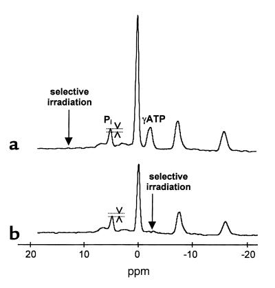
Unidirectional rates of ATP synthesis were measured using the saturation transfer experiment applied to the exchange between Pi and ATP. The steady-state Pi magnetization Mz was measured in the presence of selective irradiation of the γATP, and compared with the equilibrium Pi magnetization M0 in a control spectrum (without irradiation of γATP), as shown in Figure 1.
Figure 1.
Principle of ATP synthesis rate measurement by 31P NMR spectroscopy. (a) Control spectrum in one volunteer: the saturation pulse is applied symmetrically to the γATP Larmor frequency. (b) Spectrum with γATP saturation acquired in the same volunteer (NS = 128). Subtraction of spectrum (b) from (a) gives the fraction of Pi involved in the synthesis of ATP, as shown on the Pi peak. ppm, parts per million.
The fractional reduction of Pi magnetization upon γATP saturation (M0 – Mz)/M0 was measured from our spectra and was used in the calculation of the pseudo-first-order rate constant according to the equation of Forsen and Hoffman: k1 = [(M0 – Mz)/M0](1/T1*), where T1* is the spin-lattice relaxation time for the phosphorus nucleus of Pi when ATP is saturated.
T1* was then measured using a modified version of the inversion-recovery experiment (180°-τ-90°-acq-ID) in the presence of steady-state saturation of γATP during the total interpulse delay (ID) of 4 seconds and during the variable delay (τ). T1* was evaluated using a nonlinear least-squares fitting method. Unidirectional flux of ATP synthesis was then calculated by multiplying the constant k1 by the Pi concentration. The Pi concentration was measured from the control spectrum, assuming a constant ATP concentration of 5.5 mmol/kg in muscle (19).
Total acquisition time for 31P spectra was about 120 minutes. After completion of 31P measurements, the volunteer was removed from the magnet for a 10-minute break. The NMR radio-frequency probe was changed, and the volunteer was repositioned for measurement of the TCA cycle rate.
13C NMR measurement of TCA cycle flux.
For 13C spectroscopy, the probe consisted of one 13C circular coil and two 1H quadrature coils for 1H acquisitions and decoupling. After tuning, shimming with the FASTERMAP procedure (20), and acquiring scout images, 13C NMR spectra were acquired for 20 minutes before and during a 120-minute [2-13C]acetate infusion (350 mmol/l sodium salt, 99% 13C enriched; Isotec Inc., Miamisburg, Ohio, USA) at an infusion rate of 2.9 mg/kg/min.
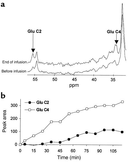
13C NMR spectra were acquired using either an image-selected in vivo spectroscopy (ISIS) sequence (TR = 2.3 seconds), or a nonlocalized sequence (TR = 1.4 seconds) with suppression of the 34.4 parts per million (ppm) lipid signal after inversion by an adiabatic pulse. Temporal resolution was 10 minutes. After Fourier transformation and baseline subtraction, glutamate C2 peaks were integrated between 55.3 and 55.8 ppm, and glutamate C4 peaks were integrated between 33.6 and 35.0 ppm (Figure 2).
Figure 2.
13C NMR data. (a) 13C spectra acquired in a volunteer before and at the end of the [2-13C]acetate infusion. (b) Time course of glutamate C4 and glutamate C2 peak areas during the same experiment. Glutamate C4 appears soon after the beginning of the [2-13C]acetate infusion. The increase in glutamate C2 is delayed in comparison to that of glutamate C4, reflecting the extra turn of the TCA cycle required to randomize the 13C-labeled carbon into this position.
Fractional enrichments and plasma acetate were measured from blood samples collected at 10-minute intervals and analyzed on a Hewlett-Packard 5890 gas chromatograph (HP-1 capillary column, 12 m × 0.2 mm × 0.33 mm film thickness) interfaced to a Hewlett-Packard 5971A mass selective detector operating in the electron impact ionization mode (12).
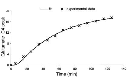
TCA cycle flux was assessed by monitoring the incorporation of 13C into glutamate from [2-13C]acetate and modeling the data using Cwave software (21, 22). An intracellular concentration of glutamate in skeletal muscle of 2.41 ± 0.28 mmol/l (as measured in the muscle biopsies described below) was used in the model. The model describes the increase in glutamate C4 labeling resulting from [2-13C]acetate conversion into [2-13C]acetyl CoA, which in turn condenses with oxaloacetate to produce [4-13C]citrate; this in turn labels α-ketoglutarate C4, which then equilibrates with glutamate C4. Following the second turn of the cycle, C2 and C3 of glutamate become labeled. Nonlinear least squares fit of glutamate C4 and C2 yielded an estimate of TCA cycle flux. A typical fit of the C4 glutamate data is shown in Figure 3. It is important to note that the TCA cycle flux calculated from our mathematical model is independent of both the amount of acetate incorporated into the TCA cycle (flux through acetyl CoA synthetase) and the substrate source (glucose, fatty acids, or acetate). This approach is similar to the methodology developed and validated by Chatham et al. in the perfused heart, which found a 1:1 relationship between 13C NMR–determined rates of oxygen consumption and measured rates of oxygen consumption (23).
Figure 3.
Typical model fit of the 13C NMR data of glutamate C4 in skeletal muscle (R2 = 0.93).
Muscle biopsies.
Muscle tissue (50–100 mg) was obtained from the vastus lateralis muscles of three additional subjects by a punch biopsy procedure using a 5-mm Bergstrom biopsy needle (Life Medical Equipment, Miami, Florida, USA); this was done before and after T3 treatment in order to determine muscle glutamate concentration for the TCA cycle flux calculations. The muscle tissue was quickly blotted on filter paper and immediately frozen in liquid nitrogen. The time from obtaining the tissue specimen until freezing was less than 30 seconds. Glutamate concentrations were measured using a 2300 STAT Plus analyzer (Yellow Springs Instrument Co., Yellow Springs, Ohio, USA).
Statistical analysis.
All data are expressed as mean ± SE. Comparisons of values measured before and after T3 treatment were made using the paired Student’s t test.
Results
Three days of T3 treatment induced a mild hyperthyroid state in these subjects, as reflected by a significant increase in plasma T3 concentration that was associated with suppression of the mean concentration of thyroid stimulating hormone (TSH) in plasma, a 30% increase in heart rate, a 17% increase in the mean rate of whole-body oxygen consumption (Table 1), and a 15% increase in whole-body energy expenditure (basal: 1819 ± 129 kcal/24 h vs. 2089 ± 141 kcal/24 h after T3 treatment; P < 0.02) as assessed by indirect calorimetry.
Table 1.
Effect of T3 treatment on clinical and metabolic parameters
The rate of ATP synthesis as assessed by 31P NMR was 5.2 ± 0.6 μmol/g/min in the basal state and it did not change significantly after T3 treatment (4.6 ± 0.8 μmol/g/min; P = 0.16 vs. basal), as shown in Figure 4.
Figure 4.
Muscle TCA cycle and ATP synthesis fluxes measured in seven volunteers before (basal) and after T3 treatment (post-T3). Thyroid hormone induces a significant increase in muscle TCA cycle flux, whereas the rate of ATP synthesis does not show any significant change. The resulting ATP/TCA ratio (normalized to the basal state) reflects a decrease in mitochondrial energy coupling following T3 treatment.
In contrast, the mean rate of the TCA cycle as assessed by 13C NMR from the rate of 13C incorporation into glutamate C4 and C2 increased by 70% after T3 treatment (P = 0.01 vs. basal). The TCA cycle flux was 0.064 ± 0.008 μmol/g/min in the basal state, and increased to 0.109 ± 0.013 μmol/g/min after T3 administration (Figure 4).
An estimate of mitochondrial energy coupling was obtained by expressing the ratio of ATP synthesis to TCA cycle flux, normalized to the basal state and this was found to decrease by 45% after T3 treatment (P = 0.001 vs. basal) (Figure 4).
Discussion
While it is well established that thyroid hormone regulates metabolic rate in humans, the mechanism by which it does so remains unknown. Although T3 has been shown to increase energy needs by activating ion pumping across membranes (2), substrate cycling (3, 4), and protein synthesis (5), regulation of basal metabolic rate by uncoupling substrate oxidation from ATP synthesis has been postulated (6) but never before demonstrated in humans.
In order to address this question, we used a novel 13 C/31 PNMR approach to measure T3-induced changes in mitochondrial energy coupling in human skeletal muscle. We found that three days of T3 treatment increased skeletal muscle TCA cycle flux by 70% that was associated with no significant change in the rate of ATP synthesis. Since there was a disproportionate increase in TCA cycle flux compared with ATP synthesis, these results suggest that T3 promotes increased thermogenesis in part by promoting mitochondrial energy uncoupling in skeletal muscle.
The TCA cycle is coupled to muscle oxygen consumption via a stoichiometric relationship with substrate utilization (24). Therefore it is likely that the 70% T3-induced increase in TCA cycle flux reflects a similar increase in muscle oxygen consumption. In contrast, whole-body oxygen consumption increased by only 17% following T3 treatment. The difference in magnitude between the T3-induced increase in muscle versus whole-body oxygen consumption can probably be ascribed to the limited contribution of resting skeletal muscle to whole-body oxygen consumption (25), and demonstrates the important advantage of this tissue-specific NMR method over whole-body indirect calorimetry. Furthermore, if one extrapolates the observed T3-induced increase in muscle energy metabolism to the whole body, it can be estimated to account for an increase of approximately 7% in whole-body oxygen consumption. The remainder (∼10%) can most likely be explained by the contribution of other tissues such as liver, kidney, and heart to whole-body oxygen consumption (26).
Uncoupling effects of high doses of thyroid hormone have previously been reported in vitro (6, 7), and more recently T3-induced proton leaks in the inner mitochondrial membrane have been demonstrated in vitro in rat liver (27) and rat skeletal muscle (28, 29). However, no evidence of a similar uncoupling effect in humans has yet been demonstrated, due to the difficulty in extrapolating in vitro measurements of isolated mitochondrial membrane permeability to the in vivo situation. Another difficulty in extrapolating the in vitro data to in vivo conditions is that the in vitro measurements are usually performed on isolated mitochondria under nonlimiting concentrations of ADP (state 3 respiration), in which proton leaks play a minor role (30). Under in vivo basal conditions, skeletal muscle respiration is likely to be operating under conditions of limited ADP (state 4 respiration), where proton leaks may account for a more important fraction of oxygen consumption (6, 31). The in vivo NMR method used in this study circumvents these limitations by assessing mitochondrial energy coupling in intact skeletal muscle under ambient ADP concentrations.
The mechanism by which thyroid hormone might promote increased mitochondrial uncoupling is unknown. However, a skeletal muscle–specific uncoupling protein (UCP-3) has recently been cloned in humans, and has been shown to be unregulated by thyroid hormone (9–18). This protein is thought to create a pathway that allows exothermic movement of protons through the inner mitochondrial membrane, and it has been speculated that UCP-3 might be involved in regulating energy expenditure and body weight in humans. Whether UCP-3 mediates mitochondrial uncoupling or not remains unclear. It has been reported recently that UCP-3 null mice have a normal response to thyroid hormone (32). On the other hand, transgenic mice with UPC-3 overexpression in skeletal muscle are hyperphagic and lean (33). However, the very high level of UCP-3 overexpression reported in these mice (∼60-fold) does not argue in favor of UCP-3 being an efficient mitochondrial uncoupler. Taken together, these data suggest that an increase in UCP-3 expression may not be the only factor responsible for the observed T3-induced increase in mitochondrial uncoupling.
Given the increasing prevalence of obesity in the industrialized nations (34), there is considerable interest in the regulation of uncoupling protein expression and activity as a means of promoting nonshivering thermogenesis and weight loss. Although thyroid hormone promotes UCP-3 expression, the ubiquitous distribution of thyroid hormone receptor and action throughout the body, especially in the heart and central nervous system, limits its use in this regard. In contrast, UCP-3 is expressed almost exclusively in skeletal muscle, and the present data would support UCP-3 as a possible pharmacological target to promote weight loss through selective increases in mitochondrial energy uncoupling in this tissue. Furthermore, conversion of intramyocellular lipid into heat by this mechanism would also be predicted to reverse fat-induced insulin resistance (35).
In summary, we used a novel NMR approach to measure T3-induced changes in mitochondrial energy coupling in human skeletal muscle. We found that three days of T3 treatment induced a disproportionate increase in TCA cycle flux compared with ATP synthesis in skeletal muscle. These data demonstrate that T3 can increase thermogenesis in skeletal muscle by promotion of mitochondrial uncoupling that may be partly mediated by an increase in UCP-3 protein expression. This in vivo NMR approach should be useful for exploring the roles of mitochondrial energy uncoupling processes in regulating energy metabolism and obesity in humans.
Acknowledgments
The authors would like to thank Joan Boyer, Laura Burden, Damien Nuzzo, and the staff of the Yale–New Haven Hospital General Clinical Research Center for expert technical assistance, Terry Nixon for maintenance and upgrades to the spectrometer, Peter Brown for coil construction, Jun Shen for assistance in pulse sequence implementation, Graeme Mason for assistance in modeling, and Gerard Burrow for helpful discussions. This work was supported by grants from the United States Public Health Service (R01 DK-49230, P30 DK-45735, and M01 RR-00125) and a K-23 award to K.F. Petersen. V. Lebon and S. Dufour are research associates and G.I. Shulman is an investigator of the Howard Hughes Medical Institute.
Footnotes
Vincent Lebon, Sylvie Dufour, and Kitt Falk Petersen contributed equally to this work.
References
- 1.Magnus-Levi A. Uber den respiratorischen Gaswechsel unter dem Einfluss des Thyroidea sowie unter vershiedenen pathalogishen Zustanden. Berl Klin Wochenschr. 1895;34:650. [Google Scholar]
- 2.Izmail-Beigi F, Edelman IS. Mechanism of thyroid calorigenesis: role of active sodium transport. Proc Natl Acad Sci USA. 1970;67:1071–1078. doi: 10.1073/pnas.67.2.1071. [DOI] [PMC free article] [PubMed] [Google Scholar]
- 3.Newsholme EA, Crabtree B. Substrate cycles in metabolic regulation and in heat generation. Biochem Soc Symp. 1976;41:61–109. [PubMed] [Google Scholar]
- 4.Shulman GI, Ladenson PW, Wolfe MH, Ridgway EC, Wolfe RR. Substrate cycling between gluconeogenesis and glycolysis in euthyroid, hypothyroid, and hyperthyroid man. J Clin Invest. 1985;76:757–764. doi: 10.1172/JCI112032. [DOI] [PMC free article] [PubMed] [Google Scholar]
- 5.Gustafsson R, Tata JR, Lindberg O, Ernster L. The relationship between the structure and activity of rat skeletal muscle mitochondria after thyroidectomy and thyroid hormone treatment. J Cell Biol. 1965;26:555–578. doi: 10.1083/jcb.26.2.555. [DOI] [PMC free article] [PubMed] [Google Scholar]
- 6.Freake HC, Oppenheimer JH. Thermogenesis and thyroid function. Annu Rev Nutr. 1995;15:263–291. doi: 10.1146/annurev.nu.15.070195.001403. [DOI] [PubMed] [Google Scholar]
- 7.Lardy HA, Feldcott G. Metabolic effects of thyroxine in vitro. Ann NY Acad Sci. 1951;54:636–648. doi: 10.1111/j.1749-6632.1951.tb46619.x. [DOI] [PubMed] [Google Scholar]
- 8.Martius C, Hess B. The mode of action of thyroxin. Arch Biochem Biophys. 1951;33:486–487. doi: 10.1016/0003-9861(51)90137-3. [DOI] [PubMed] [Google Scholar]
- 9.Boss O, et al. Uncoupling protein-3: a new member of the mitochondrial carrier family with tissue-specific expression. FEBS Letters. 1997;408:39–42. doi: 10.1016/s0014-5793(97)00384-0. [DOI] [PubMed] [Google Scholar]
- 10.Gong DW, He Y, Karas M, Reitman M. Uncoupling protein-3 is a mediator of thermogenesis regulated by thyroid hormone, β3-adrenergic agonists, and leptin. J Biol Chem. 1997;272:24129–24132. doi: 10.1074/jbc.272.39.24129. [DOI] [PubMed] [Google Scholar]
- 11.Jekabsons MB, Gregoire FM, Schonfeld-Warden NA, Warden CH, Horwitz BA. T3 stimulates resting metabolism and UCP-2 and UCP-3 mRNA but not nonphosphorylating mitochondrial respiration in mice. Am J Physiol. 1999;277:E380–E389. doi: 10.1152/ajpendo.1999.277.2.E380. [DOI] [PubMed] [Google Scholar]
- 12.Jucker BM, et al. Assessment of mitochondrial energy coupling in vivo using a novel 13C/31P NMR approach. Proc Natl Acad Sci USA. 2000;97:6880–6884. doi: 10.1073/pnas.120131997. [DOI] [PMC free article] [PubMed] [Google Scholar]
- 13.Masaki T, Yoshimatsu H, Sakata T. Expression of rat uncoupling protein family mRNA levels by chronic treatment with thyroid hormone. Int J Obes Relat Metab Disord. 2000;24(Suppl. 2):S162–S164. doi: 10.1038/sj.ijo.0801315. [DOI] [PubMed] [Google Scholar]
- 14.Cusin I, Rouru J, Visser T, Burger AG, Rohner-Jeanrenaud F. Involvement of thyroid hormones in the effect of intracerebroventricular leptin infusion on uncoupling protein-3 expression in rat muscle. Diabetes. 2000;49:1101–1105. doi: 10.2337/diabetes.49.7.1101. [DOI] [PubMed] [Google Scholar]
- 15.Larkin S, et al. Regulation of the third member of the uncoupling protein family, UCP3, by cold and thyroid hormone. Biochem Biophys Res Commun. 1997;240:222–227. doi: 10.1006/bbrc.1997.7636. [DOI] [PubMed] [Google Scholar]
- 16.Gong DW, He Y, Karas M, Reitman M. Uncoupling protein-3 is a mediator of thermogenesis regulated by thyroid hormone, B3-adrenergic agonists, and leptin. J Biol Chem. 1997;272:24129–24132. doi: 10.1074/jbc.272.39.24129. [DOI] [PubMed] [Google Scholar]
- 17.Pedersen SB, et al. Regulation of uncoupling protein-2 and -3 by growth hormone in skeletal muscle and adipose tissue in growth hormone-deficient adults. J Clin Endocrinol Metab. 1999;84:4073–4078. doi: 10.1210/jcem.84.11.6109. [DOI] [PubMed] [Google Scholar]
- 18.Barbe P, et al. Triidothyronine-mediated upregulation of UCP2 and UCP3 mRNA expression in human skeletal muscle without coordinated induction of mitochondrial chain genes. FASEB J. 2001;15:13–15. doi: 10.1096/fj.00-0502fje. [DOI] [PubMed] [Google Scholar]
- 19.Harris RC, Hultman E, Nordesjo LO. Glycogen, glycolytic intermediates and high energy phosphates determined in biopsy samples of musculus quadriceps femoris of man at rest. Scand J Clin Lab Invest. 1974;33:109–120. [PubMed] [Google Scholar]
- 20.Shen J, Rycyna RE, Rothman DL. Improvements on an in vivo automatic shimming method (FASTERMAP) Magn Reson Med. 1997;38:834–839. doi: 10.1002/mrm.1910380521. [DOI] [PubMed] [Google Scholar]
- 21.Mason GF, Rothman DL, Behar KL, Shulman RG. NMR determination of the TCA cycle rate and alpha-ketoglutarate/glutamate exchange rate in rat brain. J Cereb Blood Flow Metab. 1992;12:434–447. doi: 10.1038/jcbfm.1992.61. [DOI] [PubMed] [Google Scholar]
- 22.Mason GF. CWave: software for the design and analysis of 13C labeling studies performed in vivo. Proc Intl Soc Mag Reson Med. 2000;3:1870 . (Abstr.) [Google Scholar]
- 23.Chatham JC, Forder JR, Glickson JD, Chance EM. Calculation of absolute metabolic flux and the elucidation of the pathways of glutamate labeling in perfused rat heart by 13C NMR spectroscopy and nonlinear least squares analysis. J Biol Chem. 1995;270:7999–8008. doi: 10.1074/jbc.270.14.7999. [DOI] [PubMed] [Google Scholar]
- 24.Stryer, L. 1995. Biochemistry. W.H. Freeman and Company. New York, New York, USA. 507–558.
- 25.Field J, Beldings HS, Martin AW. An analysis of the relation between basal metabolism and summated tissue respiration in the rat. Journal of Cellular and Comparative Physiology. 1939;14:143–157. [Google Scholar]
- 26.Barker SB, Klitgaard HM. Metabolism of tissues excised from thyroxine-injected rats. Am J Physiol. 1952;170:81–86. doi: 10.1152/ajplegacy.1952.170.1.81. [DOI] [PubMed] [Google Scholar]
- 27.Brand MD, Steverding D, Kadenbach B, Stevenson PM, Hafner RP. The mechanism of the increase in mitochondrial proton permeability induced by thyroid hormones. Eur J Biochem. 1992;15:775–781. doi: 10.1111/j.1432-1033.1992.tb16984.x. [DOI] [PubMed] [Google Scholar]
- 28.Porter RK, et al. Indirect measurement of mitochondrial proton leak and its application. Int J Obes Relat Metab Disord. 1999;23:S12–S18. doi: 10.1038/sj.ijo.0800937. [DOI] [PubMed] [Google Scholar]
- 29.Lanni A, et al. Expression of uncoupling protein-3 and mitochondrial activity in the transition from hypothyroid to hyperthyroid state in rat skeletal muscle. FEBS Lett. 1999;444:250–254. doi: 10.1016/s0014-5793(99)00061-7. [DOI] [PubMed] [Google Scholar]
- 30.Groen AK, Wanders RJ, Westerhoff HV, Van der Meer R, Tager JM. Quantification of the contribution of various steps to the control of mitochondrial respiration. J Biol Chem. 1982;25:2754–2757. [PubMed] [Google Scholar]
- 31.Hafner RP, Nobes CD, McGown AD, Brand MD. Altered relationship between protonmotive force and respiration rate in non-phosphorylating liver mitochondria isolated from rats of different thyroid hormone status. Eur J Biochem. 1988;15:511–518. doi: 10.1111/j.1432-1033.1988.tb14477.x. [DOI] [PubMed] [Google Scholar]
- 32.Gong DW, et al. Lack of obesity and normal response to fasting and thyroid hormone in mice lacking uncoupling protein-3. J Biol Chem. 2000;275:16251–16257. doi: 10.1074/jbc.M910177199. [DOI] [PubMed] [Google Scholar]
- 33.Clapham JC, et al. Mice overexpressing human uncoupling protein-3 in skeletal muscle are hyperphagic and lean. Nature. 2000;406:415–418. doi: 10.1038/35019082. [DOI] [PubMed] [Google Scholar]
- 34.Flegal KM, Carroll MD, Kiczmarski RJ, Johnson CL. Overweight and obesity in the United States: prevalance and trends, 1960-1994. Int J Obes Relat Metab Disord. 1998;22:39–47. doi: 10.1038/sj.ijo.0800541. [DOI] [PubMed] [Google Scholar]
- 35.Shulman GI. Cellular mechanisms of insulin resistance. J Clin Invest. 2000;106:171–176. doi: 10.1172/JCI10583. [DOI] [PMC free article] [PubMed] [Google Scholar]


