The incidence of conjunctival melanoma is about 0.2–0.8/1 000 000 cases each year.1,2 As a consequence our knowledge about this tumour is limited, but cell lines derived from these tumours will give more information about them. So far only three conjunctival melanoma cell lines have been described.3,4 The first cell line, IPC 292, was described in 1993 by Aubert et al.4 Recently, Nareyeck et al developed two additional conjunctival melanoma cell lines (CRMM1 and CRMM2).3 We wish to report a further, fourth, conjunctival melanoma cell line, CM2005.1.
The conjunctival melanoma cell line CM2005.1 was established from tumour material derived from a 84‐year‐old male. The primary tumour was located at the medial side of the inner upper eyelid adjacent to the cornea. The tumour was treated initially by local excision together with adjuvant iridium 192 brachytherapy. After 3 years the tumour reappeared on the inner side of the lower eyelid. The tumour extended into the nasal cavity and disseminated further. The patient died 1 year later from the melanoma which had originated in the conjunctiva. Both the primary tumour and the local recurrence were histologically proven conjunctival melanomas.
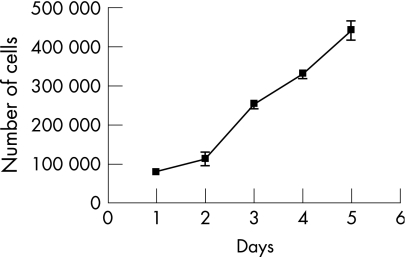
From the local recurrence two small tumour specimens were available for cell culture. The tumour material was cut into small pieces with scalpels and transferred to several culture plates. Culture plates contained 10 ml/dish RPMI 1640 (Invitrogen‐Gibco, Groningen, the Netherlands) supplemented with 10% FCS (Hyclone, Logan, UT), 100 IU/ml penicillin (Invitrogen‐Gibco) and 100 μg/ml streptomycin (Invitrogen‐Gibco). Cultures were incubated at 37°C in a humidified atmosphere and a CO2 content of 5% in air and the culture medium was refreshed every 72–96 h. After 4 months a stable cell line had been developed, which was grown for over 22 passages. Cell doubling time was measured three times by culturing 100 000 cells/plate. The cell count was measured again at days 1, 2, 3, 4 and 5, which showed a cell doubling time of around 35 h (fig 1).
Figure 1 Growth curve of conjunctival melanoma cell line CM2005.1 showing the number of cells at various time points during culture. Cell doubling time was around 35 h.
To establish the melanocytic origin of the cultured cells, cytospins were undertaken and immunohistochemically stained for S100, MelanA, NKI‐C3 and HMB 45.
Immunohistochemical reactions were performed using the streptavidin‐biotin method. The melanocytic origin of the cultured cells was proved by the fact that 95–100% were positive for all four primary antibodies. MelanA, HMB 45 and NKI‐C3 strongly stained the cultured cells, while the S100 labelling was slightly less intense.
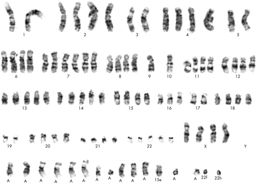
Cytogenetic analysis was performed to assess the karyotype of cell line CM2005.1. Chromosome preparations were obtained according to standard procedures and stained to produce R or Q banding. Cytogenetic abnormalities were described in accordance with the ISCN.5
The following karyogram was found in most cells: 83∼96, XX, +X, −Y, −Y, −1, −1, −3, −3, −4, −5, −5, −6, +7, +7, +del (7) (q22), −8, −9, −9, −10, −10, del (11) (q23)x2, −12, +13, +13, +14, −16, −17, −17, −18, −19, +20, −21, −21, −21, −22, +12∼16 mar (fig 2).
Figure 2 Karyogram of cell line CM2005.1. A very complex karyogram with gains, deletions and rearrangements of almost all chromosomes was observed in the majority of cultured cells.
Cell line CM2005.1 was derived from a recurrent conjunctival melanoma that reoccurred after excision and brachytherapy, which could explain the distorted karyogram and the relatively high cell doubling time of this cell line. However, the cytogenetic analysis was performed 4 months after start of the culture, which could have influenced the karyogram and may therefore not represent the original tumour. Nareyeck et al found cell doubling times of around 60 h for their conjunctival melanoma cell lines CRMM‐1 and CRMM‐2.3
This fourth conjunctival melanoma cell line is now available for research, and can hopefully contribute to expansion of our knowledge of conjunctival melanomas.
Footnotes
Funding: None.
Competing interests: None declared.
References
- 1.Missotten G S, Keijser S, De Keizer R J.et al Conjunctival melanoma in the Netherlands: a nationwide study. Invest Ophthalmol Vis Sci 20054675–82. [DOI] [PubMed] [Google Scholar]
- 2.Seregard S. Conjunctival melanoma. Surv Ophthalmol 199842321–350. [DOI] [PubMed] [Google Scholar]
- 3.Nareyeck G, Wuestemeyer H, von der Haar D.et al Establishment of two cell lines derived from conjunctival melanomas. Exp Eye Res 200581361–362. [DOI] [PubMed] [Google Scholar]
- 4.Aubert C, Rouge F, Reillaudou M.et al Establishment and characterization of human ocular melanoma cell lines. Int J Cancer 199354784–792. [DOI] [PubMed] [Google Scholar]
- 5.Mitelman F. ed. ISCN:an International System for Human Cytogenetic Nomenclature. Basel, Switzerland: Karger, 1995


