

Abstract
Undifferentiated rat pheochromocytoma (PC12) cells extend neurites when cultured in the presence of nerve growth factor (NGF). Extracellular guanosine synergistically enhances NGF-dependent neurite outgrowth. We investigated the mechanism by which guanosine enhances NGF-dependent neurite outgrowth. Guanosine administration to PC12 cells significantly increased guanosine 3-5-cyclic monophosphate (cGMP) within the first 24 h whereas addition of soluble guanylate cyclase (sGC) inhibitors abolished guanosine-induced enhancement of NGF-dependent neurite outgrowth. sGC may be activated either by nitric oxide (NO) or by carbon monoxide (CO).  -Nitro-l-arginine methyl ester (l-NAME), a non-isozyme selective inhibitor of nitric oxide synthase (NOS), had no effect on neurite outgrowth induced by guanosine. Neither nNOS (the constitutive isoform), nor iNOS (the inducible isoform) were expressed in undifferentiated PC12 cells, or under these treatment conditions. These data imply that NO does not mediate the neuritogenic effect of guanosine. Zinc protoporphyrin-IX, an inhibitor of heme oxygenase (HO), reduced guanosine-dependent neurite outgrowth but did not attenuate the effect of NGF. The addition of guanosine plus NGF significantly increased the expression of HO-1, the inducible isozyme of HO, after 12 h. These data demonstrate that guanosine enhances NGF-dependent neurite outgrowth by first activating the constitutive isozyme HO-2, and then by inducing the expression of HO-1, the enzymes responsible for CO synthesis, thus stimulating sGC and increasing intracellular cGMP.
Keywords: carbon monoxide, cyclic GMP, guanosine, heme oxygenase, neurite outgrowth, NGF, nitric oxide synthase, PC12 cells
Introduction
The roles of extracellular purine nucleosides and nucleotides as neurotransmitters and modulators are well documented [1]. Extracellular purines also exert trophic effects on cells; influencing growth, division [2, 3] differentiation, and even apoptosis [4–6]. Purines are released from cells under physiological conditions, acting as neurotransmitters and neuromodulators [7–9]. Trauma and other insults to the central nervous system are also potent stimuli that cause the release of purines [3], most of which are converted by ectoenzymes to adenosine and guanosine [3, 5]. After insults to cells, more guanine-based than adenine-based purines are released [10]. Furthermore, the extracellular concentration of guanosine remains elevated for prolonged periods in vitro [10] and for up to a week after central nervous system (CNS) injury [11], implying that extracellular guanosine may exert trophic effects in vivo.
Sprouting of neurites, which later become axons and dendrites, is an important change associated with neural development and differentiation [12, 13]. Neurite sprouting through regeneration or collateral sprouting also plays an important role in the functional recovery following injury to the central or peripheral nervous systems [14]. Amongst its trophic effects, guanosine stimulates neurite outgrowth and enhances NGF-dependent neurite outgrowth [3, 15].
PC12 cells serve as a useful model for studying cell signaling [16]. They respond to many growth factors, neurotrophins and hormones, which initiate multiple signaling pathways [17–20]. The specific cellular targets of these signaling pathways mediate the distinct responses of differentiation, proliferation and survival, all of which can be assessed [16]. The addition of NGF to PC12 cells causes a sustained activation of ERK, a mitogen-activated protein kinase, through activation of the TrkA receptor [16, 19]. This leads to the development of many phenotypic characteristics in PC12 cells, which are also associated with mature sympathetic neurons [13, 21].
Extracellular guanosine not only stimulates neurite outgrowth from primary cultures of rat hippocampal neurons [3], and PC12 cells [15] but also enhances the neuritogenic effects of NGF on PC12 cells [15, 22, 23]. Both in astrocytes [24] and in PC12 cells [22], guanosine increases intracellular adenosine 3′,5′-cyclic monophosphate (cAMP). Some of the neuritogenic effects of guanosine in PC12 cells appeared to be mediated through this mechanism [22]. However, guanosine also has cAMP-independent effects, synergistic with NGF, because its effects are not abolished by an adenylate cyclase inhibitor [22] and are synergistic with substances that increase intracellular cAMP [15].
The cAMP-independent component of the signal transduction mechanism through which guanosine synergistically enhances NGF-dependent neurite outgrowth is unknown. However, several lines of evidence indicate a role for guanosine 3′,5′-cyclic monophosphate (cGMP). First, extracellular guanosine increases intracellular cGMP in rat mesenteric artery [25]. Second, nitric oxide (NO) donors enhance NGF-dependent PC12 cell neurite outgrowth via a cGMP-dependent mechanism; and thirdly, the neuritogenic effects of the NO donors are abolished by guanylyl cyclase inhibitors and mimicked by cGMP analogs [26]. The mechanism by which cGMP enhances neurite outgrowth is currently unclear.
Nitric oxide synthases are a family of enzymes, which synthesize NO through the catalytic conversion of l-arginine to l-citrulline. The constitutively expressed forms are the endothelial (eNOS) or neuronal (nNOS), which are regulated by the cytosolic concentration of Ca2+ [27]. The inducible isoform (iNOS) is widely distributed, and becomes active only hours after an inducing event [28]. NO is involved in cell communication and signal transduction in many systems including the CNS [29–31]. NGF induced different isoforms of NOS in PC12 cells after 4 days in vitro [32, 33]. Peunova and Enikolopov [32] reported that the NO produced preceding the development of the differentiated phenotype is due predominantly to iNOS.
The diffusible gas carbon monoxide (CO) is a putative neurotransmitter [34]. Heme oxygenase (HO) synthesizes CO from the biologically active substrate biliverdin, which is rapidly reduced to bilirubin, and iron from intracellular heme [34]. CO can modulate activities attributed to cGMP in the nervous system [35–38]. It has been proposed that CO production is responsible for baseline cGMP levels in the hippocampus [36]. In cerebellar granule cell cultures, CO produced by HO affects intracellular cGMP concentrations by modulating the NO-soluble guanylate cyclase signaling system [39]. Conversely, NO, synthesized by NOS may induce the expression of HO-1 [40–42], indicating a close and reciprocal interaction between the NOS-NO and the HO-CO signaling systems [35, 36]. Based on these findings it has been proposed that a possible role for HO-1 is to counteract NO toxicity [43].
Together, these data led us to question whether guanosine enhanced NGF-dependent neurite outgrowth is through a mechanism involving cGMP, and if so, whether it was attributable to the stimulation of either NO or CO synthesis.
Materials and methods
Cell culture and treatments
Tissue culture supplies were from Life Technologies. All other supplies were obtained from Sigma RBI unless otherwise stated. 2.5S NGF was a generous gift from Dr. M. Coughlin, Department of Medicine, McMaster University, 6-(phenylamino)-5,8-quinolinedione (LY83583) was obtained from (Calbiochem), copper protoporphyrin from (Porphyrin Products), and zinc protoporphyrin IX from (Research Biochemical).
PC12 cells were maintained in either RPMI 1640 medium supplemented with 5% heat-inactivated (HI) fetal calf serum (FCS), 5% HI-horse serum (HS) and 1% antibiotic-antimycotic (Anti-Anti; 10,000 units of penicillin, 10,000 µg of streptomycin, 25 µg amphotericin B/ml in 0.85% saline) [15] or F-12K (Kaighn's Modification) medium supplemented with 15% HI-HS, 2.5% HI-FCS, and 1% Anti-Anti at 37 °C in a 5% CO2 environment.
Neurite outgrowth assay
To evaluate the effect of test compounds on neurite outgrowth in PC12 cells, they were added to cultures for 48 h as previously described [15]. Briefly, PC12 cells were plated onto poly-d,l-ornithine (PORN)-coated 24-well plates at a density of 2.5 × 10⁴ cells/well. Cells were cultured in RPMI 1640 supplemented with 1.5% HI-HS, 1.5% HI-FCS and 1% Anti-Anti. Guanosine was dissolved in 10% sodium hydroxide (1N NaOH) and when added to the culture medium the final concentration was 0.01% sodium hydroxide. In experiments in which guanosine (300 µM) was added to each well first, followed within 15 min by the addition of 2.5S NGF (40 ng/ml). Methylene blue, LY83583, hemoglobin, Nω-nitro-l-arginine methyl ester (l-NAME), copper protoporphyrin-IX (CuPP) and zinc protoporphyrin-IX (ZnPP), were added 15 min prior to the addition of guanosine. All test substances remained in the medium throughout the 48-h test period. Cells were maintained at 37 °C in a humidified atmosphere of 95% air, 5% CO2. After 48 h, cells were fixed with 10% formalin in phosphate buffered saline (PBS), pH 7.4 for 10 min and stored at 4 °C in PBS (pH 7.4) containing sodium azide (0.1%, w/v) until counted (usually within 2 weeks). Total cell number was determined by counting two randomly selected areas in each well. The observer was blind to the treatments the cells had received. At least 50 cells per well were counted, and the number of cells bearing one or more neurites determined using a NIKON Diaphot microscope equipped with phase contrast optics. Neurites were defined as processes extending at least one cell body diameter from the cell with growth cones at their tips [44].
Western immunoblot analysis
The expression of HO-1, HO-2, iNOS, nNOS, and β-actin in PC12 cells was determined by Western immunoblot analysis. PC12 cells were seeded onto PORN-coated plates at a density of 1 × 10⁶ cells/plate and maintained at 37° for 48 h prior to treatments. Cells were placed in a medium containing 3% HI-FCS and 3% HI-HS 12 h before the start of treatments. Cells were then treated with various agents for 0, 6, 12, 24, or 48 h. At the end of the treatment period cells were washed once with PBS and then harvested at 4 °C using a lysis buffer (25 µM Tris/HCl pH 7.4, 10 µM NaCl, 10 µM EDTA, 100 µl/10 ml Tween 20, 10 µM sodium pyrophosphate decahydrate, 10 mM sodium orthovanadate, 5 µg/ml leupeptin, 10 mM glycerophosphate). Cells were disrupted by sonication and aliquots (25 µl) were removed for the determination of protein concentrations using the bicinchoninic acid (BCA) protein assay (Pierce, Illinois, USA). Cells lysates were separated on 12% SDS-polyacrylamide gels and electrophoretically transferred to nitrocellulose membranes (PALL, Michigan, USA). Membranes were incubated with a specific primary antibody overnight at 4 °C then were exposed to a secondary antibody for 1 h at room temperature. Positive control peptides for nNOS and iNOS (obtained from Cayman Chemical, Ann Arbor. MI) were used as electrophoresis standard. The following antibodies were used: monoclonal antibody to brain NOS (Sigma, dilution: 1:1,000), monoclonal antibody to the inducible NOS (Sigma, dilution: 1:1,000), anti-iNOS/bNOS, goat anti-mouse IgG-HRP (StressGen Biotechnologies, dilution 1:100,000), monoclonal antibody to β-actin (abCAM, dilution: 1:20,000), monoclonal anti-β-actin, goat anti-mouse IgG-HRP (Novus Biological, dilution 1:100,000), monoclonal antibody to HO-1 (StressGen Biotechnologies, dilution 1:2,000), anti-HO-1, goat anti-mouse IgG-HRP (StressGen Biotechnologies, dilution 1:200,000), rabbit polyclonal antibody to HO-2/HO-1 (StressGen Biotechnologies, dilution 1:1,000), anti HO-2/HO-1, goat anti-rabbit IgG-HRP (StressGen Biotechnologies, dilution 1:200,000). Immunocomplexes were then visualized using a chemiluminescence substrate (Sigma-Aldrich). Immunoblots were quantified by densitometric analyses, using the Northern Eclipse program (EPIX). All bands on the immunoblots were normalized to their corresponding β-actin bands prior to statistical analysis.
Determination of cyclic GMP
PC12 cells were seeded onto PORN-coated 12-well plates at a density of 5 × 10⁵ cells/well and maintained at 37 °C for 48 h prior to treatments. Cells were placed in a medium containing 3% HI-FCS and 3% HI-HS 12 h before the start of treatments. Cells were then treated with various agents for different times (0, 6, 12, 24, or 48 h). At the end of the treatment period cGMP was extracted and analyzed using a cGMP enzyme immunoassay (EIA) kit (Amersham Biosciences). The assay is based on a competition between unlabeled cGMP in the sample or (standard) and a fixed quantity of peroxidase-labeled cGMP for a limited number of binding sites on a cGMP specific antibody. With fixed amounts of antibody and cGMP-peroxidase conjugate, the amount of bound cGMP-peroxidase conjugate is inversely proportional to the concentration of unlabeled cGMP.
Statistical analysis
Statistical analysis was carried out using a two-way ANCOVA, when applicable, or a two-way ANOVA followed by Fischer's LSD test for multiple comparisons.
Results
Guanosine enhances NGF-dependent neurite outgrowth via activation of soluble guanylate cyclase
As we have reported previously, we have evaluated the effect of guanosine on NGF-dependent neurite outgrowth during the first 48 h, as guanosine most likely exerted its effects within the first 1–2 h after treatment [23]. Therefore, all experiments reported in this manuscript were performed during the first 48 h of PC12 cell treatment with various agents.
PC12 cells contain both soluble, and particulate isoforms of guanylate cyclase (GC) [26, 45]. To determine whether activation of GC was necessary for the neuritogenic effect of guanosine, inhibitors of GC were added for 48 h to PC12 cells treated with NGF plus guanosine. Methylene blue at concentrations ranging from 0.1 to 1 mM was added to PC12 cell cultures that contained NGF alone (40 ng/ml), or guanosine (300 mM) plus NGF (40 ng/ml). Cultures were then evaluated for neurite outgrowth. At the concentrations used in these experiments, methylene blue inhibits sGC but not particulate GC [46, 47]. Methylene blue had no effect on the outgrowth of neurites induced by NGF alone (Figure 1). However, in cultures treated with both guanosine and NGF, the addition of methylene blue reduced significantly (P < 0.01) the proportion of neurite-bearing cells in a concentration-dependent manner (Figure 1). LY83583 inhibits both, particulate GC and sGC [48]. When LY83583 (10 nM) was added to PC12 cell cultures, it inhibited neurite outgrowth elicited by guanosine plus NGF but had no effect on neurite outgrowth in cultures treated with NGF alone (data not shown). These data support the hypothesis that cGMP plays a role in enhancing the effect of guanosine on NGF-mediated neurite outgrowth. Moreover, since methylene blue at the concentrations used inhibits soluble but not particulate GC, these data imply that guanosine activates sGC.
Figure 1.
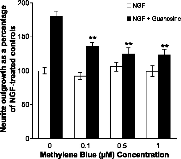
Methylene blue attenuates guanosine-enhanced nerve growth factor-dependent neurite outgrowth in PC12 cells. PC12 cells were cultured in RPMI 1640 medium supplemented with 5% heat-inactivated fetal calf serum, 5% heat-inactivated horse serum and 1% antibiotic-antimycotic for 48 h. Cultures were then treated with NGF (40 ng/ml) or NGF (40 ng/ml) plus guanosine (300 µM) and with increasing concentrations of methylene blue (0–1 µM). After 48 h, the total cell number and number of cells bearing one or more neurites were determined by counting two random areas in each well. The mean proportion of neurite-bearing cells in cultures treated with NGF was approximately 25%–35%. Because this value varied slightly between experiments, all experimental values are expressed relative to the NGF treated cultures, which was defined as 100%. Open bars: NGF treatment, closed bars: NGF plus guanosine treatment. Cultures treated with guanosine plus NGF had a significantly (P < 0.01, two-way independent ANOVA) greater proportion of neurite-bearing cells than those treated with NGF alone. Methylene blue (0.1 to 1.0 µM) had no effect on the proportion of neurite-bearing cells in cultures treated with NGF alone, but at concentrations from 0.1 to 1.0 µM, it significantly (**P < 0.01, two-way independent ANOVA) reduced the proportion of neurite-bearing cells in cultures treated with guanosine plus NGF. Data represent the mean ± SEM of 12 determinations from two replicate experiments.
Guanosine may activate sGC either directly, or indirectly by stimulating the formation of NO, CO or hydroxyl radicals, which are all physiological activators of sGC [49]. To distinguish between these possibilities we added hemoglobin (100 nM) to cultures of PC12 cells. Extracellular hemoglobin directly scavenges NO and CO because these substances avidly bind to the heme moiety, thereby inhibiting their GC activating activity [46]. Hemoglobin reduced the synergistic effects of guanosine on NGF-mediated neurite outgrowth but had no effect on neurite outgrowth elicited by NGF alone (data not shown). Although these data imply that guanosine stimulates the synthesis of a sGC-activating factor, they do not show whether this factor is NO, CO or hydroxyl radical.
Inhibition of nitric oxide synthase (NOS) has no effect on guanosine-enhanced NGF-dependent neurite outgrowth
In the rat mesenteric artery, guanosine stimulates NO formation [25]. Therefore, we investigated if guanosine could also stimulate NO synthesis in PC12 cells. To examine whether NO was involved in the signal transduction pathway that mediated the enhancement of neurite outgrowth by guanosine through sGC, we inhibited the enzyme NOS, which catalyzes the conversion of l-arginine to l-citrulline plus NO. The l-arginine analog, Nω-nitro-l-arginine methyl ester hydrochloride (l-NAME) [50] inhibits NOS competitively [25, 51]. In this experiment, we reduced the concentration of l-arginine in the culture medium from 1 mM to 80 µM, thus permitting more effective competition of the NOS inhibitor with l-arginine. Under these conditions, L-NAME inhibits NOS at 10 µM [25, 51]. When added to the culture medium of PC12 cells at 10 µM l-NAME did not inhibit NGF-dependent neurite outgrowth, and had no effect on the guanosine-mediated enhancement of NGF-dependent neurite outgrowth (Figure 2a). To confirm that NO was not involved in the signal transduction pathway that mediates the enhancement of neurite outgrowth by guanosine, we determined the expression of two subtypes of NOS isoenzymes: iNOS, the inducible isoform (Figure 2b), and nNOS, the constitutive isoform (Figure 2c). PC12 cultures were exposed to guanosine alone (300 µM), or NGF alone (40 ng/ml), or to the combination of guanosine plus NGF for 48 h, and NOS expression was determined by Western immunoblot analysis. We analyzed various protein concentrations (5–50 µg/ml) and determined β-actin expression on the same Western immunoblots. Using protein concentrations as high as 50 µg/ml neither nNOS nor iNOS expression could be detected in PC12 cells treated with guanosine, or NGF, or the combination of the two agents. These data indicate that since NOS is not expressed during the first 48 h of treatment, thus NO cannot mediate the effect of guanosine on neurite outgrowth.
Figure 2.

(a) Inhibition of nitric oxide synthase has no effect on the proportion of neurite-bearing PC12 cells cultured for 48 h in the presence of NGF, or NGF plus guanosine. PC12 cells were cultured with NGF (40 ng/ml) or NGF (40 ng/ml) plus guanosine (300 µM) as described in Figure 1. Cultures were treated with the general nitric oxide synthase inhibitor, l-NAME (0.1–10 mM) for 48 h and the number of cells bearing one or more neurites were determined as described in Figure 1. Open bars: NGF treatment, closed bars: NGF plus guanosine treatment. Neurite outgrowth in cultures treated with guanosine plus NGF was significantly (P < 0.01, two-way independent ANOVA) greater than in cultures treated with NGF alone. l-NAME (0.1–10 µM) had no effect on the proportion of neurite bearing cells in cultures treated with NGF alone, or in combination with guanosine. Data represent the mean ± SEM of 12 determinations from two replicate experiments. (b) Inducible nitric oxide synthase is not expressed in PC12 cells cultured for 48 h in the presence of guanosine, or NGF, or guanosine plus NGF. PC12 cells were cultured on plates coated with poly-d,l-ornithine for 72 h. Cells were then grown in serum-reduced medium (3% heat-inactivated fetal calf serum and 3% heat-inactivated horse serum) for 12 h, and were treated with guanosine (G, 300 µM) or NGF (N, 40 ng/ml) or guanosine (300 µM) plus NGF (40 ng/ml) (G + N), or with no added treatments (C). The expression of inducible nitric oxide synthase was determined at various time points (6, 12, 24 and 48 h) by Western immunoblot analysis. Recombinant inducible nitric oxide synthase protein (50 ng) was used as a positive control (P). Immunoblots were quantified by densitometric analysis and were normalized to the corresponding β-actin bands as described in the Materials and methods. Statistical analysis was performed using a two-way ANCOVA followed by Fischer's LSD post-hoc comparison test. Data presented are representative of at least three independent experiment. (c) Neuronal nitric oxide synthase is not expressed in PC12 cells cultured for 48 h in the presence of guanosine, or NGF, or guanosine plus NGF. PC12 cells were cultured as described in panel (b), and the expression of neuronal nitric oxide synthase was determined at various time points (6, 12, 24 and 48 h) by Western immunoblot analysis. Recombinant neuronal nitric oxide synthase protein (50 ng) was used as a positive control (P). Immunoblots were quantified by densitometric analysis and were normalized to the corresponding β-actin bands as described in the Materials and methods. Statistical analysis was performed using a two-way ANCOVA followed by Fischer's LSD post-hoc comparison test. Data presented are representative of at least three independent experiment.
Inhibition of heme oxygenase (HO) attenuates guanosine-enhanced NGF-dependent neurite outgrowth
Since NO apparently did not mediate the effects of guanosine, we examined the possibility that guanosine stimulated CO synthesis. Heme oxygenase (HO), the enzyme which synthesizes CO, has been detected in rat adrenal glands [52]. Zinc protoporphyrin-IX (ZnPP) inhibits both the constitutive HO (HO-2) and the inducible HO (HO-1) isoforms of this enzyme [52, 53]. We, therefore, tested the effect of increasing concentrations of ZnPP on the ability of NGF or, NGF plus guanosine to induce neurite outgrowth. Addition of ZnPP (0.01, 0.1, 1 µM) to PC12 cell cultures did not significantly reduce outgrowth of neurites induced by NGF (Figure 3), when compared to control cultures. In contrast, ZnPP attenuated the ability of guanosine to enhance NGF-stimulated neurite outgrowth (Figure 3). As a control for non-specific effects of metalloporphyrins we used copper protoporphyrin-IX (CuPP), which does not inhibit heme oxygenase [54]. The addition of CuPP to PC12 cells did not inhibit either NGF-dependent neurite outgrowth or the ability of guanosine to enhance NGF-dependent neurite outgrowth (data not shown).
Figure 3.
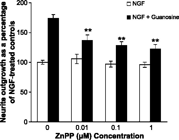
Inhibition of heme oxygenase attenuates guanosine-enhanced NGF-dependent neurite outgrowth in PC12 cells. PC12 cells were cultured with NGF (40 ng/ml) or NGF (40 ng/ml) plus guanosine (300 µM) as described in Figure 1. Some cultures were treated with the selective inhibitor of heme oxygenase zinc protoporphyrin-IX (0.01Y1 µM) for 48 h and the number of cells bearing one or more neurites was determined as described in Figure 1. Open bars: NGF treatment, closed bars: NGF plus guanosine treatment. Cultures treated with guanosine plus NGF had a significantly (P < 0.01 two-way independent ANOVA) greater proportion of neurite-bearing cells than cultures treated with NGF alone. Zinc protoporphyrin-IX significantly decreased (**P < 0.01) the neurite growth from cultures treated with guanosine plus NGF, but had no significant effect on neurite outgrowth in cultures treated with NGF alone. Data represent the mean ± SEM of 12 determinations from two replicate experiments.
Guanosine induces heme oxygenase-1 (HO-1) expression
PC12 cells have been previously shown to express both HO-1 and HO-2 proteins under basal conditions [55]. Since the addition of ZnPP to PC12 cultures decreased NGF-dependent neurite outgrowth, we attempted to establish whether guanosine treatment had any effect on the expression of the HO-1 protein during the first 48 h of this process. We treated PC12 cultures with guanosine alone (300 µM), or with NGF alone (40 ng/ml), or with a combination of guanosine plus NGF for 48 h, and determined HO-1 expression by Western immunoblot analysis. We used the monoclonal antibody directed against rat HO-1, which recognizes a single protein band, at 32 kDa, the reported molecular weight for this enzyme [36] (Figure 4b). Western immunoblots were quantified and normalized against their corresponding β-actin band. Statistical analysis revealed no differences in β-actin expression for any of the treatments at the different time points (data not shown); therefore, this was used as a control for normalizing HO-1 protein expression. There was no significant change in HO-1 expression in untreated PC12 cells during the 48-h period. In cells exposed to guanosine alone HO-1 expression, unexpectedly, was significantly reduced at 6 h compared to untreated cells at 0 time (P < 0.05), and at 6 h (P < 0.01) (Figure 4). A possible explanation for this result may stem from the observation that HO-1 gene expression is suppressed by elevated intracellular calcium concentrations [56] and guanosine has been shown to increase intracellular calcium concentrations in astrocytes [57]. Since increases in calcium are rapid and transient, this may account for the reduced HO-1 expression at 6 h. The addition of NGF, however, is sufficient to override this effect and so at later time points HO-1 expression is up-regulated by the combination of NGF and guanosine. The addition of guanosine plus NGF significantly increased HO-1 expression at 12 h (P < 0.05), compared to that of control cells at 0 time (Figure 4). After 24 h all three treatments (guanosine alone, or NGF alone or guanosine plus NGF) led to a significant increase in HO-1 expression compared to control cells at 0 time (P < 0.01) (Figure 4). In cells exposed to guanosine alone the enhanced HO-1 expression at 24 h was also significantly different from that detected in untreated cells at 24 h (P < 0.05). Whereas in cells treated with NGF alone, or with the combination of NGF plus guanosine this difference was significant at P < 0.01 (Figure 4). After 48 h, HO-1 expression was elevated significantly only in cells exposed to NGF alone when compared to untreated cells at 0 time, and at 48 h (P < 0.05) for both comparisons. In cells exposed to guanosine alone or to NGF plus guanosine HO-1 expression by 48 h declined and was similar to untreated control values at 0 time and at 48 h.
Figure 4.
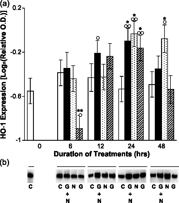
Guanosine induces the expression of heme oxygenase-1 protein in PC12 cells. PC12 cells were cultured on plates coated with poly-d,l-ornithine for 72 h. Cells were then grown in serum-reduced medium (3% heat-inactivated fetal calf serum and 3% heat-inactivated horse serum) for 12 h, and were treated with guanosine (G, 300 µM) or NGF (N, 40 ng/ml) or guanosine (300 µM) plus NGF (40 ng/ml) (G + N), or with no added treatments (C) as described in Figure 2b. The expression of heme oxygenase-1 was determined at various time points (6, 12, 24 and 48 h) by Western immunoblot analysis. Immunoblots were quantified by densitometric analysis and were normalized to the corresponding β-actin bands as described in the Materials and methods. Open bars: untreated controls (C), closed bars: guanosine plus NGF (G + N) treatment, stippled bars: NGF (N) treatment, hatched bars: guanosine (G) treatment. Statistical analysis was performed using a two-way ANCOVA followed by Fischer's LSD post-hoc comparison test (○ P < 0.05 compared with control time point 0); (○○ P < 0.01 compared with time point 0); (* P < 0.05 compared with control at same time point); (** P < 0.01 compared with control at same time point). (a) Data represent the mean optical density ± SEM obtained in six independent experiments. (b) Results are representative Western immunoblots obtained in these experiments.
In parallel experiments, we determined whether guanosine had an effect on the expression of the constitutive isoform, HO-2 in these cells. We treated PC12 cultures with guanosine alone (300 µM), or NGF alone (40 ng/ml), or in combination guanosine plus NGF for 48 h and determined HO-2 expression by Western immunoblot analysis. Using the monoclonal antibody directed against rat HO-2 we detected a single protein band, of molecular weight 36 kDa, as described for HO-1 [36] (Figure 5b). Western immunoblots were quantified and normalized against their corresponding β-actin band. Again, statistical analysis revealed no differences in β-actin expression for any of the treatments at the different time points (data not shown), therefore, this was used as a control for normalizing HO-2 protein bands. HO-2 expression was detectable at 0 time in untreated PC12 cells, but none of the treatments had any significant effect on its expression at any time point determined. In the guanosine-treated cells HO-2 expression was elevated slightly at 12 h, but this was not statistically significant.
Figure 5.
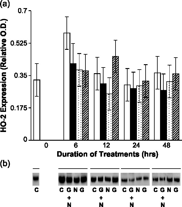
Guanosine has no effect on heme-oxygenase-2 protein expression in PC12 cells. PC12 cells were cultured on plates coated with poly-d,l-ornithine for 72 h. Cells were then grown in serum-reduced medium (3% heat-inactivated fetal calf serum and 3% heat-inactivated horse serum) for 12 h, and were treated with guanosine (G, 300 µM) or NGF (N, 40 ng/ml) or guanosine (300 µM) plus NGF (40 ng/ml) (G + N), or with no added treatments (C) as described in Figure 2b. The expression of heme oxygenase-2 was determined at various time points (6, 12, 24 and 48 h) by Western immunoblot analysis. Immunoblots were quantified by densitometric analysis and were normalized to the corresponding β-actin bands as described in the Materials and methods. Open bars: untreated controls (C), closed bars: guanosine plus NGF (G + N) treatment, stippled bars: NGF (N) treatment, hatched bars: guanosine (G) treatment. Statistical analysis was performed using a two-way ANOVA. (a) Data represent the mean optical density T SEM obtained in three independent experiments. (b) Results are representative Western immunoblots obtained in these experiments.
Guanosine increases intracellular cGMP concentrations in PC12 cells during guanosine-enhanced NGF-dependent neurite outgrowth
CO is a known activator of sGC [36]. Since we have shown that HO-1 expression is elevated in PC12 cells exposed to guanosine, we next investigated whether this increased HO-1 expression is accompanied by elevation in intracellular cGMP concentrations. PC12 cell cultures were treated with guanosine alone (300 µM), or NGF alone (40 ng/ml), or in combination guanosine plus NGF for 48 h. Intracellular cGMP concentrations were determined after 6, 12, 24 and 48 h of treatment. In cells exposed to guanosine alone, cGMP concentrations increased significantly after 6 h compared to untreated cells at this time point (P < 0.05). In PC12 cells treated with guanosine plus NGF, cGMP concentrations increased significantly after 6 h compared to untreated cells at 0 time (P < 0.05), remained significantly elevated at 12 h (P < 0.05) and at 24 h (P < 0.05), and declined to values comparable to control by 48 h.
Inhibition of heme oxygenase (HO) attenuates intracellular cGMP concentrations in PC12 cells during guanosine enhanced NGF-dependent neurite outgrowth
Previous experiments have demonstrated that the HO inhibitor, ZnPP, blocked guanosine enhanced NGF-dependent neurite outgrowth of PC12 cells (Figure 3). We have also shown that HO-1 expression and cGMP concentrations were elevated during the first 12–24 h of this neurite outgrowth process (Figures 4 and 6, respectively). We therefore examined whether pre-treatment of cells with the HO inhibitor ZnPP had any effect on cGMP concentrations. Although metalloporphrins, such as ZnPP-IX have been used widely to demonstrate the role for this enzyme in numerous physiological situations, their selectivity for HO has been questioned; as they may also inhibit haemoproteins such sGC and NOS [58]. However, metalloporphrins used at a concentration below 10 µM are more selective for HO [59]. Since we found that ZnPP, at 100 nM concentration significantly attenuated guanosine-enhanced neurite outgrowth (Figure 3), we used this concentration of ZnPP to test its effect on intracellular cGMP concentration. PC12 cell cultures were exposed to guanosine alone (300 µM), or NGF alone (40 ng/ml), or in combination guanosine plus NGF for 12 h. ZnPP (100 nM) was added to some cultures prior to the addition of guanosine, or NGF, or NGF plus guanosine, and intracellular cGMP concentrations were determined. In cells exposed to guanosine alone, or to guanosine plus NGF, cGMP concentrations were significantly elevated compared to untreated cells (P < 0.01) and to cells treated with NGF alone (P < 0.01) (Figure 7). In cultures pretreated with ZnPP, cGMP concentrations were significantly reduced in cells exposed to guanosine alone (P < 0.05), or guanosine plus NGF (P < 0.05) compared to the corresponding treatments without the inhibitor. Treatment of PC12 cells with NGF did not increase intracellular cGMP concentrations, and the addition of ZnPP had no effect on cGMP concentrations.
Figure 6.
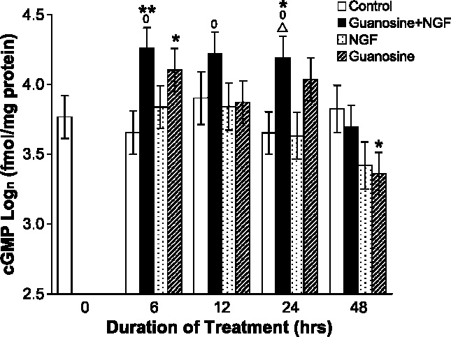
Guanosine increases cGMP concentrations during guanosine-enhanced NGF-dependent neurite outgrowth in PC12 cells. PC12 cells were cultured on plates coated with poly-d,l-ornithine for 72 h. Cells were then grown in serum-reduced medium (3% heat-inactivated fetal calf serum and 3% heat-inactivated horse serum) for 12 h, and were treated with guanosine (G, 300 µM) or NGF (N, 40 ng/ml) or guanosine (300 µM) plus NGF (40 ng/ml) (G + N), or with no added treatments (C) as described in Figure 2b. Cells were lysed at time points 0, 6, 12, 24, and 48 h and cGMP concentrations were determined by a competitive enzyme immunoassay. Open bars: untreated controls (C), closed bars: guanosine plus NGF (G + N) treatment, stippled bars: NGF (N) treatment, hatched bars: guanosine (G) treatment. Statistical analysis was performed using a two-way ANCOVA followed by Fischer's LSD post-hoc comparison test (○ P < 0.05, compared to time point 0); (* P < 0.05, relative to control); (** P < 0.01, relative to control); (Δ P < 0.05, relative to NGF). Data presented represent the mean relative optical density T SEM obtained in six independent experiments.
Figure 7.
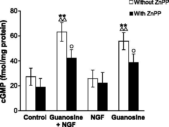
Inhibition of heme oxygenase attenuates cGMP concentrations during guanosine enhanced NGF-dependent neurite outgrowth in PC12 cells. PC12 cells were cultured on plates coated with poly-d,l-ornithine for 72 h. Cells were then grown in serum-reduced medium (3% heat-inactivated) fetal calf serum and 3% heat-inactivated horse serum) for 12 h, and were treated with guanosine (G, 300 µM) or NGF (N, 40 ng/ml) or guanosine (300 µM) plus NGF (40 ng/ml) (G + N), or with no added treatments (C) as described in Figure 2b. The selective inhibitor of heme oxygenase, zinc protoporphyrin-IX (100 nM), was added to some cultures prior to the addition of guanosine, or NGF, or guanosine plus NGF. Cells were lysed after 12 h and cGMP concentrations were determined by a competitive enzyme immunoassay. Open bars: no zinc protoporphyrin-IX added, closed bars: zinc protoporphyrin-IX added. Statistical analysis was performed using a one-way ANOVA followed by Fischer's LSD post-hoc comparison test (** P < 0.01, relative to control); (ΔΔ P < 0.01, relative to NGF); (○ P < 0.05, relative to treatment without zinc protoporphyrin-IX). Data presented represent the mean relative optical density T SEM obtained in six independent experiments.
Discussion
Addition of guanosine to cultures of undifferentiated, NGF-naïve PC12 cells modestly increases the proportion of cells with neurites after 48 h and, in cultures treated with maximally effective concentrations of NGF, addition of guanosine produces a significant further increase in the proportion of neurite-bearing cells [15]. This implied that the effects of NGF and guanosine are mediated through distinct signaling systems. We reported previously that, whereas some of the neuritogenic effects of guanosine were mediated by increases in intracellular cAMP [22, 23], there was also a cAMP-independent component [22]. Several points led us to consider that a signaling pathway involving cGMP may be responsible for the cAMP-independent component. Vuorinen et al. [25] had shown that extracellular guanosine increases intracellular cGMP in rat mesenteric artery through a NO-dependent mechanism; nitric oxide donors enhance NGF-dependent PC12 cell neurite outgrowth via a cGMP-dependent mechanism [26]; and the neuritogenic effects of the NO donors are abolished by guanylyl cyclase inhibitors and mimicked by cGMP analogs [26]. Our data indicated that indeed cGMP is involved in the neuritogenic effects of guanosine in PC12 cells, but, surprisingly, that NO is not.
Increases in intracellular cGMP are often due to activation of GC by NO [35, 45]. Schulick et al. [60] demonstrated that activation of GC by NO donors or atrial natriuretic peptide enhanced NGF-dependent neurite outgrowth in PC12 cells. Further, in PC12 cells NGF upregulates NOS expression, and NO plays an important role in their cellular differentiation [32]. However, NGF-induced differentiation of undifferentiated NGF-naïve PC12 cells could not be abolished by inhibitors of sGC, and NO alone is not sufficient to induce PC12 cell differentiation [26, 33].
In the experiments reported here, pre-treating undifferentiated, NGF-naïve PC12 cells with the NOS inhibitor l-NAME, had no effect on the guanosine-enhanced NGF-induced neurite outgrowth. Moreover, like others [61, 62], we found that neither nNOS nor iNOS isoforms were expressed during the first 48 h of cellular differentiation of PC12 cells. Therefore, NO cannot mediate the neuritogenic effects of guanosine on PC 12 cells during the first 48 h.
Rather than NO, our data indicated that CO might activate sGC in response to guanosine in undifferentiated PC12 cells. The heme oxygenase inhibitor, ZnPP attenuated both the ability of guanosine to enhance NGF-dependent neurite outgrowth, and its ability to increase intracellular cGMP, alone or in the presence of NGF. This led us to conclude that guanosine either activated constitutive HO-2 or induced the expression of either HO-1 and/or the HO-2 isoforms of this enzyme.
Our data indicated that, as others have reported [55], both HO-1 and HO-2 were expressed constitutively in PC12 cells under basal cell culture conditions. Guanosine induced a significant increase in the expression of HO-1 protein at 24 h in undifferentiated PC12 cells, but had no significant effect on HO-2 expression. Guanosine, in the presence of NGF, produced an even more rapid and pronounced enhancement of HO-1 expression, which was detectable after 12 h. Treatment of PC12 cells with NGF alone also increased the expression of HO-1. Several authors reported recently that NGF promotes HO-1 gene transcription in PC12 cells [63, 64] via activation of the MEK pathway. HO-1 mediates the anti-oxidative and antiapoptotic effects of NGF in serum-deprived PC12 cells [63]. In PC12 cells exposed to oxidative stress induced by 6-hydroxy dopamine, NGF promotes HO-1 upregulation, in this case via the PI3K/Akt/PKB pathway [64].
Others [63, 64] have reported increased HO-1 protein by 6–9 h after exposure to NGF, whereas in our study we observed no change in HO-1 protein concentration until 24 h after NGF addition. This discrepancy may be due to a difference in the PC12 cell line, or to the experimental conditions to which the cells were exposed.
We did not expect to find inhibition of HO-1 protein expression by guanosine at 6 h. It has been reported however, that the HO-1 gene expression is suppressed by the elevation of intracellular calcium concentration [56]. Since we have found that guanosine increases intracellular calcium concentrations in astrocytes [57], similar mechanisms may downregulate HO-1 expression in PC12 cells at the 6-h time point. As the guanosine-stimulated calcium increases are rapid and transient, at later time points (12–24 h) other intracellular pathways, such as the MAPK and PI-3 kinase pathways are activated, and these are known to promote HO-1 gene expression. Most likely, by 48 h these pathways are no longer activated.
In contrast to HO-1, neither guanosine nor NGF, alone or in combination with guanosine, affected HO-2 expression. This confirms the results of others who have found that HO-2 is not regulated by NGF [63, 64]. The HO-1 isozyme is induced by diverse intracellular signals (reviews: Maines [36], Immenshchuh and Ramadori [65]), that include some of the pathways which activate protein kinases, such as PKA [66], or PKC [67, 68], or the second messenger cGMP [42] most likely via PKG [69]. These pathways likely converge on nuclear effectors such as the nuclear factor-κB (NF-κB), activator protein-1 (AP-1) and heat shock factor, to enhance HO-1 gene transcription by interacting with the regulatory elements in the promoter region the gene [36, 65].
We reported previously that inhibition of transport of guanosine into PC12 cells did not affect its neuritogenic activity [23], raising the possibility that its effects are mediated by activation of a cell surface, membrane-bound receptor. Indeed, recently we have identified and pharmacologically characterized a specific binding site for guanosine in rat brain membranes [70], in cultured astrocytes [71] and in PC12 cells (F. Caciagli et al., unpublished observations), with characteristics of a Gi-protein coupled receptor [5, 70]. Occupation of this putative receptor by guanosine activates the MAPK pathway [5] and the PI3K/Akt/PKB pathway [72, 73]. These effects are most likely mediated by the β,γ-subunit of the Gi-protein, coupled to the guanosine receptor [74–76]. Guanosine has also been shown to increase intracellular cAMP concentrations in several cell types [22–24] and phosphorylate the cAMP binding protein, CREB (F. Caciagli et al., unpublished observations).
In PC12 cells NGF-promoted induction of HO-1 expression requires the activation of the MEK/ERK pathway [63] and the PI3K/Akt/PKB pathway [64]. Since guanosine, via its putative receptor may also stimulate these intracellular pathways, HO-1 expression in PC12 cells may be regulated by similar mechanisms.
Guanosine plus NGF in combination induce HO-1 expression to a similar extent than NGF alone at 6, 12 and 24 h. At 48 h, however the effect of guanosine plus NGF on HO-1 expression is comparable to that of control and lower than NGF alone. Since the mechanisms that induce HO-1 expression are complex, the roles played by guanosine and NGF, and the time course of these effects are unclear. We have shown, however that guanosine alone is insufficient to cause major neurite outgrowth. It does however enhance the NGF-induced neuritogenesis. Neuritogenesis is not solely due to HO-1 expression. Either HO-1 or HO-2 could be responsible for these early events since ZnPP, a non-selective HO inhibitor reduces the concentration of cGMP elicited by guanosine or guanosine plus NGF by 12 h.
NGF activates a number of signalling pathways in PC12 cells, including the MAP kinase and PI3-kinase pathway [18] and the c-JUN N-terminal kinases (JNKs.) [20]. Induction of the HO-1 gene by these pathways seems to override the inhibitory effect of guanosine on HO-1 expression. HO-1 expression at 12 and 24 h is upregulated to a similar extent by guanosine alone, or by NGF alone or by guanosine plus NGF, so there is no additive effect of the combination treatment. As the mechanisms involved in HO-1 gene induction are complex and not clearly defined, our results suggest that the activation of these processes by the three treatments is comparable.
At 48 h HO-1 expression in cells treated with guanosine alone, or guanosine plus NGF is similar to control. In the NGF-treated cells, however HO-1 expression is still significantly upregulated compared to control. We attribute this to a difference in the time course of HO-1 gene activation by guanosine, and NGF. Since NGF-activated intracellular signalling pathways are complex and manifold [18, 20] these effects may persist for a longer time. These results are different from those we obtained at 6 h. At this time point, the only significant effect on HO-1 expression is its inhibition by guanosine alone, and this may be due to downregulation of HO-1 expression by guanoisne-stimulated elevations in intracellular Ca2+ concentrations as described above.
In several types of cells, HO-1 expression is regulated by the NO-cGMP pathway (reviewed by Maines [36]). In the case of guanosine this mechanism is not likely to play a significant role, as neither iNOS, nor nNOS was detected in NGF-treated PC12 cells during the first 48 h of guanosine-promoted neurite outgrowth.
In contrast, upregulation of HO-1 expression by activation of the constitutive HO-2 via the transcription factor NF-κB, is a very plausible alternative mechanism [36]. HO-2 may be activated by PKC, which in turn is stimulated by increased intracellular calcium concentrations [77]. PKC may also activate sGC directly, by phosphorylation, thus providing a receptor-mediated elevation of cGMP concentrations in PC12 cells [78]. We have shown that guanosine increases intracellular calcium concentrations under certain conditions [57], and this in turn may activate PKC. Thus, guanosine may activate HO-2 in a calcium-dependent manner. The time course of intracellular cGMP elevation elicited by guanosine, or by guanosine plus NGF, is consistent with this hypothesis. In PC12 cells exposed to guanosine alone, or to guanosine and NGF, cGMP concentrations were significantly elevated by 6 h and until 12 h in cells exposed to guanosine plus NGF. Although the addition of NGF to PC12 cells induces HO-1 expression at 24 and 48 h, this treatment does not increase cGMP concentrations. Initially we were also puzzled by these data, as we expected a parallel increase in the cGMP concentrations with this treatment. It has been reported by Liu et al. [79] that NGF treatment of PC12 cells rapidly decreases sGC mRNA and protein in a transcription- and translation-dependent manner. So our interpretation of these results is that although HO-1 protein is upregulated by NGF, there is a concurrent suppression of sGC expression, so no increase in cGMP is detected under these conditions. In addition, NGF may also inhibit the activities of some phosphodiesterase isoforms, and thus decrease intracellular cGMP concentrations [80].
Since HO-1 expression in the guanosine treated cells was not observed until 24 h, and since neither iNOS nor nNOS was detectable at these times, our data is most parsimoniously explained by activation of HO-2.
Although we have not determined HO-2 activity, we have shown, using Western immunoblot analysis that this isozyme is expressed in PC12 cells constitutively, throughout the time course of the experiment (from 0 to 48 h) during all treatment conditions.
CO synthesized by HO-1 and HO-2 will bind and activate sGC, increasing intracellular cGMP [35–38] and this in turn may activate protein kinase G (PKG) [81, 82] and regulate phosphodiesterases (PDE) [81, 82]. Our data are compatible with the neuritogenic effects of guanosine being mediated by cGMP, possibly from early activation of HO-2, and later the induction of HO-1, leading to CO generation, and activation of sGC. But how these molecular events enhance neuritogenesis is unknown. CO itself may directly activate several intracellular signaling pathways, including the MAPK pathway [83] and p38 MAPK [84], the transcription factor NF-κB [85] and induce the transcription of immediate early genes [37, 38].
Recently, Xiang et al. [86] proposed that navigation of growing axons in response to guidance cues, is mediated by Gi protein coupled receptors, via elevation of cytosolic cGMP [86]. This is another mechanism by which guanosine may promote differentiation of PC12 cells.
In conclusion, we have demonstrated the importance of HO, CO and cGMP in mediating the neuritogenic effect of guanosine. The relationship of this signaling mechanism to the putative Gi-protein-linked receptor for guanosine is currently the subject of active investigation in our laboratories.
Acknowledgement
The authors thank Dr. Michael Coughlin for the gift of NGF. We thank Dr. Raelene Kinlough-Rathbone for critical appraisal of the manuscript. This work was supported by grants from the Canadian Spinal Research Organization (CSRO) (MPR, ESW) and M.U.R.S.T. (FC, RC, PDI). S.J. is a recipient of the Brian Keown Investigator Award of CSRO.
References
- 1.Burnstock G. Physiological and pathological roles of purines: An update. Drug Dev Res 1993; 28: 195–206. [DOI]
- 2.Neary JT, Rathbone MP, Cattabeni F et al. Trophic actions of extracellular nucleotides and nucleosides on glial and neuronal cells. Trends Neurosci 1996; 19: 13-. [DOI] [PubMed]
- 3.Rathbone MP, Middlemiss PJ, Gysbers JW et al. Trophic effects of purines in neurons and glial cells. Prog Neurobiol 1999; 59: 663-0. [DOI] [PubMed]
- 4.Abbracchio MP, Ceruti S, Barbieri D et al. A novel action for adenosine: Apoptosis of astroglial cells in rat brain primary cultures. Biochem Biophys Res Commun 1995; 213: 908-5. [DOI] [PubMed]
- 5.Di Iorio P, Caciagli F, Giuliani P et al. Purine nucleosides protect injured neurons and stimulate neuronal regeneration by intracellular and membrane receptor-mediated mechanisms. Drug Dev Res 2001; 52: 303-5. [DOI]
- 6.Di Iorio P, Kleywegt S, Ciccarelli R et al. Mechanisms of apoptosis induced by purine nucleosides in astrocytes. Glia 2002; 38: 179-0. [DOI] [PubMed]
- 7.Abbracchio MP, Burnstock G. Purinergic signalling: Pathophysiological roles. Jpn J Pharmacol 1998; 78: 113-5. [DOI] [PubMed]
- 8.Burnstock G. Purinergic nerves. Pharmacol Rev 1972; 24: 509-1. [PubMed]
- 9.Burnstock G. Purinergic nerves and receptors. Prog Biochem Pharmacol 1980; 16: 141-4. [PubMed]
- 10.Ciccarelli R, Di Iorio P, Giuliani P et al. Rat cultured astrocytes release guanine-based purines in basal conditions and after hypoxia/hypoglycemia. Glia 1999; 25: 93-. [DOI] [PubMed]
- 11.Uemura Y, Miller JM, Matson WR, Beal MF. Neurochemical analysis of focal ischemia in rats. Stroke 1991; 22: 1548-3. [DOI] [PubMed]
- 12.Da Silva JS, Dotti CG. Breaking the neuronal sphere: Regulation of the actin cytoskeleton in neuritogenesis. Nat Rev Neurosci 2002; 3: 694–704. [DOI] [PubMed]
- 13.Fujita K, Lazarovici P, Guroff G. Regulation of the differentiation of PC12 pheochromocytoma cells. Environ Health Perspect 1989; 80: 127-2. [DOI] [PMC free article] [PubMed]
- 14.Kapfhammer JP. Axon sprouting in the spinal cord: Growth promoting and growth inhibitory mechanisms. Anat Embryol 1997; 196: 417-6. [DOI] [PubMed]
- 15.Gysbers JW, Rathbone MP. Guanosine enhances NGF-stimulated neurite outgrowth in PC12 cells. NeuroReport 1992; 3: 997–1000. [DOI] [PubMed]
- 16.Vaudry D, Chen Y, Hsu CM, Eiden LE. PC12 cells as a model to study the neurotrophic activities of PACAP. Ann NY Acad Sci 2002; 971: 491-. [DOI] [PubMed]
- 17.D’Arcangelo G, Halegoua S. A branched signaling pathway for nerve growth factor is revealed by Src-, Ras-, and Raf-mediated gene inductions. Mol Cell Biol 1993; 13: 3146-5. [DOI] [PMC free article] [PubMed]
- 18.Klesse LJ, Meyers KA, Marshall CJ, Parada LF. Nerve growth factor induces survival and differentiation through two distinct signaling cascades in PC12 cells. Oncogene 1999; 18: 2055-8. [DOI] [PubMed]
- 19.Rakhit S, Pyne S, Pyne NJ. Nerve growth factor stimulation of p42/p44 mitogen-activated protein kinase in PC12 cells: Role of G(i/o), G protein-coupled receptor kinase 2, beta-arrestin I, and endocytic processing. Mol Pharmacol 2001; 60 (1): 63–70. [DOI] [PubMed]
- 20.Waetzig V, Herdegen T. The concerted signaling of ERK1/2 and JNKs is essential for PC12 cell neuritogenesis and converges at the level of target proteins. Mol Cell Neurosci 2003; 24 (1): 238-9. [DOI] [PubMed]
- 21.Greene LA, Tischler AS. Establishment of a noradrenergic clonal line of rat adrenal pheochromocytoma cells which respond to nerve growth factor. Proc Natl Acad Sci USA 1976; 73: 2424-. [DOI] [PMC free article] [PubMed]
- 22.Gysbers JW, Rathbone MP. Neurite outgrowth in PC12 cells is enhanced by guanosine through both cAMP-dependent and -independent mechanisms. Neurosci Lett 1996; 220: 175-. [DOI] [PubMed]
- 23.Gysbers JW, Rathbone MP. GTP and guanosine synergistically enhance NGF-induced neurite outgrowth from PC12 cells. Int J Dev Neurosci 1996; 14: 19–34. [DOI] [PubMed]
- 24.Rathbone MP, Middlemiss PJ, Deluca B, Jovetich M. Extracellular guanosine increases astrocyte cAMP: Inhibition by adenosine A2 antagonists. NeuroReport 1991; 2: 661-. [DOI] [PubMed]
- 25.Vuorinen P, Porsti I, Metsa-Ketela T et al. Endothelium-dependent and -independent effects of exogenous ATP, adenosine, GTP and guanosine on vascular tone and cyclic nucleotide accumulation of rat mesenteric artery. Br J Pharmacol 1992; 105: 279-4. [DOI] [PMC free article] [PubMed]
- 26.Hindley S, Juurlink BH, Gysbers JW et al. Nitric oxide donors enhance neurotrophin-induced neurite outgrowth through a cGMP-dependent mechanism. J Neurosci Res 1997; 47: 427-9. [DOI] [PubMed]
- 27.Forstermann U, Boissel JP, Kleinert H. Expressional control of the ‘constitutive-isoforms of nitric oxide synthase (NOS I and NOS III). FASEB J 1998; 12: 773-0. [PubMed]
- 28.Galea E, Feinstein DL. Regulation of the expression of the inflammatory nitric oxide synthase (NOS2) by cyclic AMP. FASEB J 1999; 13: 2125-7. [DOI] [PubMed]
- 29.Bredt DS, Snyder SH. Nitric oxide: A physiologic messenger molecule. Annu Rev Biochem 1994; 63: 175-5. [DOI] [PubMed]
- 30.Esplugues JV. NO as a signalling molecule in the nervous system. Br J Pharmacol 2002; 135: 1079-5. [DOI] [PMC free article] [PubMed]
- 31.Garthwaite J, Boulton CL. Nitric oxide signaling in the central nervous system. Annu Rev Physiol 1995; 57: 683–706. [DOI] [PubMed]
- 32.Peunova N, Enikolopov G. Nitric oxide triggers a switch to growth arrest during differentiation of neuronal cells. Nature 1995; 375: 68–73. [DOI] [PubMed]
- 33.Phung YT, Bekker JM, Hallmark OG, Black SM. Both neuronal NO synthase and nitric oxide are required for PC12 cell differentiation: A cGMP independent pathway. Mol Brain Res 1999; 64: 165-8. [DOI] [PubMed]
- 34.Marks GS, Brien JF, Nakatsu K, McLaughlin BE. Does carbon monoxide have a physiological function? Trends Pharmacol Sci 1991; 12: 185-. [DOI] [PubMed]
- 35.Baranano DE, Snyder SH. Neural roles for heme oxygenase: Contrasts to nitric oxide synthase. Proc Natl Acad Sci 2001; 98: 10996–11002. [DOI] [PMC free article] [PubMed]
- 36.Maines MD. The heme oxygenase system: A regulator of second messenger gases. Annu Rev Pharmacol Toxicol 1997; 37: 517-4. [DOI] [PubMed]
- 37.Ryter SW, Otterbein LE. Carbon monoxide in biology and medicine. BioEssays 2004; 26(3): 270-0. [DOI] [PubMed]
- 38.Ryter SW, Morse D, Choi AMK. Carbon Monoxide: To Boldly Go Where No Has Gone Before. Science’s STKE, 2004; 2004: re6. Retrieved from http://www.stke.org/cgi/content/full/sigtrans;2004/230/re6. [DOI] [PubMed]
- 39.Ingi T, Cheng J, Ronnett GV. Carbon monoxide: An endogenous modulator of the nitric oxide-cyclic GMP signaling system. Neuron 1996; 16: 835-2. [DOI] [PubMed]
- 40.Bouton C, Demple B. Nitric oxide-inducible expression of heme oxygenase-1 in human cells. J Biol Chem 2000; 275: 32688-3. [DOI] [PubMed]
- 41.Kitamura Y, Matsuoka Y, Nomura Y, Taniguchi T. Induction of inducible nitric oxide synthase and heme oxygenase-1 in rat glial cells. Life Sci 1998; 62: 1717-1. [DOI] [PubMed]
- 42.Polte T, Oberle S, Schröder H. The nitric oxide donor SIN-1 protects endothelial cells from tumor necrosis factor-α mediated cytotoxicity: Possible role for cyclic GMP and heme oxygenase. J Mol Cell Cardiol 1997; 29: 3305-0. [DOI] [PubMed]
- 43.Demple B. Signal transduction by nitric oxide in cellular stress responses. Mol Cell Biochem 2002; 234-35: 11–18. [DOI] [PubMed]
- 44.Gunning PW, Landreth GE, Bothwell MA, Shooter EM. Differential and synergistic actions of nerve growth factor and cyclic AMP in PC12 cells. J Cell Biol 1981; 89: 240-. [DOI] [PMC free article] [PubMed]
- 45.Whalin ME, Scammell JG, Strada SJ, Thompson WJ. Phosphodiesterase II, the cGMP-activatable cyclic nucleotide phosphodiesterase, regulates cyclic AMP metabolism in PC12 cells. Mol Pharmacol 1991; 39: 711-. [PubMed]
- 46.Martin W, Villani GM, Jothianandan D, Furchgott RF. Selective blockade of endothelium-dependent and glyceryl trinitrate-induced relaxation by hemoglobin and by methylene blue in the rabbit aorta. J Pharmacol Exp Ther 1985; 232: 708-6. [PubMed]
- 47.Mayer B, Brunner F, Schmidt K. Inhibition of nitric oxide synthesis by methylene blue. Biochem Pharmacol 1993; 45: 367-4. [DOI] [PubMed]
- 48.O’Donnell ME, Owen NE. Role of cyclic GMP in atrial natriuretic factor stimulation of Na+, K+, Cl- cotransport in vascular smooth muscle cells. J Biol Chem 1986; 261: 15461-. [PubMed]
- 49.Schmidt HH. NO, CO and OH. Endogenous soluble guanylyl cyclase-activating factors. FEBS Lett 1992; 307: 102-. [DOI] [PubMed]
- 50.Bogle RG, Moncada S, Pearson JD, Mann GE. Identification of inhibitors of nitric oxide synthase that do not interact with the endothelial cell l-arginine transporter. Br J Pharmacol 1992; 105: 768-0. [DOI] [PMC free article] [PubMed]
- 51.Dawson VL, Dawson TM, Bartley DA et al. Mechanisms of nitric oxide-mediated neurotoxicity in primary brain cultures. J Neurosci 1993; 13: 2651-1. [DOI] [PMC free article] [PubMed]
- 52.Maines MD. Heme oxygenase: Function, multiplicity, regulatory mechanisms, and clinical applications. FASEB J 1988; 2: 2557-8. [PubMed]
- 53.Verma A, Hirsch DJ, Glatt CE et al. Carbon monoxide: A putative neural messenger. Science 1993; 259: 381-. [DOI] [PubMed]
- 54.Glaum SR, Miller RJ. Zinc protoporphyrin-IX blocks the effects of metabotropic glutamate receptor activation in the rat nucleus tractus solitarii. Mol Pharmacol 1993; 43: 965-. [PubMed]
- 55.Leon A, Le Foll I, Charriault-Marlangue C et al. Level of haem oxygenase does not obligatorily reflect the sensitivity of PC12 cells to an oxidative shock induced by glutathione depletion. J Neurochem 2003; 84: 459-0. [DOI] [PubMed]
- 56.Ishizaka N, Griendling KK. Heme oxygenase-1 is regulated by angiotensin II in rat vascular smooth muscle cells. Hypertension 1997; 29: 790-5. [DOI] [PubMed]
- 57.Chen Y, Rathbone MP, Hertz L. Guanosine-induced increase in free cytosolic calcium concentration in mouse astrocytes in primary cultures: Does it act on an A3 adenosine receptor? J Neurosci Res 2001; 65: 184-. [DOI] [PubMed]
- 58.Grundemar L, Ny L. Pitfalls using metalloporphyrins in carbon monoxide research. Trends Pharmacol Sci 1997; 18: 193-. [DOI] [PubMed]
- 59.Appleton SD, Chretien ML, McLaughlin BE et al. Selective inhibition of heme oxygenase, without inhibition of nitric oxide synthase or soluble guanylyl cyclase, by metalloporphyrins at low concentrations. Drug Metab Dispos 1999; 27: 1214-. [PubMed]
- 60.Schulick A, Gusovsky F, Yasumoto T, Daly JW. Effects of maitotoxin on atrial natriuretic factor-mediated accumulation of cyclic GMP in PC12 cells. Life Sci 1990; 46: 671-. [DOI] [PubMed]
- 61.Kalisch BE, Demeris CS, Ishak M, Rylett RJ. Modulation of nerve growth factor-induced activation of MAP kinase in PC12 cells by inhibitors of nitric oxide synthase. J Neurochem 2003; 87: 1321-2. [DOI] [PubMed]
- 62.Sheehy AM, Phung YT, Riemer RK, Black SM. Growth factor induction of nitric oxide synthase in rat pheochromocytoma cells. Brain Res Mol Brain Res 1997; 52: 71-. [DOI] [PubMed]
- 63.Liu H, Nowak R, Chao W, Bloch KD. Nerve growth factor induces anti-apoptotic heme oxygenase-1 in rat pheochromocytoma PC12 cells. J Neurochem 2003; 86: 1553-3. [DOI] [PubMed]
- 64.Salinas M, Diaz R, Abraham NG et al. Nerve growth factor protects against 6-hydroxydopamine-induced oxidative stress by increasing expression of heme oxygenase-1 in a phosphatidylinositol 3-kinase-dependent manner. J Biol Chem 2003; 278: 13898-04. [DOI] [PubMed]
- 65.Immenschuh S, Ramadori G. Gene regulation of heme oxygenase-1 as a therapeutic target. Biochem Pharmacol 2000; 60: 1121-. [DOI] [PubMed]
- 66.Immenschuh S, Kietzmann T, Hinke V et al. The rat heme oxygenase-1 gene is transcriptionally induced via the protein kinase A signaling pathway in rat hepatocyte cultures. Mol Pharmacol 1998; 53: 483-1. [DOI] [PubMed]
- 67.Matsuoka Y, Kitamura Y, Kakimura J, Taniguchi T. Expression of heme oxygenase-1 mediated by non-NMDA and metabotropic receptors in glial cells: Possible involvement of reactive oxygen species production and protein kinase C activation. Neuropharmacology 1999; 38: 825-4. [DOI] [PubMed]
- 68.Terry CM, Clikeman JA, Hoidal JR, Callahan KS. TNF-alpha and IL-1alpha induce heme oxygenase-1 via protein kinase C, Ca2+, and phospholipase A2 in endothelial cells. Am J Physiol 1999; 276: H1493-01. [DOI] [PubMed]
- 69.Gudi T, Hong GK, Vaandrager AB et al. Nitric oxide and cGMP regulate gene expression in neuronal and glial cells by activating type II cGMP-dependent protein kinase. FASEB J 1999; 13: 2143-2. [PubMed]
- 70.Traversa U, Bombi G, Di Iorio P et al. Specific [3H]-guanosine binding sites in rat brain membranes. Br J Pharmacol 2002; 135: 969-6. [DOI] [PMC free article] [PubMed]
- 71.Traversa U, Di Iorio P, Palmieri C et al. Identification of a guanosine receptor linked to the modulation of adenylate cyclase and MAPK activity in primary cultures of rat astrocytes. Italian Purine Club Meeting, 9, 2002.
- 72.Di Iorio P, Ballerini P, Traversa U et al. The antiapoptotic effect of guanosine is mediated by the activation of the PI 3-kinase/AKT/PKB pathway in cultured rat astrocytes. Glia 2004; 46: 356-8. [DOI] [PubMed]
- 73.Pettifer KM, Kleywegt S, Bau CJ et al. Guanosine protects SH-SY5Y cells against beta-amyloid-induced apoptosis. NeuroReport 2004; 15: 833-. [DOI] [PubMed]
- 74.Della Rocca GJ, Maudsley S, Daaka Y et al. Pleiotropic coupling of G protein-coupled receptors to the mitogen-activated protein kinase cascade. Role of focal adhesions and receptor tyrosine kinases. J Biol Chem 1999; 274: 13978-4. [DOI] [PubMed]
- 75.Selbie LA, Hill SJ. G protein-coupled-receptor cross-talk: The fine-tuning of multiple receptor-signalling pathways. Trends Pharmacol Sci 1998; 19: 87–93. [DOI] [PubMed]
- 76.Wymann MP, Zvelebil M, Laffargue M. Phosphoinositide 3-kinase signalling–which way to target? Trends Pharmacol Sci 2003; 24: 366-6. [DOI] [PubMed]
- 77.Dore S, Takahashi M, Ferris CD et al. Bilirubin, formed by activation of heme oxygenase-2, protects neurons against oxidative stress injury. Proc Natl Acad Sci USA 1999; 96: 2445-0. [DOI] [PMC free article] [PubMed]
- 78.Louis JC, Revel MO, Zwiller J. Activation of soluble guanylate cyclase through phosphorylation by protein kinase C in intact PC12 cells. Biochim Biophys Acta 1993; 1177: 299–306. [DOI] [PubMed]
- 79.Liu H, Force T, Bloch KD. Nerve growth factor decreases soluble guanylate cyclase in rat pheochromocytoma PC12 cells. J Biol Chem 1997; 272: 6038-3. [DOI] [PubMed]
- 80.Laasberg T, Pihlak A, Neuman T et al. Nerve growth factor increases the cyclic GMP level and activates the cyclic GMP phosphodiesterase in PC12 cells. FEBS Lett 1988; 239: 367-0. [DOI] [PubMed]
- 81.Fiscus RR. Involvement of cyclic GMP and protein kinase G in the regulation of apoptosis and survival in neural cells. NeuroSignals 2002; 11: 175-0. [DOI] [PubMed]
- 82.Lucas KA, Pitari GM, Kazerounian S et al. Guanylyl cyclases and signaling by cyclic GMP. Pharmacol Rev 2000; 52: 375–414. [PubMed]
- 83.Otterbein LE, Bach FH, Alam J et al. Carbon monoxide has anti-inflammatory effects involving the mitogen-activated protein kinase pathway. Nat Med 2000; 6: 422-. [DOI] [PubMed]
- 84.Zhang X, Shan P, Otterbein LE et al. Carbon monoxide inhibition of apoptosis during ischemia-reperfusion lung injury is dependent on the p38 mitogen-activated protein kinase pathway and involves caspase 3. J Biol Chem 2003; 278: 1248-8. [DOI] [PubMed]
- 85.Brouard S, Berberat PO, Tobiasch E et al. Heme oxygenase-1-derived carbon monoxide requires the activation of transcription factor NF-kappa B to protect endothelial cells from tumor necrosis factor-alpha-mediated apoptosis. J Biol Chem 2002; 277: 17950-1. [DOI] [PubMed]
- 86.Xiang Y, Li Y, Zhang Z et al. Nerve growth cone guidance mediated by G protein-coupled receptors. Nat Neurosci 2002; 5: 843-8. [DOI] [PubMed]