Abstract
This paper reports the availability of a high-resolution atlas of the adult rat. The atlas is composed of 9475 cryosectional images captured in 4600 × 2580 × 24-bit TIFF format, constructed using serial cryosection-milling techniques. Cryosection images were segmented, labelled and reconstructed into three-dimensional (3D) computerized models. These images, 3D models, technical details, relevant software and further information are available at our website, http://vchibp.vicp.net/vch/mice/.
Keywords: anatomical atlas, imaging, rat
The rat is extensively used in the scientific research of conditions that have a serious impact on our general population. A detailed anatomical rat atlas will benefit rat studies by providing precise structural information on the whole rat. The rat atlas also provides a framework for integrative study of the physiological and pathological phenotype of the rat, which may provide great insight into the relationship between the structure and function of the rat (Bard, 2005). Some scientists and groups of scientists have made progress on the anatomical rat atlas. But until now, these rat atlases have looked only at regional anatomy (Toga et al. 1995; Bard et al. 1998), or are of low resolution such as the computer tomography atlas (Montgomery et al. 2001). In this study, we have developed a high-resolution Sprague–Dawley (SD) rat atlas that covers the anatomy of the whole rat.
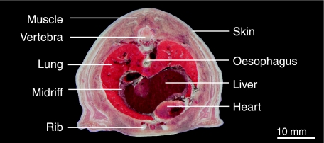
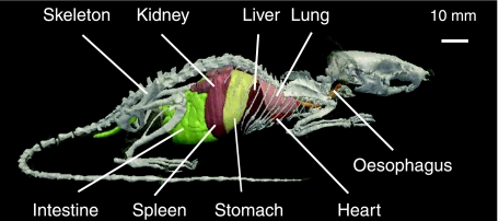
Serial cryosection-milling and digital imaging techniques were employed to create the SD rat anatomical atlas. Healthy adult male SD rats were used for the study. After being anaesthetized, depilated and killed via suffocation, the rats were frozen completely in a −85 °C ultra freezer for a minimum of 24 h, ensuring that the specimens remained in the correct anatomical posture with both sides symmetrical. The rats were then embedded with a 3% gelatin solution in an embedding box and frozen to −85 °C for 48 h. Serial cryosection images of the rats were acquired using the cryosection-milling imaging system. Thereafter, 9475 horizontal section images (4600 × 2580 × 24-bit TIFF format) of rat anatomy (at 20 µm thickness) were obtained (Fig. 1). Coronal and sagittal section images were computerized from a horizontal section. Anatomical structures and arterial vessels were segmented using an auto-segmentation program, but most of the inner organs were segmented manually by tracing digital images from these serial cryosection images using Photoshop software (Adobe, USA) under the guidance of an experienced anatomist. Interactive two-dimensional (2D) atlas-viewing software was developed to visualize three orthoslices, featuring zoom, anatomical labelling and organ measurement. A 3D computerized model of rat anatomy was generated using a parallel reconstruction algorithm (Fig. 2) (Liu et al. 2005). An interactive Internet 3D organs browser based on virtual reality modelling language technique was deployed on our website. The cryosection images, 3D models, technical details, relevant software and further information are available on our website at http://vchibp.vicp.net/vch/mice/. An ftp server is also provided for full access of the data with permission.
Fig. 1.
Cryosection images of rat thorax.
Fig. 2.
Three-dimensional visualization of the rat.
Acknowledgments
This work was supported by grants from the National High-Tech Research and Development Program of China (2003AA231011) and Program of Science and Technology Research of the Ministry of Education (505010).
References
- Bard JL. Anatomics: the intersection of anatomy and bioinformatics. J Anat. 2005;206:1–16. doi: 10.1111/j.0021-8782.2005.00376.x. [DOI] [PMC free article] [PubMed] [Google Scholar]
- Bard JL, Kaufman MH, Dubreuil C, et al. An internet-accessible database of mouse developmental anatomy based on a systematic nomenclature. Mech Dev. 1998;74:111–120. doi: 10.1016/s0925-4773(98)00069-0. [DOI] [PubMed] [Google Scholar]
- Liu Q, Gong H, Luo QM. Parallel visualization of visible chinese human with extremely large datasets. In: Zhang YT, editor. Proceedings of the 2005 IEEE, Engineering in Medicine and Biology 27th Annual Conference; 1–4 September 2005; Shanghai. pp. 5172–5175. IEEE. [DOI] [PubMed] [Google Scholar]
- Montgomery K, Bruyns C, Wildermuth S. A virtual environment for simulated rat dissection: a case study of visualization for astronaut training. IEEE Computer Soc. 2001;C6:509–514. [PubMed] [Google Scholar]
- Toga AW, Santori EM, Hazani R, Ambach K. A 3D digital map of rat brain. Brain Res Bull. 1995;38:77–85. doi: 10.1016/0361-9230(95)00074-o. [DOI] [PubMed] [Google Scholar]


