Abstract
Recently published reports on Korea’s medieval mummies have been regarded as an invaluable source for studies into the physical characteristics of medieval Koreans. However, even though the mummified tissues have been investigated histologically on various previous occasions, there are many unanswered questions relating to their tissue preservation. The aim of this study was to obtain new data on the ultramicroscopic characteristics of the mummified skin of a fifteenth-century mummy found recently in Daejeon – one of the oldest ever found in Korea. Electron microscopy revealed that much of the epidermis had decayed; what remained of the dermis was filled with collagen fibres and melanin granules or invading bacterial spores present within the mummified epidermis. Considering the histological characteristics shared by naturally formed mummies in different parts of the world, we concluded that the ultramicroscopic patterns of the Daejeon mummy were more comparable with those naturally formed mummies than with artificially formed ones. This is the first full description of the morphological characteristics of the skin collected from this recently found medieval mummy from Daejeon, South Korea.
Keywords: Daejeon, electron microscope, medieval, mummy
Introduction
Well-preserved medieval mummies have been found in several tombs from the Chosun Dynasty (1392–1910) (Shin et al. 2003a). Mummies have been found in the cities or counties of Gwangju (1968), Cheongwon (1977), Cheongyang (1982), Ulsan (1986), Paju (1995), Andong (1998), Okcheon (2000), Yangju (2001), Paju (2002), Buan (2004) and Daejeon (2004). In the earlier period of the Chosun Dynasty, tombs were constructed with large stone blocks; however, the practice of sealing tombs with a lime–soil mixture was subsequently adopted among the royal families of the dynasty because the neo-confucianist ruling elites were afraid that the excessively hard work involved in the more traditional construction methods would cause unrest among the people. The novel burial system in tombs with a lime–soil mixture barrier was thereafter adopted increasingly by the ruling elite, and has even influenced the funeral customs of modern Korea (Chung, 1994).
The medieval mummies in Korea, the subject of the present study, were found exclusively in tombs with a lime–soil mixture barrier. As the burial system was adopted primarily by the ruling elite, one by-product has been that abundant grave goods have been found along with these medieval mummies, providing another invaluable source of information about medieval Korean society.
Our previous report on the Yangju mummy (Shin et al. 2003b) described the use of light and electron microscopy to determine that the mummified skin was partially preserved. The corneal layer and the anucleated cells within it were thought to be preserved in the epidermis, while the dermis was filled with collagen fibres running in various different directions. However, although we were able to present the histological characteristics of Korean mummified skin for the first time, the description was not detailed enough for us to be able to compare medieval Korean mummies with those reported from other countries.
When a fifteenth-century mummy was recently found in Daejeon we therefore thought it might reveal the preservation qualities of mummified skin in Korean mummies; although this was one of the oldest mummies ever found in Korea, the morphology of the mummified skin showed a much better preservation pattern than those of any other cases in Korea. The aim of this study was to present ultramicroscopic findings of the skin of the Daejeon mummy, in order to provide additional data on the histological characteristics of medieval Korean mummies. As our electron microscopic data yielded better images than those we had obtained previously, it seemed likely that we would be able to assess whether there were histological similarities between the mummified skin of medieval Korean mummies and that of other mummies previously reported from other countries.
Materials and methods
Skin sampling and electron microscopy
Our study was performed in accordance with the Vermillion Accord on Human Remains, World Archaeological Congress, South Dakota, 1989. The skin samples used in this study (1 × 1 cm2) were collected from the skin covering the back muscle of the mummy. Sections were stained using the haematoxylin–eosin, Masson’s trichrome, Fontana-Masson, orcein and silver-staining methods for light microscopic observations (Sheehan & Hrapchak, 1980). Counter-staining with haematoxylin–eosin was also performed on silver-stained sections. A control section of normal adult skin was selected from the educational slides for medical students at Seoul National University College of Medicine, Korea. Transmission electron microscopy (TEM) was performed in accordance with previously described methods (Hayat, 1970; Bozzola & Russell, 1992). Hair samples were immersed in 2.5% paraformaldehyde/glutaraldehyde in neutral 0.1 m phosphate buffer for 1 h. Tissues were post-fixed for 1 h in 1% (w/v) osmic acid dissolved in phosphate-buffered saline (PBS), dehydrated in a graded series of ethanol and embedded in Epon812 (EMS, Fort Washington, PA, USA). Ultrathin sections were cut and mounted on nickel grids coated with Formvar film and viewed under a JEOL 100 CX-II TEM (Tokyo, Japan) after uranyl–lead counterstaining. To clean the surface of the skin, additional sonication treatment was performed on samples with an ultrasonic cleaner (Branson, 2510R-DTH, USA) for 10 min.
Scanning electron microscopy (SEM) was also performed in accordance with previously reported methods (Hayat, 1970; Bozzola & Russell, 1992). Hair samples were prefixed by immersion in 4% paraformaldehyde/0.1% glutaraldehyde in neutral 0.1 m phosphate buffer, and post-fixed for 2 h in 1% (w/v) osmic acid dissolved in PBS. Samples were treated in a graded series of ethanol and isoamyl acetate, dried in a critical-point dryer (Hitachi SCP-II), gold coated using an ion coater (JFC-1100), and observed under a JSM-840A SEM.
Electron dispersive X-ray spectroscopy (EDS)
EDS was used to analyse materials making up the surface coat of the skin. After collecting surface-coating samples from the mummified skin, the surfaces of randomized particles and of pressed discs of the samples were scanned, and the elemental compositions were analysed using a scanning electron microscope equipped with an energy-dispersive X-ray spectrometer.
Radiocarbon dating
We performed radiocarbon dating to determine the exact age of the mummy (although we also tried to identify the mummy based on genealogical records). The samples acquired for carbon dating were 5.22 mg of mummy intestine. Accelerator mass spectroscopy (AMS) was performed to measure the amount of 14C in the sample. Graphite samples prepared from the reduction procedure were put to the Cs-sputtering ion source of the tandem-type electrostatic accelerator (Model 4130 Tandetron, High Voltage Engineering Europa) at the Inter-Universities Research Facility of Seoul National University, Korea.
Results
Profile
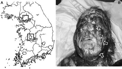
This male mummy was found in Daejeon, Korea (Fig. 1A) on 20 May 2004 during the reburial process for ancient tombs, which was performed by the descendants. The tomb in which the mummy was found consisted of double-layered wooden ‘inner coffins’ and the ‘outer coffin’ comprising a lime–soil mixture. As the reburial process is usually performed to transfer an ancestor’s tomb to a new graveyard for reasons such as urban development, the body was easily identified by a search of the family’s genealogical records. According to these records, the mummy was that of an ancestor, who had lived in the fifteenth century and served as a high-ranking general at the royal court of King Sejong (1418–1450), Chosun Dynasty. The mummy was found together with various items of clothing from the period. As the AMS radiocarbon age, estimated from mummy intestine, was found to be 540 ± 40 years bp, the genealogical records were deemed to be reliable.
Fig. 1.
(A) The mummy we investigated previously (Shin et al. 2003a) was found in Yangju, Korea (rectangle marked ‘a’). The current case was found in Daejeon, Korea (rectangle marked ‘b’). (B) Face of the mummy. Note that white coating materials (arrows) were observed on the skin.
General histology of the skin
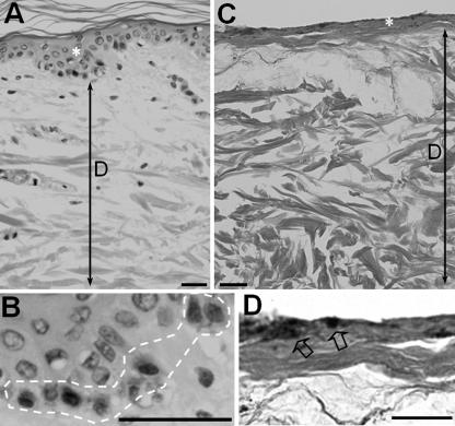
We examined the general structure of the mummified skin using light microscopy. The relative ratio of each skin layer was markedly different from that of normal skin tissue (Fig. 2A,B). Among the mummified skin layers observed, the epidermis showed a marked decrease in thickness; this decrease was not as marked in the dermis. In addition, within the epidermis of the mummified skin, we found black granules (Fig. 2C,D) which appeared to be of melanin. Because, in normal adult skin, melanin granules are localized in the basal cell layer of the epidermis (Fig. 2A), we suggest that the layer in which the black granules were localized – if these granules are in fact melanin pigment – may be the remaining mummified layer of the epidermis.
Fig. 2.
Comparison of the skin structure collected from an adult individual (A,B) and the fifteenth-century mummy (C,D). Both sections were stained with haematoxylin–eosin. Comparing the relative ratio of epidermis (asterisks) and dermis (D) with that of normal skin tissue (A,B), mummified skin exhibited an epidermis with remarkably decreased thickness, whereas the thickness of the dermis had not changed as much (C,D). Melanin granules were observed in the basal layer of epidermis (the region within the white dotted line in B) in modern adult skin, while black granules (arrows in D), which also seem to be melanin granules, were localized within the presumptive remaining epidermis of the mummified skin. Bars, 50 µm.
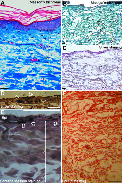
We also suggest that the remaining structures in this layer may be collagen fibres, based on our previous study of the Yangju mummy, which showed the exclusive presence of collagen fibres within the dermis. This speculation is supported by additional histochemical staining specific for collagen or elastic fibres. As seen in Fig. 3(A–C), the dermis of the mummified skin was intensely stained green by Masson’s trichrome technique for collagen fibres, while the same mummified skin tissue was negative for silver staining of reticular fibres, except for the counterstaining performed on the slide. In the magnified silver staining image, melanin pigments in the epidermis could be observed (Fig. 3D). The melanin pigments in the epidermis were confirmed by Fontana-Masson staining (Fig. 3E), which demonstrates melanin in skin (Drury & Wallington, 1980). Orcein staining, which stains elastic fibres dark brown (Sheehan & Hrapchak, 1980), was negative (Fig. 3F).
Fig. 3.
Comparison of skin collected from a modern adult individual (A) and the fifteenth-century mummy (B–D). (A,B) Sections stained by Masson’s trichrome techniques. (A) The dermis (D) was stained green owing to the presence of collagen fibres. (B) The dermis of the mummified skin was also intensely stained green by Masson’s trichrome method; red staining due to the presence of cytoplasm, which could be observed in epidermis (Epi) in A, could not be localized in B. (C) Silver staining for reticular fibres was negative in the mummified skin tissue. (D) Magnified image of silver staining. Melanin pigments (indicated by arrows) are present in the epidermis (Epi). (E) Fontana-Masson staining for melanin pigments (indicated by white open arrows) in the epidermis (Epi) overlying the much thicker dermis (D). (F) Orcein staining for elastic fibres. Dark brown-staining structures for elastic fibres were not detected. Bars, 50 µm.
The surface coating on the epidermis
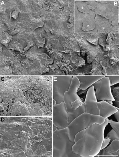
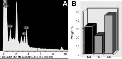
During SEM observation of the surface of the skin, we observed white substances on the surface of the epidermis (Fig. 4A). After the tissue was washed by the sonication method, the underlying keratinized epithelium was exposed, clearly revealing the keratinized scales (Fig. 4B). Figure 4(C–E) are images of the surface coating on the epidermis at different magnification. In Fig. 4(E), we observed that the surface coating material was composed of crystaline debris. Using EDS, we detected the presence of sodium (Na), potassium (K) and calcium (Ca) (Fig. 5A,B).
Fig. 4.
SEM observation on the surface of the skin. (A) A white coating was present on the surface of the epidermis. (B) After the tissue was washed using sonication, the underlying keratinized epithelium was exposed, in which we could clearly observe keratinized scales (asterisks). (C–E) Images of the surface coat on the epidermis, magnified sequentially. The surface coating material was observed to be composed of crystaline debris. Bars: A–C,) 100 µm; D, 50 µm; E, 5 µm.
Fig. 5.
(A) Use of EDS to analyse the coating substance found on the surface of the epidermis. (B) Relative percentage weight of the materials in the debris. Sodium (Na), potassium (K) and calcium (Ca) were detected.
Ultramicroscopic findings of the epidermis and dermis
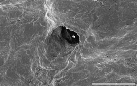
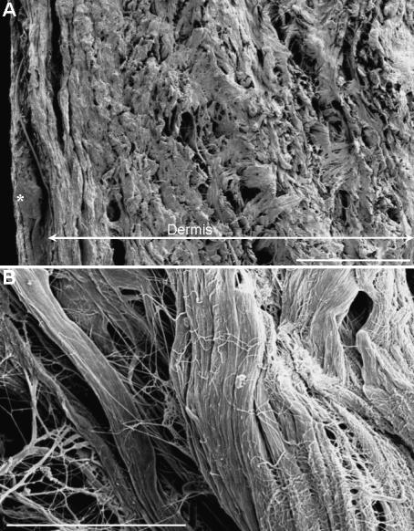
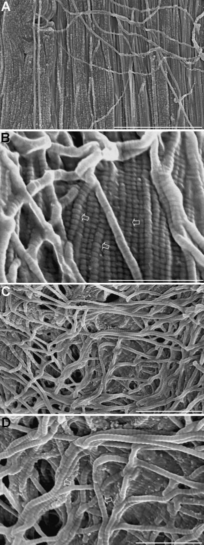
On the surface of the skin, which was washed using a sonicator treatment, we observed the hair shaft holes using SEM (Fig. 6). In the other SEM images of the lateral surface of skin, we observed thread-like structures, which are most likely to be collagen fibres in the dermis (Fig. 7A,B). In higher-magnification images of presumptive dense connective tissue, collagen fibres were aligned in parallel (Fig. 8A,B). In the magnified image (Fig. 8B), the mummified collagen fibres showed a ‘banding pattern’ that is known to be formed by the overlapping of collagen molecules. As the ultramicroscopic images of the mummified collagen fibres were found to be similar to those observed in living mouse dermis (left inset of Fig. 8B), the mummified skin in this case could be said to be well preserved. A similar pattern was also observed in presumptive loose connective tissue. The collagen fibres in this region (Fig. 8C,D), which similarly were not arranged regularly as in Fig. 8(A,B), also exhibited a marked ‘banding pattern’.
Fig. 6.
SEM data showing holes for hair shafts (asterisk) on the surface of the skin, which was washed with sonicator treatment. Bar, 200 µm.
Fig. 7.
(A) SEM data on the lateral cut surface of the skin sample. Thread-like structures, which seem to be collagen fibres, filled the dermis. Asterisk indicates the epidermis. (B) Magnified image of presumptive collagen fibres in the dermis. Bars: A, 500 µm; B, 30 µm.
Fig. 8.
(A,B) SEM data on collagen fibres within the region that might be filled with dense connective tissue. The collagen fibres were aligned in parallel. (B) Magnified image of A. In B, the mummified collagen fibres showed a ‘banding pattern’ (arrows), which resembles those observed in the collagen fibres within dermis from living mouse. (C,D) SEM data on collagen fibres within the mummified dermis in which loose connective tissue might be present. The collagen fibres were loosely arranged in various directions. (D) Magnified image of C. Banding patterns (arrows in D) in the collagen fibres were also identified. Bars: A, 4 µm; B, 1 µm; C, 2 µm; D, 1 µm.
Melanin granules in epidermis
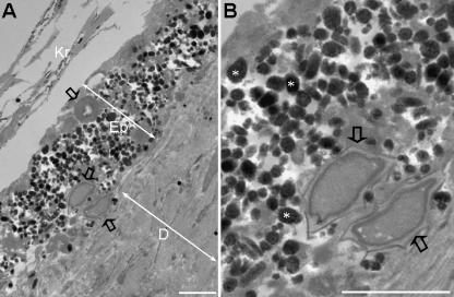
As we speculated that the black pigments in the mummified epidermis (Fig. 2C) might be melanin granules, we performed a TEM study focusing mainly on the epidermis. Figure 9(A,B) shows a number of black granules, which were aggregated within the mummified epidermis, and whose ultramicroscopic morphology led us to believe that they were of melanin.
Fig. 9.
(A,B) TEM data on the presence of melanin pigment in the epidermis of mummified skin. (B) Magnified image of epidermis in A. In A, a number of melanin granules (asterisks in B) were aggregated within the remaining epidermis (Epi) in which epithelial cells were not clearly discernible, even though the keratinized layers (Kr) were visible on the surface of the skin epithelium. D, dermis. Bar, 2 µm.
Discussion
As discussed in our previous study, the medieval mummies recently found in Korea were not intended to be preserved; in fact their descendants hoped that the bodies would disintegrate completely after interment. In addition, as the climate of Korea was not conducive to natural mummification (which is usually observed in dry or permafrost environments), the exact reasons why mummification had been able to take place Korea appeared to be a mystery (Shin et al. 2003b). During the Chosun Dynasty (1392–1920), the bodies of the dead were laid within double-layered wooden ‘inner coffins’ that were sealed by an external coffin made of a lime–soil mixture. As mummies were only found in cases where the lime–soil mixture barrier had not been broken until their discovery – making it possible that the inner space of the coffins was separated completely from the surrounding area – it is possible that the completely sealed inner environment provided conditions conducive for mummification. As yet, however, we have not been able to confirm whether or not the conditions inside the Korean coffins are similar to those where natural mummification has taken place (i.e. in dry or permafrost conditions), because in the Korean cases the mummification process seems to have resulted from the distinctive burial practices of medieval Korea, which are not directly comparable with situations in which mummification has occurred naturally.
Previous histological studies on mummified skin have identified two different types of preserved structure, which seem to reflect differences in the mummification process (natural and artificial). In the case of skin samples collected from 2300- to 1600-year-old bodies preserved in the bogs of northern Germany, collagen bundles were observed profusely in the dermis, whereas the epidermis had almost disappeared. Under electron microscopy, the naturally mummified dermis of the bog bodies, in which no cellular elements were observed, also exhibited collagen fibrils with a typical periodicity (Stucker et al. 2001). A similar histological pattern was also observed in naturally formed 2500-year-old Altai mummies, in which collagen fibres were found to be a predominant protein (Romakov et al. 2002). Those reports might be taken to indicate that a dermis rich in collagen bundles but with a scarcity of cellular elements is characteristic of naturally mummified bodies.
By contrast, much well-preserved structure has been observed in the skin of artificially embalmed Egyptian mummies. In the case of Egyptian mummies buried between 150 BCE and 90 CE (Perrin et al. 1994), even the epidermis – the skin layer which had completely disappeared in most of the naturally mummified cases – was reported to be sufficiently well preserved to show the subcellular structures of desmosomes or tonofilament bundles. The superior skin preservation of the Egyptian mummies was also evident in the dermis, where elastic fibres – which were not found in naturally mummified bodies – were found together with collagen fibres (Montes et al. 1985; Perrin et al. 1994). Another example of the well-preserved embalmed skin of Egyptian mummies was reported by Hino et al. (1982a,b). In an ultramicroscopic study of the skin of a female Egyptian mummy (embalmed between about 100 and 300 CE), subcellular structures such as mitochondria and desmosomes were observed in epidermal cells, while in the dermis, elastic fibres as well as collagen fibres were localized. Although the epidermis may not be in such a good state of preservation in all Egyptian mummies (Perrin et al. 1994), it is possible to make some generalizations about the skin histology of artificially embalmed mummies in Egypt: these mummies show preservation of the epidermis, the additional presence of elastic fibres in the dermis and various subcellular structures observed in the epidermal cells, all of which are different from those in naturally formed mummies. Although it remains unclear whether the better preservation of mummified skin in Egypt is correlated with embalming of the skin, it is undeniable that artificially formed mummies exhibited better preservation of skin histology than naturally formed mummies. In this regard, because medieval Korean mummification may have been induced by the combined effects of both natural and artificial mummification processes (Shin et al. 2003a), the current study on mummified skin in Korea may be useful for confirming which preservation pattern (natural or artificial) was responsible for the cases of mummification in Korea.
For information regarding the characteristics of the mummified skin discussed here, we refer the reader to our earlier findings on the Yangju mummy (Shin et al. 2003a,b), in which we noted the relative thickness change in each skin layer, the good state of preservation of the collagen fibres in the dermis, and the very thin epidermis, with its paucity of cellular elements. However, as our previous reports on the histological characteristics of the mummified skin from Yangju could not provide sufficient data, the present ultramicroscopic study on the skin of a recently discovered Daejeon mummy was undertaken in the hope of providing good additional data on the state of preservation of the skin of Korean mummies.
In this regard, as we observed a change in the relative ratio of the thickness of each mummified skin layer, including a marked decrease in the thickness of the epidermis (unlike the dermis, which maintained its thickness), the current ultramicroscopic data may support earlier findings concerning the skin of Korean mummies. In addition, because the existing structures in the dermis in the Daejeon mummies were mainly composed of well-preserved collagen fibres, as confirmed by silver or Orcein staining techniques (to eliminate the possibility of the presence of elastic or reticular fibres within the dermis), it is easy to demonstrate similarities in the histological findings between the Yangju and the Daejeon cases. However, electron microscopy indicated that the collagen fibres in the recently found Daejeon mummy were much better preserved than those in the Yangju case, even though the former is thought to have been buried earlier. In addition, the identification of melanin pigments in the mummified epidermis – not easily observed in the previous case, but here confirmed by Fontana-Masson staining – may be regarded as one of the unique findings of the present study.
Although some of our findings were unique to the present case, overall we believe that our histological findings show more similarities with naturally formed mummies than with artificially embalmed ones: namely, the loss of epidermis, the fact that the dermis retained only collagen fibres, and the lack of any specific subcellular structure except for melanin granules within the mummified epidermis. Therefore, although the Korean mummies were formed through a very exceptional mummification process, which cannot strictly be classified as either ‘artificial’ or ‘natural’ based on comparison with the techniques used in other countries, the general pattern of preservation of the mummified skin in the medieval Korean mummy indicates that the mummification process was more natural than artificial.
Finally, we should also comment on the white coating on the surface of the mummified skin, which was clearly visible to the naked eye. Using EDS to analyse the composition of surface materials on the mummified skin, calcium was found to be the most prominent material. As calcium is not commonly found in the soil around the tomb, we suggest that it may have come from the lime–soil barrier mixture, which included large quantities of calcium. Before the lime–soil mixture began the hardening process around the coffin, calcium may have infiltrated the inner space of the coffin, and then been included on the surface coat on the skin. It seems possible that the calcium-rich coating may have sealed the skin of the mummy from the air trapped inside the coffin, helping to preserve the mummified skin in the same way as the lime–soil barriers preserved the inner coffins.
Conclusion
In studies from other countries, light and electron microscopy have demonstrated that naturally mummified skin exhibits a typical histological pattern, with loss of the epidermis, and the dermis being filled with collagen fibres but with no cellular components. In the present study we observed that the preservation pattern of the mummified skin corresponded well to those of other cases of natural mummification from other countries, even though the medieval Korean mummies were formed by a unique process that could not easily be classified within the mummification processes previously reported. This is the first full description of the morphological characteristics of mummified skin collected from the recently found fifteenth-century mummy in Daejeon, Korea.
Acknowledgments
The investigations on the recently found mummy from Daejeon were superintended by the Research Team for Medieval Mummies in Daejeon, Korea University, and we are particularly grateful to them for their generous decision to supply the samples to our lab. The mummy is currently displayed in the Gyeryongsan Natural History Museum, Korea. We thank Mr Edward Fenton for his help with the English language version of the manuscript. This study was supported by Korea Research Foundation Grant (KRF-2003-I01485-E00006).
References
- Bozzola JJ, Russell LD. Electron MicroscopyPrinciples and Techniques for Biologists. Boston: Jones and Bartlett; 1992. pp. 14–62. [Google Scholar]
- Chung J. A Study on Funeral Customs in the Early Chosun Dynasty. Seoul: Chung-Ang University (in Korean); 1994. [Google Scholar]
- Drury RAB, Wallington EA. Carleton’s Histological Technique. Oxford: Oxford University Press; 1980. [Google Scholar]
- Hayat MA. Principles and Techniques of Electron Microscopy, Biological Applications. Vol. 1. New York: van Nostrand Reinhold; 1970. pp. 109–179. [Google Scholar]
- Hino H, Ammitzboll T, Moller R, Asboe-Hansen G. The ultrastructure of bacterial spores in skin of an Egyptian mummy. Acta Pathol Microbiol Immunol Scand [B] 1982a;90:21–24. doi: 10.1111/j.1699-0463.1982.tb00077.x. [DOI] [PubMed] [Google Scholar]
- Hino H, Ammitzboll T, Moller R, Asboe-Hansen G. Ultrastructure of skin and hair of an Egyptian mummy. Transmission and scanning electron microscopic observations. J Cutan Pathol. 1982b;9:25–32. doi: 10.1111/j.1600-0560.1982.tb01038.x. [DOI] [PubMed] [Google Scholar]
- Montes GS, Krisztan RM, Junqueira LC. Preservation of elastic system fibers and of collagen molecular arrangement and stainability in an Egyptian mummy. Histochemistry. 1985;83:117–119. doi: 10.1007/BF00495140. [DOI] [PubMed] [Google Scholar]
- Perrin C, Noly V, Mourer R, Schmitt D. Preservation of cutaneous structures of egyptian mummies. An ultrastructural study. Ann Dermatol Venereol. 1994;121:470–475. (in French) [PubMed] [Google Scholar]
- Romakov IuA, Bykov VA, Rebrova GA, Vasilevskii VK, Ho SS, Rebrov LB. Evaluation of preservation and the collagen structure state in the mummy tissues. Vopr Med Khim. 2002;48:357–360. (in Russian) [PubMed] [Google Scholar]
- Sheehan DC, Hrapchak BB. Theory and Practice of Histotechnology. St Louis: C.V. Mosby; 1980. [Google Scholar]
- Shin DH, Choi YH, Shin KJ, et al. Radiological analysis on a mummy from a medieval tomb in Korea. Ann Anat. 2003a;185:377–382. doi: 10.1016/S0940-9602(03)80065-1. [DOI] [PubMed] [Google Scholar]
- Shin DH, Youn M, Chang BS. Histological analysis on the medieval mummy in Korea. Forensic Sci Int. 2003b;137:172–182. doi: 10.1016/s0379-0738(03)00335-9. [DOI] [PubMed] [Google Scholar]
- Stucker M, Bechara FG, Bacharach-Buhles M, Pieper P, Altmeyer P. What remains of the skin after 2000 years in a bog? Hautarzt. 2001;52:316–321. doi: 10.1007/s001050051314. (in German) [DOI] [PubMed] [Google Scholar]


