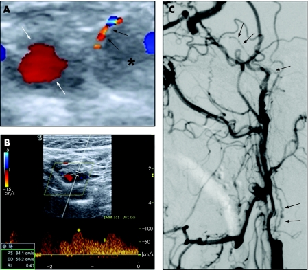
A 52‐year‐old man with obesity, type 2 diabetes mellitus and hypertension was admitted because of sudden onset of left hemiparesis and somnolence. Brain MRI disclosed a recent right thalamic stroke and multiple encephaloclastic lesions, suggestive of vascular sequelae. Carotid ultrasound demonstrated occlusion of the right internal carotid artery (ICA) and residual flow in the periphery of the left ICA lumen (fig 1A, B). Subocclusive stenosis was suspected. Carotid arteriography confirmed atherosclerotic occlusion of both bulbar ICAs. On the left side, however, filiform collateralisation with a spiral configuration was seen, extending from the ICA bulb to the cavernous segment (fig 1C). The patient was offered medical treatment.
Figure 1 (A) Colour coded duplex ultrasonography, transverse section, 1 cm above the carotid bifurcation, shows the occluded internal carotid artery (ICA) lumen (*) and a thin segment of flow within its wall (black arrows), outlining the perimeter of the artery. External carotid artery is normal (white arrows). (B) Spectral tracing from the same segment as in (A), reveals a normal waveform and velocity. (C) Contrast arteriogram of the left ICA, arterial phase, lateral view, demonstrates a rounded proximal stump. Spiral vasa vasorum (black arrows) originate from the bulb and contribute to the filling of the cavernous segment. The true lumen of the ICA is never filled.
Collateralisation through the vasa vasorum of an atheromatous occlusion of the ICA is a rare finding. Growth of collaterals in the wall of the vessel is a slow process, stimulated by the proangiogenic properties of the plaque.1
Differentiating carotid occlusion with collateralisation from pre‐occlusive stenosis is of the utmost importance because there is no benefit of endarterectomy or angioplasty in the first situation.
Carotid ultrasound is a reliable method in the assessment of ICA stenosis and occlusion. However, the findings of residual flow with normal velocities and waveform may erroneously be attributed to subocclusive carotid disease2 in which it has been shown that velocity measurements start to decrease. Therefore, corroboration with arteriography is recommended. High resolution transverse colour coded sections showing segments of flow within the ICA wall itself, outlining the circumference of the vessel may be a clue to the correct diagnosis.
Footnotes
Competing interests: None.
References
- 1.Bo W J, Mercuri M, Tucker R.et al The human carotid atherosclerotic plaque stimulates angiogenesis on the chick chorioallantoic membrane. Atherosclerosis 19929471–78. [DOI] [PubMed] [Google Scholar]
- 2.Kriegshauser J S, Patel M D, Nelson K D.et al Carotid pseudostring sign from vasa vasorum collaterals. J Ultrasound Med 200322959–963. [DOI] [PubMed] [Google Scholar]


